
High Intensity Focused Ultrasound Therapeutic System Guided by an Imaging Device

a technology of ultrasound and imaging device, applied in the field of medical devices, can solve the problems of inability to evaluate the therapeutic effect, difficult to realize the complete treatment of the entire tumor, loss of information at the ends, etc., and achieve the effects of reducing the production cost of the system, and relatively high energy focused on the diseased tissu

Inactive Publication Date: 2010-07-08
RONGHAI SUPERSONIC MEDICINE EN
View PDF25 Cites 42 Cited by
  • Summary
  • Abstract
  • Description
  • Claims
  • Application Information

AI Technical Summary

Benefits of technology

[0008]Aiming at the above-mentioned disadvantages in the prior art, the technical solutions of the present invention are to provide a high intensity focused ultrasound therapeutic system guided by an imaging device, which can image the target accurately and can perform an accurate treatment for a relatively complicated operation.

Problems solved by technology

However, the therapeutic effects are not ideal when an ultrasound therapeutic apparatus applies such multiple focuses for treatment.
This patent points out that the tissue such as tumor to be treated usually has a complicated three-dimensional shape, so it is very difficult to realize a complete treatment of the entire tumor and to evaluate the therapeutic effects simply by using the two-dimension ultrasonic tomographic images, and it presents a technical solution of utilizing the three-dimensional MRI tomographic images to image the object before and after treatment and utilizing two-dimensional ultrasound to monitor the treatment process in real time.
However, because the shape of tumor is irregular and usually is dendritic, some information at the ends will be lost when the three-dimensional images of the object to be treated are collected.
Therefore, the three-dimensional images reconstructed in this way are not real and can not show the overall original form of the tumor.
The treatment can not be carried out completely based on these distorted images.
For malignant tumors, the remaining diseased tissue will continue to expand and accordingly the hazard to the life of the patient will occur again.
So, the technical solutions of the patent mentioned above can not complete an accurate ultrasound therapy.
Also, if the MRI apparatus in that patent is not reconstructed, the patient has to be moved in and out of the MRI bore repeatedly and thereby the treatment time is prolonged.
If the patient is not moved, the structure of MRI has to be modified and thereby the treatment cost will be increased.

Method used


Image

  • High Intensity Focused Ultrasound Therapeutic System Guided by an Imaging Device
  • High Intensity Focused Ultrasound Therapeutic System Guided by an Imaging Device
  • High Intensity Focused Ultrasound Therapeutic System Guided by an Imaging Device

Examples


embodiment 1

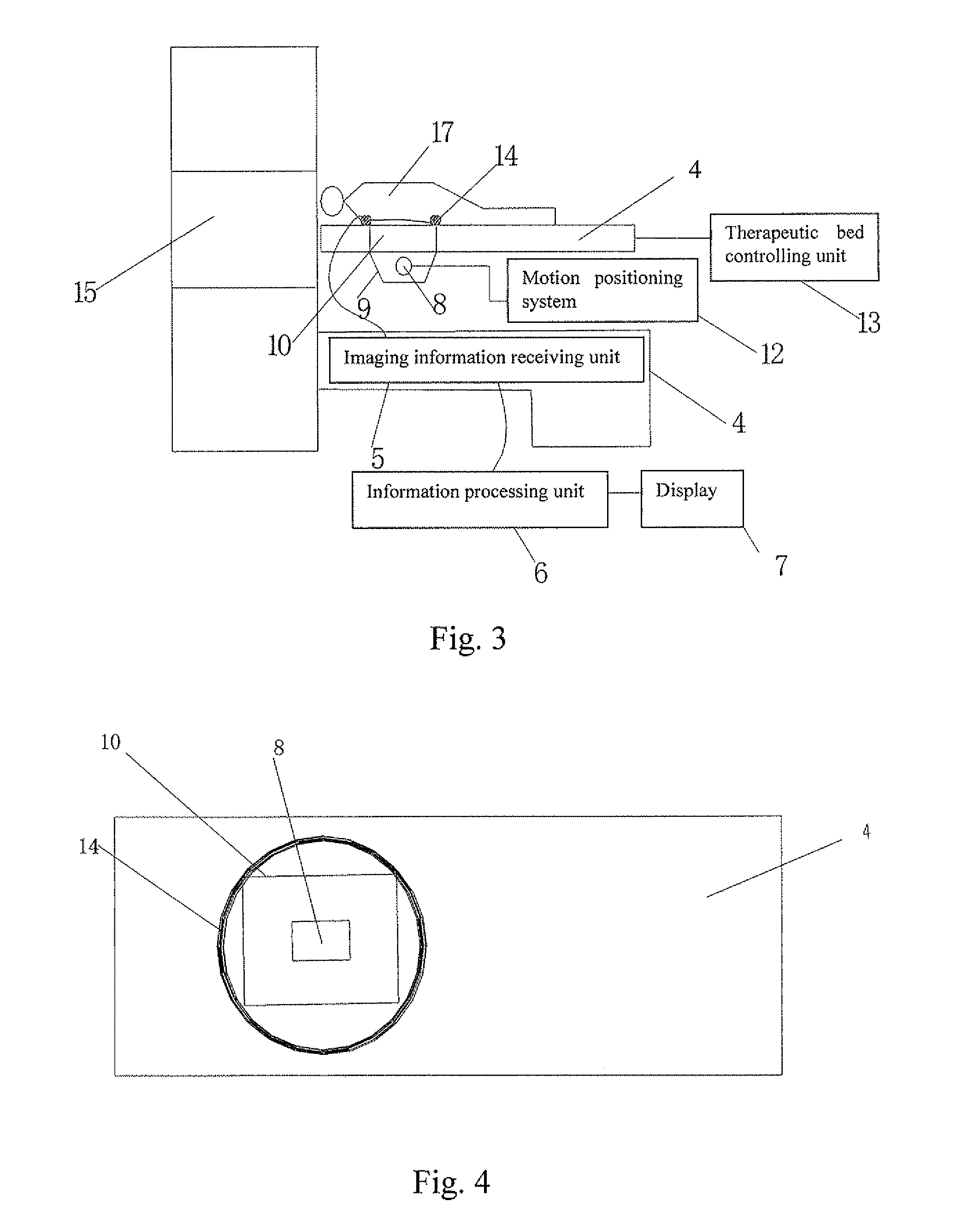
[0029]As shown in FIG. 3, the high intensity focused ultrasound therapeutic system guided by an imaging device in the present invention consists of a non-ultrasound imaging device and a high intensity focused ultrasound therapeutic device. In this embodiment, an MRI device is applied as non-ultrasound imaging device.

[0030]In the MRI device shown in FIG. 3, MRI imaging information receiving unit 5 is equipped into the MRI table 4. The imaging information receiving unit 5 is connected to information processing unit 6 and display 7 respectively.

[0031]The high intensity focused ultrasound therapeutic device comprises an ultrasound therapeutic applicator, a motion positioning system 12 of ultrasound therapeutic applicator, a therapeutic bed 11 for holding a patient and with an opening 10, and a therapeutic bed controlling unit 13 for controlling therapeutic bed 11 to move in and out of MRI bore 15.

[0032]Wherein, the ultrasound therapeutic applicator comprises an ultrasound transducer 8 a...

embodiment 2

[0041]As shown in FIG. 5, the difference between this embodiment and the embodiment 1 is that the second surface coil 16 is added at the other side of the patient and opposite to the first surface coil. The second surface coil 16 and the first surface coil 14 are integrated and then connected to the imaging information receiving unit 5 of the MRI device. The purpose of adding the second surface coil 16 is to enhance the intensity to receive MRI imaging signals. Compared with the embodiment 1, this embodiment has a higher imaging quality and it is more helpful for an operator to monitor two-dimensional images of the target scanned by the focal core 23 during treatment.

[0042]The rest of structures and procedures of application are as the same in the embodiment 1, there is no need of repeated description.


PUM


Abstract

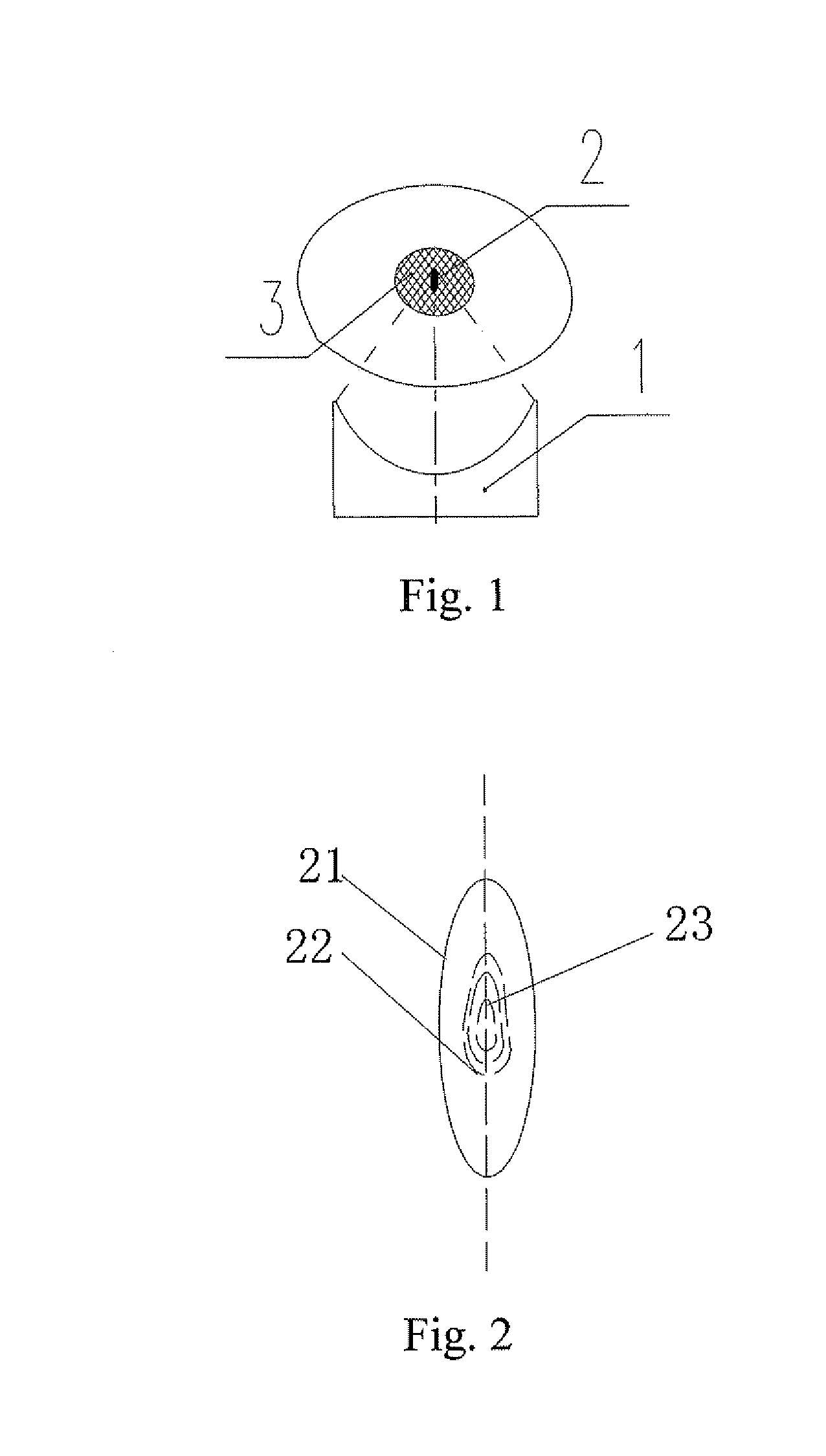
A high intensity focused ultrasound therapeutic system guided by an imaging device is provided and includes an ultrasound therapeutic applicator which contains an ultrasound transducer (8), a therapeutic bed (11), on which a patient (17) lies, and an imaging device used for imaging a diseased tissue (3). Wherein, the imaging device is a non-ultrasound imaging device with two-dimensional imaging function. The ultrasound transducer (8) with the diameter of focal core (23) ranging from 0.1 mm to 5 mm, preferably, ranging from 0.2 mm to 2 mm, in a biological focal region (2) located in the diseased tissue (3) is adopted. The non-ultrasound imaging device is a CT or an MRI device with two-dimensional imaging function. The present invention can image the target accurately, so it can be applied to all kinds of complicated surgeries and has good therapeutic effects.

Description

FIELD OF THE INVENTION[0001]The present invention pertains to the technical field of medical devices and relates to an ultrasound therapeutic system, particularly to a high intensity focused ultrasound therapeutic system guided by an imaging device.BACKGROUND OF THE INVENTION[0002]In the field of ultrasound therapy, due to technology and awareness, people do not know much about the action mechanism of the focus in vivo and always expect to use multiple focuses to achieve a fast treatment when an ultrasound thermotherapy is applied for treatment. However, the therapeutic effects are not ideal when an ultrasound therapeutic apparatus applies such multiple focuses for treatment. Meanwhile, with the development of ultrasound technology, especially when the high intensity focused ultrasound is being used widely in therapy, a fine surgical treatment apparatus is in higher demand of people because only such apparatus can be used to treat or ablate some diseases (particularly the malignant ...

Claims


Application Information

Patent Timeline
IPC IPC(8): A61N7/00A61B8/14
CPCA61B5/055A61N7/02A61B2019/5238A61B2019/5236A61B2090/376A61B2090/374A61B5/704
Inventor WANG, HAIWANG, LONGZHANG, LIANGU, YUE
Owner RONGHAI SUPERSONIC MEDICINE EN
Learn More