Method for in situ solidification of blood-polymer compositions for regenerative medicine and cartilage repair applications

Inactive Publication Date: 2010-07-15
PIRAMAL HEALTHCARE CANADA
View PDF9 Cites 21 Cited by
  • Summary
  • Abstract
  • Description
  • Claims
  • Application Information

AI Technical Summary

Benefits of technology

[0038]In accordance with another aspect of the present invention, there is thus provided a method for repair and / or regeneration in a tissue of a patient comprising administering simultaneously or sequentially into said tissue in need of repair a pro-coagulant factor and blood mixed with an effective amount of a factor capable of stimulating biological reactions that improve the spontaneous repair response, including but not limited to cell chemotaxis, angiogenesis, macrophage chemoattraction, stem cell chemotaxis, and cell survival. The composition would also be improved by the presence of components that aid in adhesion of the blood clot to the site in need of repair or regeneration.

Problems solved by technology

Some of these techniques have achieved a certain level of acceptance in clinical practice but this has mainly been so due to the absence of any practical and clearly effective method of improving the repair response compared to that found when the family of bone marrow stimulation techniques is applied.

Method used


Image

  • Method for in situ solidification of blood-polymer compositions for regenerative medicine and cartilage repair applications
  • Method for in situ solidification of blood-polymer compositions for regenerative medicine and cartilage repair applications
  • Method for in situ solidification of blood-polymer compositions for regenerative medicine and cartilage repair applications

Examples


example 1

Evaluation of the Clotting Time and Clot Tensile Strength for GP / Blood), GP / Blood and Tissue Plasminogen Activator, Chitosan-GP / Blood, Chitosan-GP / Blood and Tissue Plasminogen Activator, Chitosan-GP / Blood and rVIIa, Chitosan-GP / Blood, rVIIa and Tissue Factor

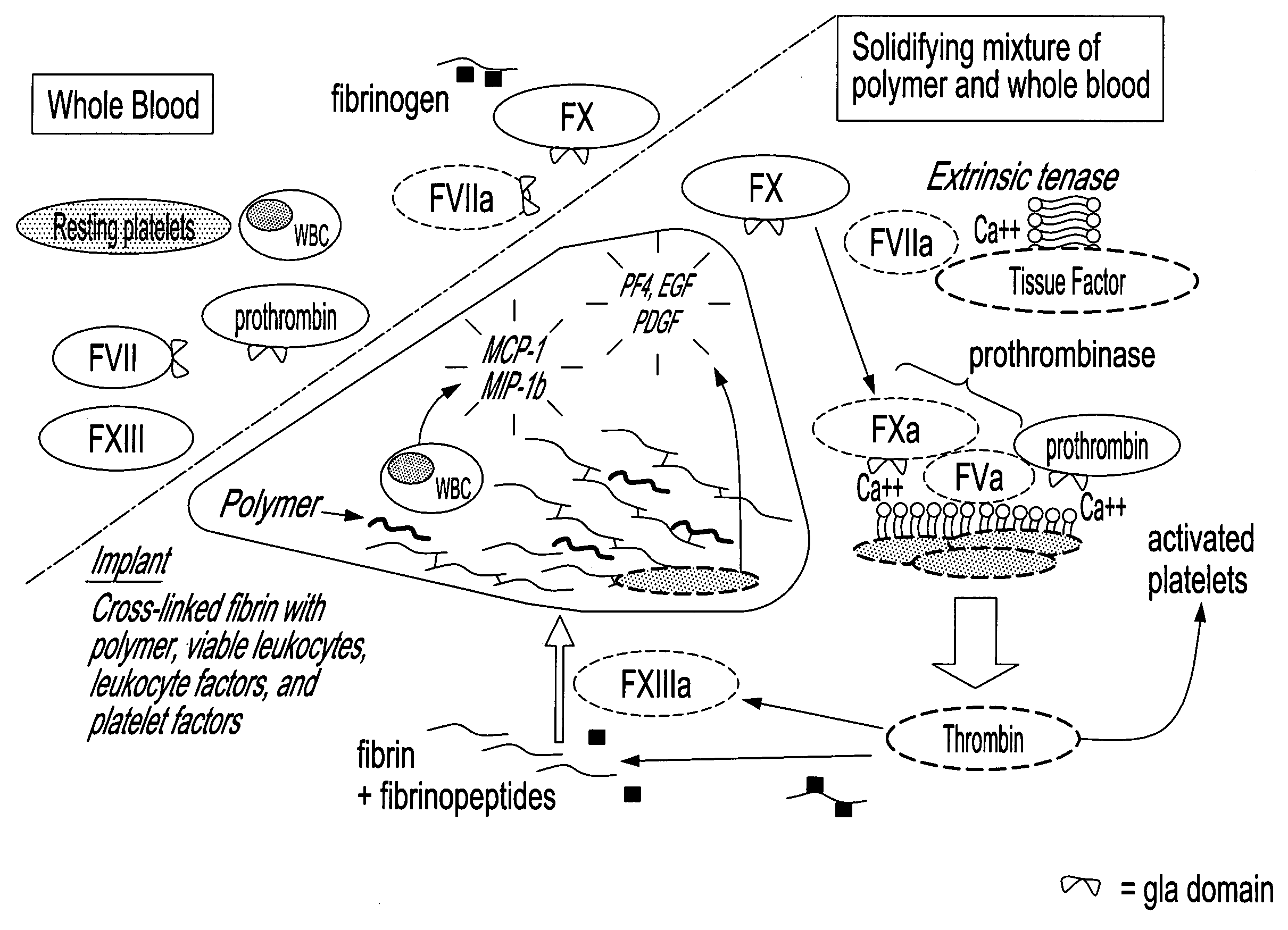
[0171]Solidification time and clot tensile strength can be evaluated using a thromboelastograph (TEG) (Bowbrick, V. A.; Mikhailidis, D. P.; and Stansby, G.: Value of thromboelastography in the assessment of platelet function. Clin Appl Thromb Hemost, 9:137-42, 2003), a type of blood rheometer. By TEG, unmodified human whole blood coagulates after a 7 to 20 minute time lapse. Samples were evaluated in a TEG set-up that allows simultaneous measurement of 8 samples, in order to evaluate the effect of clotting factors and fibrinolytic enzymes on coagulation of chitosan-GP / blood. Control samples consisted in whole blood or Glycerol Phosphate buffer (GP) without chitosan mixed with blood. Human whole blood (340 μL of freshly drawn huma...

example 2

Quantification of Levels of Thrombin Generation Via Serum Thrombin-Antithrombin (TAT) Complex Levels

[0192]Chitosan-GP / blood clots form via thrombin generation, platelet activation and Factor XIII activation. Antithrombin is in 3-fold molar excess over pro-thrombin in plasma, and binds with very high affinity to activated thrombin. Therefore, thrombin generation can be quantified via serum thrombin-antithrombin (TAT) complex levels (Rivard, G. E.; Brummel—Ziedins, K. E.; Mann, K. G.; Fan, L.; Hofer, A.; and Cohen, E.: Evaluation of the profile of thrombin generation during the process of whole blood clotting as assessed by thrombelastography. J Thromb Haemost, 3:2039-43, 2005). To measure thrombin generation in blood-polymer and blood-buffer mixtures, clotting assays were performed in plastic tubes which do not activate the contact pathway (Factor XII). Control samples were also generated in glass vials which activate the contact pathway.

[0193]To each plastic tube, 320 μL chitosan-GP...

example 3

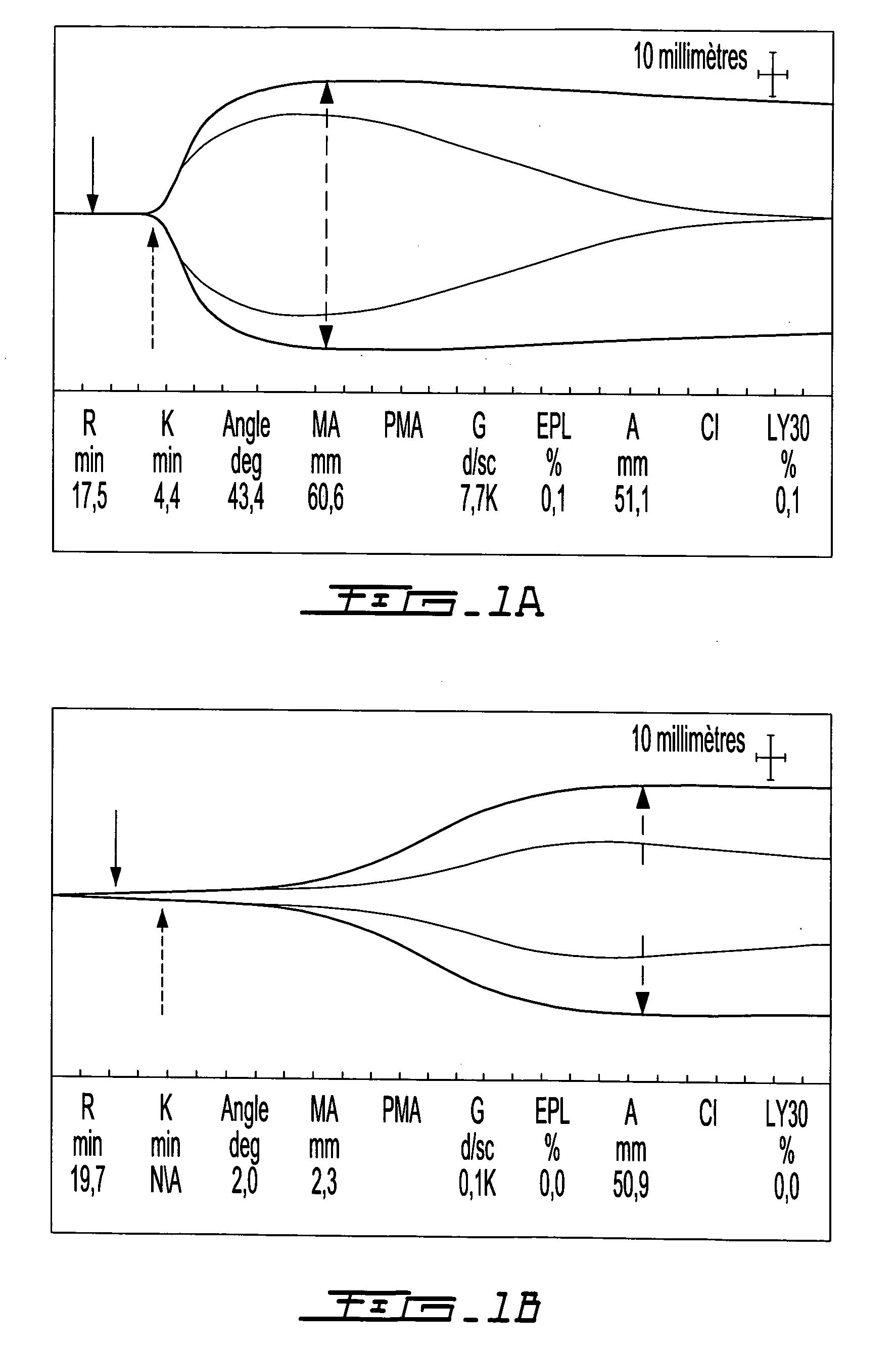
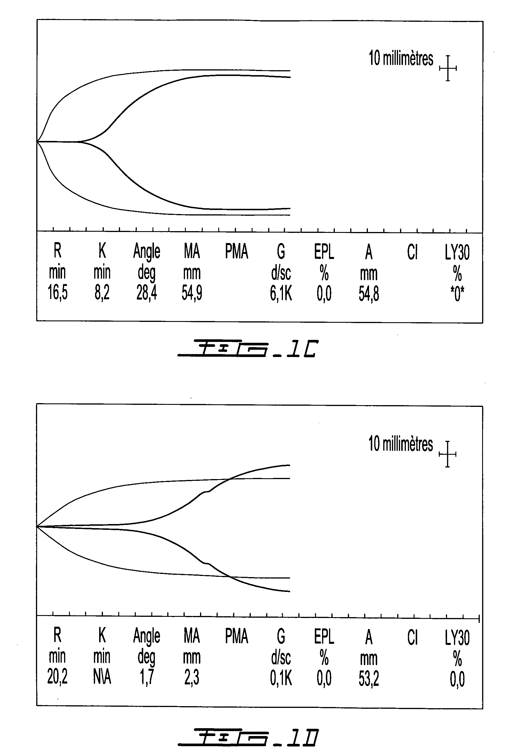
Comparison of In Situ Solidification

[0204]These tests used rabbit whole blood. In individual mixing vials, clotting factors TF-phospholipids ±rVIIa or thrombin were homogenously mixed into the blood or the chitosan-GP solution prior to combining the blood and chitosan-GP solution. FIG. 7 describes the generation of polymer-blood mixtures using chitosan-GP alone (FIG. 7A); chitosan-GP mixed with TF-rVIIa then whole blood (FIG. 7B) and chitosan-GP mixed with IIa then whole blood (FIG. 7C). FIG. 8 describes the use, on either plastic vials (the three samples on the right side of FIG. 8A and the samples on the left side of the FIG. 8B) or on glass vials (the four samples on the left side of the FIG. 8A and the samples on the right side of FIG. 8B), of the following samples:

[0205]1. Chitosan-GP mixed with blood (solid implant, arrow)

[0206]2. Chitosan-GP mixed with TF-rVIIa then blood

[0207]3a. Chitosan-GP mixed with TF then blood

[0208]3b. Chitosan-GP mixed with TF then blood-rVIIa

Immediat...


PUM

PropertyMeasurementUnit
concentrationaaaaaaaaaa

Abstract

The present invention relates to a method for repairing or regenerating tissues in a patient such as cartilage, meniscus, ligament, tendon, bone, skin, cornea, periodontal tissues, abscesses, resected tumors, cardiac tissues and ulcers. The method comprises the step of administering simultaneously or sequentially a pro-coagulant factor and an effective amount of a polymer composition comprising a biocompatible polymer and blood or a component thereof. When the polymer composition is in contact with the pro-coagulant factor it is converted into a non-liquid state such that the polymer composition will adhere to the site in need of repair to effect repair of the tissue and / or regeneration thereof.

Description

BACKGROUND OF THE INVENTION[0001](a) Field of the Invention[0002]The invention relates to a method for inducing in situ-solidification of blood containing polymers on wounds or surgical defects. The resulting solid implants stimulate the repair and regeneration of articular cartilage, joint tissues and other tissues including meniscus, ligament, tendon, bone, skin, cornea, periodontal tissues, abscesses, resected tumors, ulcers, aorta, and cardiac tissue.[0003](b) Description of Prior Art[0004]1) The Cartilage Repair Problem:[0005]Cartilage: Structure, Function, Development, Pathology[0006]Articular cartilage covers the ends of bones in diarthroidial joints in order to distribute the forces of locomotion to underlying bone structures while simultaneously providing nearly frictionless articulating interfaces.[0007]Articular cartilage is formed during the development of long bones following the condensation of prechondrocytic mesenchymal cells and induction of a phenotype switch from ...

Claims


Application Information

Patent Timeline
Patent Type & Authority Applications(United States)
IPC IPC(8): A61K35/14A61K38/18A61P19/04A61K35/16A61K35/19
CPCA61K9/00A61K31/137A61L27/3616A61K38/4833A61K38/39A61K38/36A61K38/2053A61K38/195A61K38/193A61K38/1808A61K35/19A61K35/16A61K35/14A61K31/74A61K31/722A61K31/365A61K31/661A61K31/7076A61K31/715A61K2300/00A61L27/38A61P19/04
Inventor HOEMANN, CAROLINE D.MARCHAND, CATHERINE
Owner PIRAMAL HEALTHCARE CANADA
Why Eureka
Try Eureka