
Ophthalmic compositions useful for improving visual acuity

a technology of ophthalmic compositions and compositions, applied in the field of ophthalmic compositions and methods, can solve the problems of reduced visual acuity, reduced image sharpness, and relatively sudden decrease in visual acuity, so as to improve visual acuity, prevent loss, and mitigate the damaging effect of hypertonic tear film

Inactive Publication Date: 2010-07-22
ALLERGAN INC
View PDF1 Cites 6 Cited by
  • Summary
  • Abstract
  • Description
  • Claims
  • Application Information

AI Technical Summary

Benefits of technology

[0028]It has now been discovered that novel ophthalmic compositions developed for treating eyes, afflicted or susceptible to diseases / conditions such as, without limitation, dry eye syndrome, low humidity environments, and stress / trauma, for example, due to surgical procedures, and the like, also improve visual acuity. In particular, these compositions would be useful for mitigating the damaging effects of a hypertonic tear film, regardless of cause. The present compositions can be administered, for example, topically administered, to an ocular surface of an eye of a person to prevent the loss of and / or improve visual acuity.
[0033]In each of the above-noted aspects of the invention, the present compositions for use in the method of the present invention advantageously have chemical make-ups so as the material or the mixture of organic compatible solute included in the tonicity component is effective, when the composition is administered to an eye, to allow an ocular surface of the eye to better tolerate a hypertonic condition on the ocular surface relative to an identical composition without the material or the mixture of organic compatible solute agents.
[0034]A still further broad aspect of the invention provides ophthalmic compositions for use in the method of the present invention comprising carrier component, a tonicity component and a polyanionic component. The tonicity component is present in an amount effective to provide the composition with a desired osmolality, and comprises a compatible solute component. The polyanionic component is present in an amount, when the composition is administered to a human or animal eye, to reduce at least one adverse effect of a cationic, for example, a polycationic, material on an ocular surface of a human or animal eye relative to an identical composition without the polyanionic component. This cationic material could be from any source, for example, may be endogenous, an environmental contaminant, or as an undesired consequence of applying an agent to the eye, for example a preserved solution or contact lens care product. In one very useful embodiment, hyaluronic acid is not the is sole polyanionic component. Other polyanionic components are more suited for use in the present compositions, for example, are more suited than hyaluronic acid or its salts for topical administration to an ocular surface of a human or animal eye. In another embodiment of the present invention, the composition has an osmolality in a range of about 300 to about 600 or about 1000 mOsmols / kg.
[0035]One further broad aspect of the invention provides ophthalmic compositions for use in the method of the present invention comprising a carrier component, and a polyanionic component selected from polyanionic peptides, polyanionic peptide analogs, portions of polyanionic peptide analogs, carboxymethyl-substituted polymers of sugars, including but not limited to, glucose and the like sugars and mixtures thereof. Such polyanionic components are present in an amount effective, when the compositions are administered to a human or animal eye, to reduce at least one adverse effect of a cationic, for example, polycationic, species and / or substance on an ocular surface of the eye relative to an identical composition without the polyanionic component.

Problems solved by technology

When the pupil is small (1-2 mm), image sharpness may be limited by diffraction of light by the pupil.
Any pathological process in the visual system will often cause decreases in visual acuity.
Any relatively sudden decrease in visual acuity is always a cause for concern.
As a result of this parallel organization of the visual nervous system, visual acuity measurements no longer appear to adequately describe the spatial visual abilities of a given individual.
Modern vision research has clearly demonstrated that the capacity to detect and identify spatial form varies widely as a function of target size, contrast, and spatial orientation As a consequence of the above, a simple assessment of visual acuity often does not predict an individual's ability to detect objects of larger size.
If this osmotic strength is increased, cells on the ocular surface are exposed to a hyperosmotic or hypertonic environment resulting in adverse reduction in cell volume due to trans-epithelial water loss, and other undesired changes.
The compensatory mechanisms are limited, in many respects, leading to ocular surface compromise and discomfort.
Although glycerol may serve as the sole compatible solute, it may be excessively mobile, that is, cross membranes too freely, to provide an extended benefit in certain systems.
However, as concentration in the tear falls, glycerol may be lost over time from cell to tear film, limiting the duration of benefit.
Due to the rapid mobility of water into and out of cells, however, any benefit of a hypotonic composition will be extremely short-lived.
Thus, use of a hypotonic artificial tear reduces the ability of cells to withstand hypertonicity when it returns shortly after drop instillation.
Defensins are found at increased levels in the tear film of dry eye patients, and may either directly or through interaction with other substances have adverse effects on the health of the ocular surface.
Dry eye is a multifactorial disease of the tears and ocular surface that results in symptoms of discomfort, visual disturbance, and tear film instability with potential damage to the ocular surface.
Increased viscosity of artificial tears will prolong contact time of bulk fluid on the ocular surface, but will also induce greater visual complaints in both magnitude and duration of blur associated with use of product.

Method used


Image

  • Ophthalmic compositions useful for improving visual acuity
  • Ophthalmic compositions useful for improving visual acuity
  • Ophthalmic compositions useful for improving visual acuity

Examples


example 1

[0154]In this experiment, corneal epithelial cells were isolated from the rabbit eye and grown under conditions so that they differentiate into a layered “air-lift” culture that includes basal, wing, and squamous cells. As they grow and differentiate, these cultures developed tight junctions between cells that provide the basis for a trans-epithelial electrical resistance (TEER) across the cell layers between the apical and basal surfaces. The TEER value is a sensitive measure of cell growth, differentiation and health.

[0155]After 5 days in culture during which the layered structure forms, different culture wells were exposed to hypertonic fluid (400 mOsmols / kg) with or without addition of one of 6 candidate compatible solutes at a low concentration (2 mM). The TEER was then measured after 22 hours of exposure. The TEER value was expressed as a percentage of the TEER value obtained from a similar culture under isotonic (300 mOsmol / kg) conditions. The results of these tests are shown...

example 2

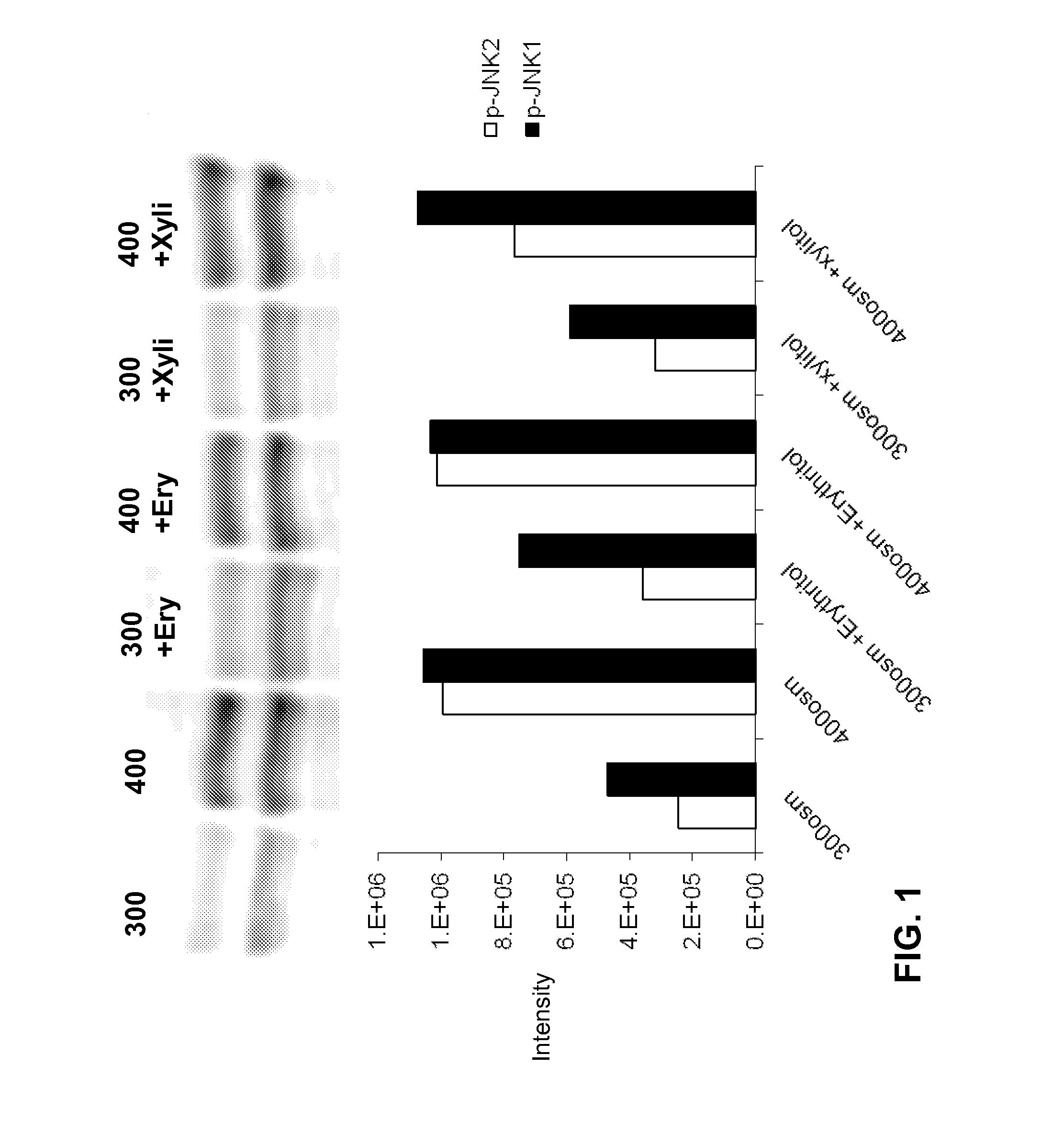
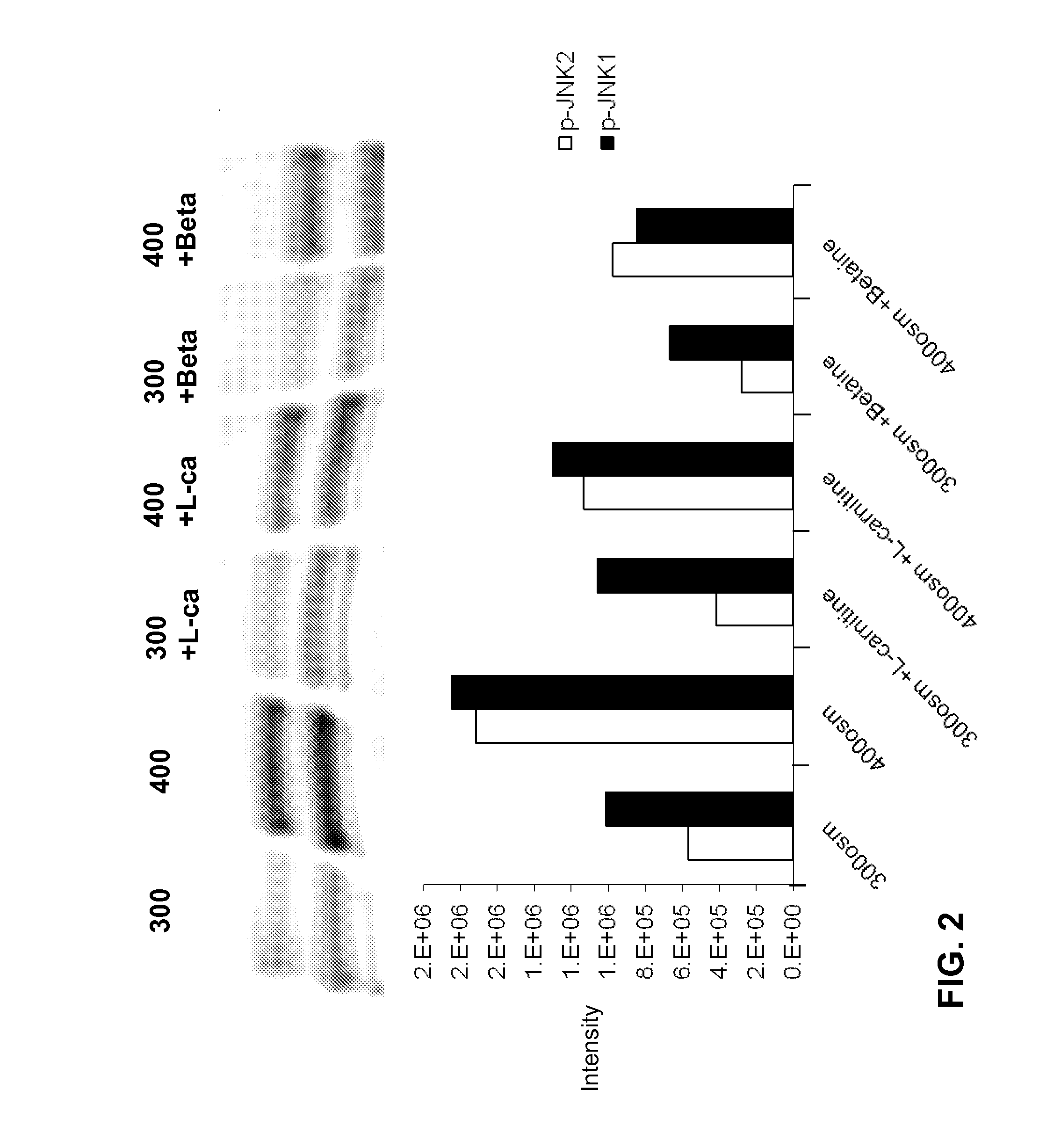
[0160]Phosphorylated JNK (the activated form of the stress associated protein kinase, SAPK) plays a key role in induction of inflammation and apoptosis in response to stress, including hyperosmolarity.

[0161]Human corneoscleral tissues, from donors aged 16-59 years were obtained from the Lions Eye Bank of Texas (Houston, Tex.). Corneal epithelial cells were grown from limbal explants. In brief, after carefully removing the central cornea, excess conjunctiva and iris and corneal endothelium, the limbal rim was cut into 12 equal pieces (about 2×2 mm size each). Two of these pieces were placed epithelial side up into each well of 6-well culture plates, and each explant was covered with a drop of fetal bovine serum (FBS) overnight. The explants were then cultured in SHEM medium, which was an 1:1 mixture of Dulbecco modified Eagle medium (DMEM) and Ham F-12 medium containing 5 ng / mL EGF, 5 μg / mL insulin, 5 μg / mL transferrin, 5 ng / mL sodium selenite, 0.5 μg / mL hydrocortisone, 30 ng / mL chol...

example 3

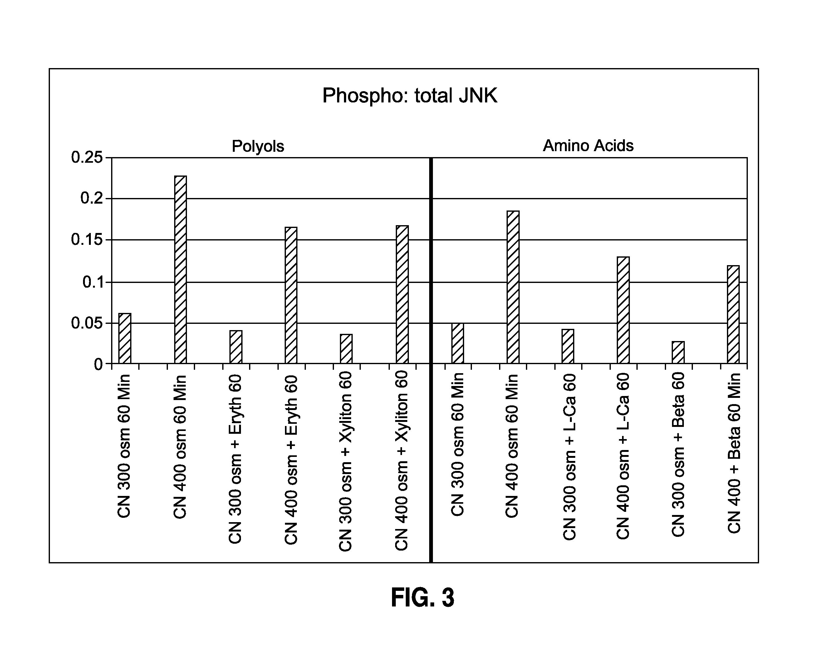
[0171]In another series of experiments, the Beadlyte® Cell Signaling Assay was used. This assay is a fluorescent bead-based sandwich immunoassay. Each sample (10 μg / 25 μL) was pipetted into a well of a 96-well plate and incubated with 25 μL of diluted 5× beads coupled to phospho-JNK, phospho-ERK, phospho-p38 or total JNK, or total ERK, or total p38 specific capture antibodies overnight. Overnight incubation was utilized for the reaction of the capture beads with the proteins from the cell lysates.

[0172]The beads were washed and mixed with biotinylated specific reporter antibodies for phospho-MAPK or total-MAPK, followed by streptavidin-phycoerythrin. The amount of total or phospho-MAPK was then quantified by the Luminex 100™ system (Luminex, Austin, Tex.). Fifty events per bead were read, and the data output obtained from the Bio-Plex Manager software were exported to Microsoft Excel® for further analysis. The results were presented as the percentage of phospho-MAPK to total-MAPK.

[0...


PUM

PropertyMeasurementUnit
diameteraaaaaaaaaa
diameteraaaaaaaaaa
heightaaaaaaaaaa

Abstract

The present invention provides a method of improving the visual acuity of a person in need thereof which comprises topically administering to said person, in an effective amount, an ophthalmic composition comprising an aqueous carrier component; and an effective amount of a tonicity component comprising a material selected from a combination of compatible solute agents, wherein said combination of compatible solute agents comprises two polyol components and one amino acid component and wherein said polyol components are erythritol and glycerol and said amino acid component is carnitine.

Description

CROSS REFERENCE[0001]This application claims the benefit of U.S. Provisional Patent Application Ser. No. 61 / 106,889, filed on Oct. 20, 2008, the entire disclosure of which is incorporated herein by this specific reference.BACKGROUND OF THE INVENTION[0002]1. Field of the Invention[0003]The present invention relates to ophthalmic compositions and methods useful for treating eyes to improve visual acuity. More particularly, the present invention relates to ophthalmic compositions including mixtures of components which are is effective in providing the desired prevention of loss in visual acuity to human or animal eyes, and to methods for treating human or animal eyes to improve and / or prevent the loss of visual acuity by using said ophthalmic compositions.[0004]2. Background of the Art[0005]To resolve detail, the eye's optical system has to project a focused image on the fovea, a region inside the macula having the highest density of cone photoreceptors, thus having the highest resolut...

Claims


Application Information

Patent Timeline
Patent Type & Authority Applications(United States)
IPC IPC(8): A61K38/16A61K31/205A61K31/717A61P27/04
CPCA61K9/0048A61K31/047A61K31/205A61K45/06A61K2300/00A61P27/02A61P27/04
Inventor SIMMONS, PETER A.VEHIGE, JOSEPH
Owner ALLERGAN INC
Learn More