Autoantibody detection systems and methods

a detection system and autoantibody technology, applied in the field of detection of autoantibodies, can solve the problems that only a few applications of autoantibodies to ct antigens have been found in diagnostics and drug development, and achieve the effect of high throughput and well-controlled multiplex assay performan

Inactive Publication Date: 2010-08-12
BONNER FERRABY PHOEBE W +2
View PDF17 Cites 20 Cited by
  • Summary
  • Abstract
  • Description
  • Claims
  • Application Information

AI Technical Summary

Benefits of technology

[0043]In certain preferred embodiments, the autoantibody detection panels of the invention employ autoantibody detection reagents that comprise recombinantly expressed full-length versions of the naturally occurring antigens with which the target autoantibody species react in vivo. With regard to autoantibody detection reagents for anti-CT antigen serum antibodies, i.e., CT antigen reagent species, in particularly preferred embodiments such detection reagents are expressed via recombinant techniques in a suitable eukaryotic expression system, for example, mammalian expression systems such as those based on recombinant CHO (Chinese Hamster Ovary) cells or the human cell line PER.C6, as well as insect cell-based expression systems, so that authentic CT antigen epitopes are exhibited. Of course, such systems, as well as solid-state synthetic processes, can also be used to produce partial proteins and protein fragments (including truncated proteins where one or more N- and / or C-terminal amino acid residues or domains are absent) and peptides of the autoantibody-specific antigens can also be used for autoantibody detection. Similarly, panels that employ one or more engineered or otherwise optimized autoantibody detection reagent species are also. For example, an autoantibody detection reagent that includes as an autoantibody-reactive moiety a polypeptide in which the antigenic epitope has been affinity matured or otherwise modified (e.g., by phage display techniques), can also be used. In certain embodiments, systems that employ automated liquid handling approaches are adapted for use in practicing the invention in order to allow for high throughput, multiplex well-controlled assay performance.

Problems solved by technology

However, to date few applications for autoantibodies to CT antigens have been found in diagnostics and drug development due to various difficulties.

Method used


Image

  • Autoantibody detection systems and methods
  • Autoantibody detection systems and methods
  • Autoantibody detection systems and methods

Examples


example 1

Automated Assays Using Autoantibody Detection Arrays

A. Introduction

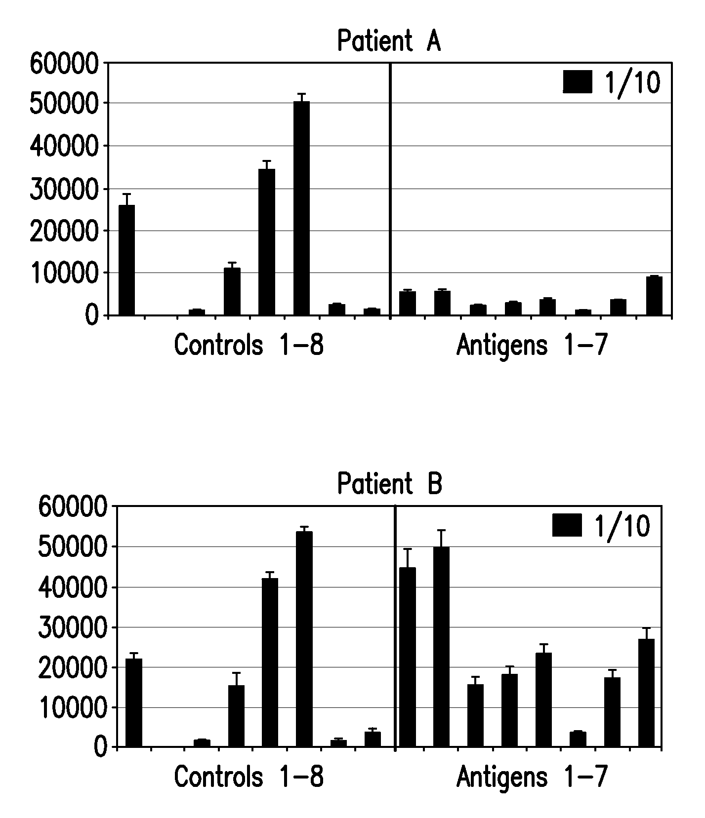
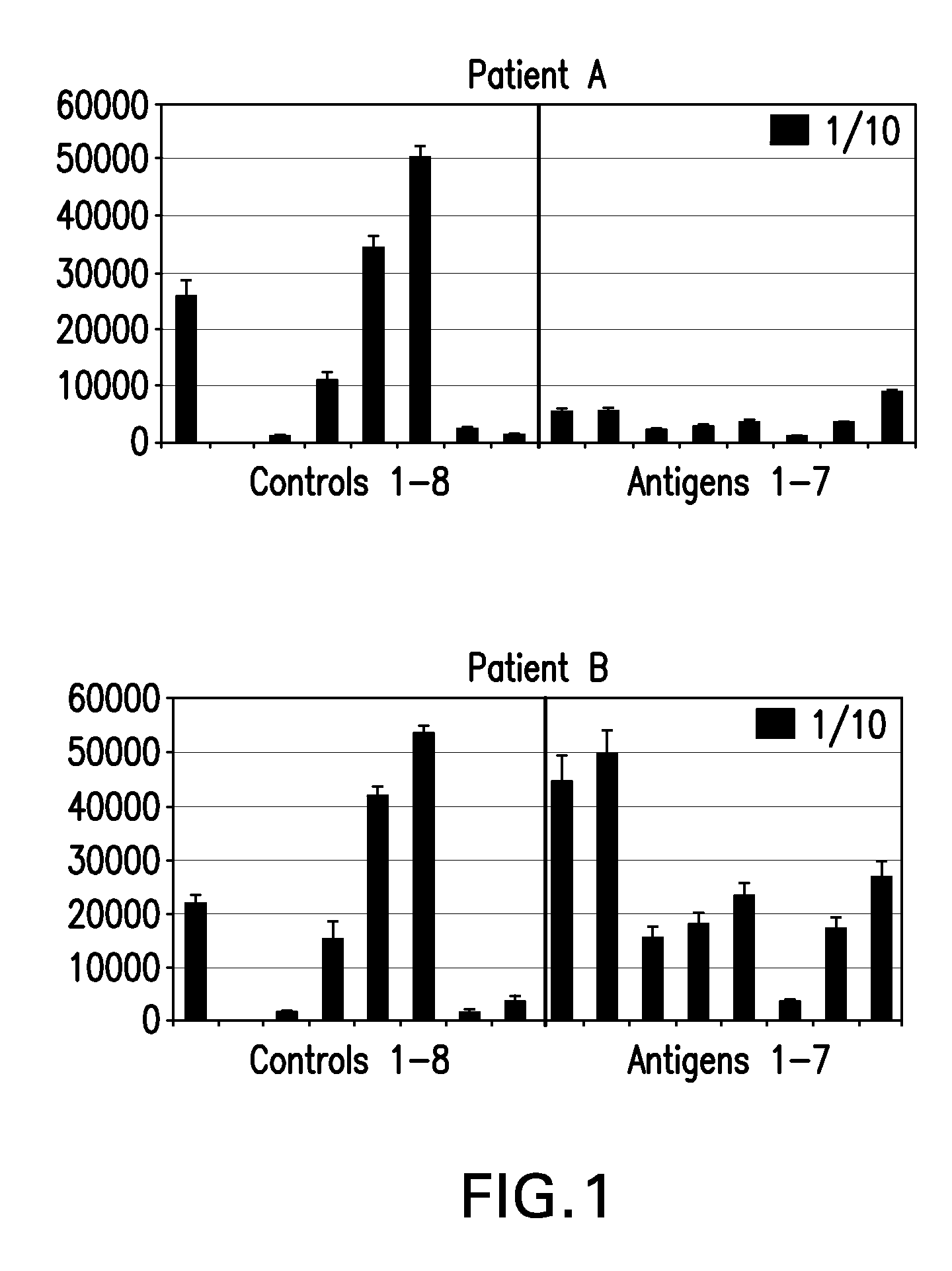
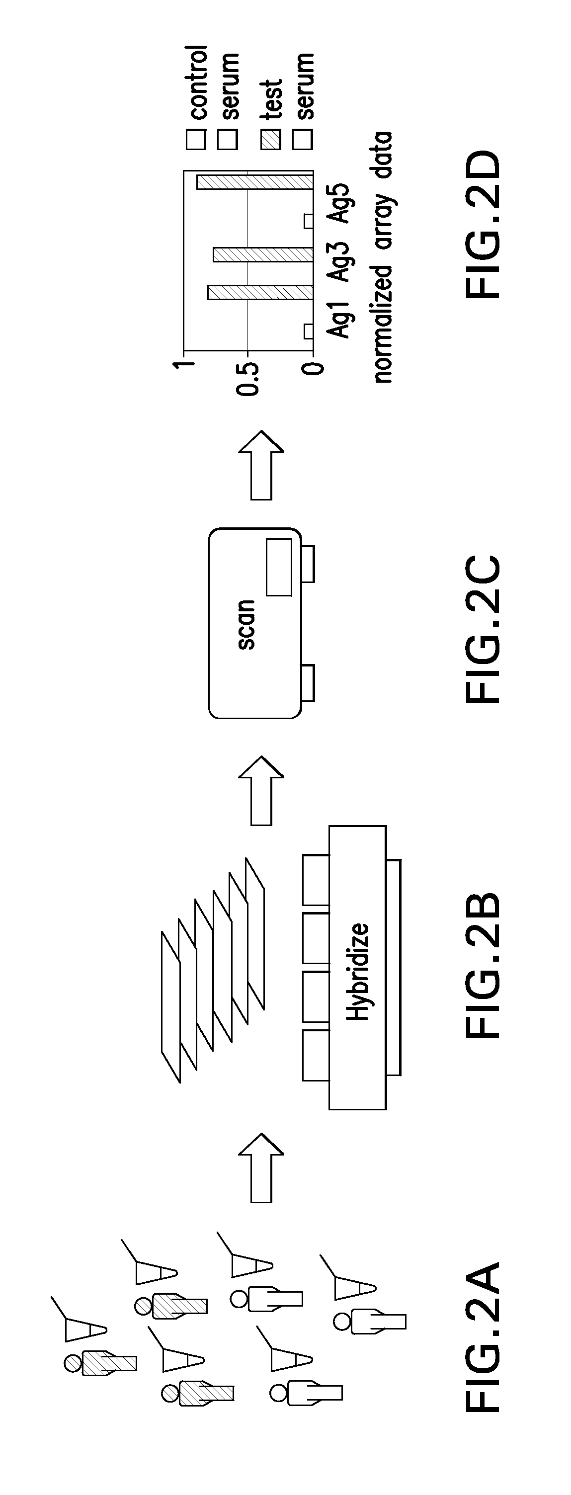
[0112]The following example addresses the problems of how to provide high content, high throughput, reliable identification of serum autoantibodies to tumor antigens through the provision of CT-antigen protein microarrays and an associated methodologies for effective assaying of human serum. Clones of full-length genes for many known CT antigens (see Table 2, above) have been obtained and expressed in insect cells. Each protein antigen is thus the product of a human gene, is full length, and has eukaryotic glycosylation by virtue of insect cell expression. Each of these factors is important in maintaining an authentic set of epitopes such recognition by serum autoantibodies is optimized. In order to assign statistical significance to candidate biomarker panels it is essential to assay as many serum samples as possible. Conventional techniques such as ELISA are limited in their throughput leading to what has been desc...

example 2

Autoantibody Detection In Melanoma Patients

[0123]Autoantibody detection arrays as described in Example 1 were used to assay serum samples from 50 patients with advanced Stage IV metastatic melanoma for antibodies to CT antigens. Patients with this disease were found to have autoantibodies to fifty CT antigens in contrast to normal healthy serum controls. Autoantibodies that were detected in the patient samples included those reactive with the following CT antigens: CTAG2, MAGEA4v2, MAGEA5, MAGEA11, NLRP4, LIP1, MAGEB6, BAGE5, MAGEB5, BAGE2, DSCR8 / MMA1, DDX53, NY-ESO-1, PBK, MICA, CXorf48.1, CT47.11, GAGE1, SSX2A, NYCO45, CSAG2, HORMAD1, ZNF165, SYCP1, GAGE5, BAGE4, SPANXD, MAGEA2, GAGE6, CEP290, NXF2, COL6A1, XAGE-2, SPANXA1, GAGE2A, SYCE1, LDHC, FTHL17, BAGE3, MAGEA4v3, MAGEB1, p53, GRWD1, MART1, MAGEA1, OIP5, CCDC33, MAGEA3, and XAGE3av2.

[0124]Results are also shown in FIG. 4, which plots relative autoantibody levels in serum versus autoantibody species for both melanoma patients ...

example 3

Autoantibody Detection In NSCLC Patients

[0125]Autoantibody detection arrays as described in Example 1 were used to assay serum samples from three patients with advanced non-small cell lung carcinoma for antibodies to CT antigens. Patients with this disease were found to have autoantibodies to numerous CT antigens in contrast to normal healthy serum controls. Autoantibodies that were detected in the patient samples included those reactive with the following CT antigens: GAGE2A, BAGE4, BAGE2, MAGEA1, DSCR8 / MMA1, CCDC33, BAGE5, CEP290, GAGE1, PBK, FTHL17, BAGE3, NLRP4, CT62, SPANXA1, DDX53, COL6A1, CSAG2, SSX2A, CT47.11, SYCP1, SPANXD, GAGE6, TSSK6, MAGEB5, ZNF165, LIP1, MICA, GAGE4, SSX4, MAGEB6, CXorf48.1, MAGEA4v2, COX6B2, MAGEA11, GRWD1, LEMD1, CTAG2, LDHC, XAGE3av2, SP011, HORMAD1, SPANXB1, TYR, MAGEB1, NYCO45, ROPN1, MAGEA5, XAGE3av1, MAGEA10, SILV, MART1, SGY-1, NXF2, MAGEA2, and RELT.


PUM

PropertyMeasurementUnit
diameteraaaaaaaaaa
concentrationsaaaaaaaaaa

Abstract

Autoantibodies in biological samples such as serum can result from changes to biomolecules (e.g., proteins, polysaccharides, and lipids) that are associated with disease. Such autoantibodies are useful biomarkers because they frequently appear early in disease and are readily accessible, particularly in biological fluids such as blood and serum. CT antigens are particularly useful for detecting autoantibodies correlated with cancer. Numerous population-based profiles for pluralities of different autoantibody species, at least some of which are specifically reactive with CT antigens, allow for simultaneous assessment of multiple disease-associated analytes is a single test, which can be more effective in diagnostics and drug development than individual profiles. The instant invention provides autoantibody detection array devices that include a plurality of independently selected autoantibody-reactive reagent species, such as full-length CT antigens or the antigenic portions thereof, disposed on a substrate. Such arrays can be used to screen biological samples taken from patients or other subjects for diagnostic, drug development, and other applications.

Description

RELATED APPLICATIONS[0001]This patent application claims priority to U.S. provisional patent application Ser. No. 61 / 120,335, filed 5 Dec. 2008 (attorney docket number SER-1001-PV), and PCT patent application serial number PCT / U.S.09 / 66902, filed 5 Dec. 2009 (attorney docket number SER-1001-PC). Each of these applications is hereby incorporated by reference in its entirety for any and all purposes.TECHNICAL FIELD[0002]This invention concerns devices and methods for the detection of autoantibodies in biological samples, as well as to ways of using data and information generated through the use of such devices and methods.BACKGROUND OF THE INVENTION1. Introduction[0003]The following description includes information that may be useful in understanding the present invention. It is not an admission that any such information is prior art, or relevant, to the presently claimed inventions, or that any publication specifically or implicitly referenced is prior art.2. Background[0004]Serum au...

Claims


Application Information

Patent Timeline
Patent Type & Authority Applications(United States)
IPC IPC(8): C40B30/04C40B40/10
CPCG01N33/57407G01N33/564
Inventor BONNER-FERRABY, PHOEBE W.HEPBURNE-SCOTT, HENRYMCCORMICK, ANN
Owner BONNER FERRABY PHOEBE W
Why Eureka
Try Eureka