Method and apparatus for breath-held mr data acquisition using interleaved acquisition

a technology of interleaved acquisition and data acquisition, which is applied in the field of magnetic resonance imaging, can solve the problems of low spatial resolution and image signal-to-noise ratio, single-shot epi acquisition (including single-shot spiral acquisition), suffer from off-resonance effects, and achieve better t1 weighting and minimize k-space transition artifacts

Inactive Publication Date: 2010-09-02
GENERAL ELECTRIC CO
View PDF8 Cites 27 Cited by
  • Summary
  • Abstract
  • Description
  • Claims
  • Application Information

AI Technical Summary

Benefits of technology

[0021]In accordance with a further aspect of the present invention, an MRI apparatus is disclosed to acquire cardiac images and near single breath-hold times. The MRI apparatus includes a magnetic resonance imaging system having a plurality of gradient coils positioned about a bore of a magnet to impress a polarizing magnetic field and an RF transceiver system and an RF switch controlled by a pulse module to transmit RF signals to an RF coil assembly to acquire MR images. The MRI apparatus further includes a computer program to segment MR data acquisition across more than one acquisition window. The proposed technique of segmenting unequal number of views minimizes k-space transition artifacts and provides better T1 weighting by beginning a data acquisition closer to the center of k-space.

Problems solved by technology

Single-shot magnetic resonance imaging using Echo Planar Imaging (EPI) techniques are able to acquire an image in 50-100 msec, thereby eliminating cardiac motion artifacts, but result in low spatial resolution and image signal-to-noise ratio.
Moreover, it is well known in the art that single-shot EPI acquisitions (including single shot spiral acquisitions) suffer from off-resonance effects which is manifested by either spatial distortion (with rectilinear read-out) or spatial blurring (with spiral acquisitions).
Images acquired using such breath-held 3D acquisitions are often characterized by low spatial resolution with only a single phase of the cardiac cycle acquired.
However, after reconstructing images with data acquired over different breath-hold periods, temporal and spatial discrepancies and inaccuracies can occur, resulting in images that are not well defined and / or blurred.
Moreover, in order to attain these shorter scan times, the acquisition window in the current 3D acquisitions are often long.
Thus, the need to accommodate a shorter breath-hold period leads to increased spatial blurring from cardiac motion as a direct consequence of a larger data acquisition window within each cardiac cycle.
In addition, respiratory-gated techniques using navigator echoes for monitoring the respiratory motion do not lend themselves to a multi-phase or CINE acquisition as a separate pulse sequence section must also be played out within each cardiac interval to interrogate the displacement of the diaphragm.
Furthermore, the acquisition of data for the different cardiac phases may not necessarily be at the same respiratory phase, leading to mixed image quality.
Repeated breath-holding, however, often results in rapid patient fatigue.
Moreover, the length of time between the acquisition of the first and last sections can also lead to varying degrees of normal myocardial suppression and hyper-enhancement of infracted tissue in the resulting images.
Repeated breath-holding may also lead to increased inconsistency of the breath-hold position.
As a result, improper registration of the individual 2D sections may occur and introduce error in the measurement of the infarct size or volume.
Notwithstanding the advantages achieved by this method, transitions in k-space can adversely affect image quality.
That is, this technique can result in a sharp, distinct discontinuity between the high spatial frequency views and the low spatial frequency views that may generate ghosting and / or artifacts in the resulting image.

Method used


Image

  • Method and apparatus for breath-held mr data acquisition using interleaved acquisition
  • Method and apparatus for breath-held mr data acquisition using interleaved acquisition
  • Method and apparatus for breath-held mr data acquisition using interleaved acquisition

Examples


Embodiment Construction

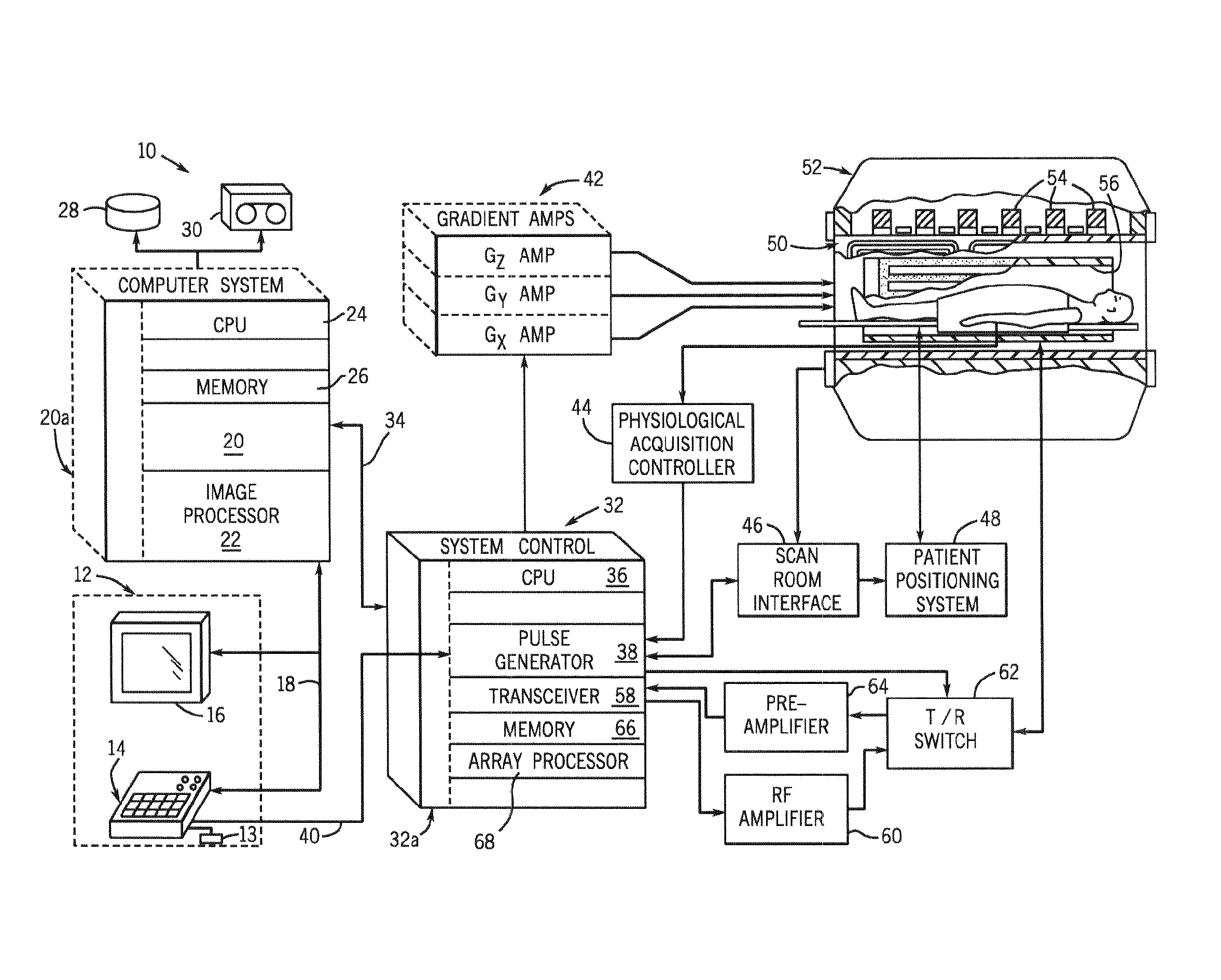
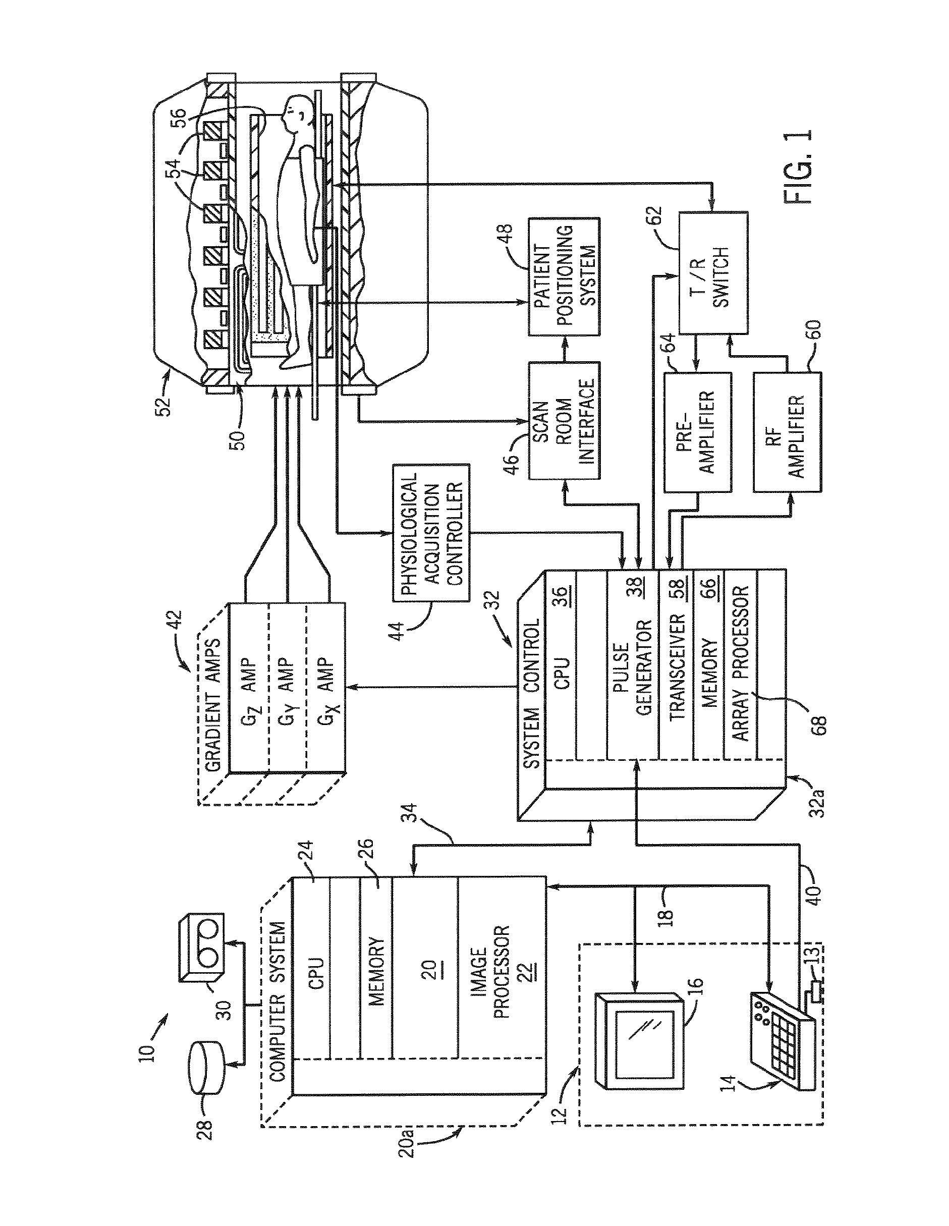
[0030]Referring to FIG. 1, the major components of a preferred MRI system 10 incorporating the present invention are shown. The operation of the system is controlled from an operator console 12 which includes a keyboard or other input device 13, a control panel 14, and a display 16. The console 12 communicates through a link 18 with a separate computer system 20 that enables an operator to control the production and display of images on the screen 16. The computer system 20 includes a number of modules which communicate with each other through a backplane 20a. These include an image processor module 22, a CPU module 24 and a memory module 26, known in the art as a frame buffer for storing image data arrays. The computer system 20 is linked to a disk storage 28 and a tape drive 30 for storage of image data and programs, and it communicates with a separate system control 32 through a high speed serial link 34. The input device 13 can include a mouse, joystick, keyboard, track ball, to...


PUM


Abstract

A method and apparatus are presented for acquiring MR cardiac images in a time equivalent to a single breath-hold. MR data acquisition is segmented across multiple cardiac cycles. MR data acquisition is interleaved from each phase of a first cardiac cycle with MR data from each phase of a subsequent cardiac cycle. Preferably, low spatial frequency data are interleaved between multiple cardiac cycles, and the subsequent cardiac cycle acquisition includes sequential acquisition of high spatial frequency data towards the end of the acquisition window. An MR image can then be reconstructed with data acquired from each of the acquisitions that reduce ghosting and artifacts. Volume images of the heart can be produced within a single breath-hold. Images can be acquired throughout the cardiac cycle to measure ventricular volumes and ejection fractions. Single phase volume acquisitions can also be performed to assess myocardial infarction.

Description

CROSS-REFERENCE TO RELATED APPLICATIONS[0001]The present application claims the benefit of U.S. Ser. No. 10 / 249,914 filed May 16, 2003, which claims the benefit of U.S. Ser. No. 60 / 319,254 filed May 17, 2002.BACKGROUND OF THE INVENTION[0002]The present invention relates generally to magnetic resonance imaging (MRI), and more particularly, to a method and apparatus for cardiac MR imaging using an interleaved variable sampling-in-time scheme for data acquisition.[0003]When a substance such as human tissue is subjected to a uniform magnetic field (polarizing field B0), the individual magnetic moments of the spins in the tissue attempt to align with this polarizing field, but precess about it in random order at their characteristic Larmor frequency. If the substance, or tissue, is subjected to a magnetic field (excitation field B1) which is in the x-y plane and which is near the Larmor frequency, the net aligned moment, or “longitudinal magnetization”, MZ, may be rotated, or “tipped”, i...

Claims


Application Information

Patent Timeline
Patent Type & Authority Applications(United States)
IPC IPC(8): A61B5/055
CPCA61B5/055A61B5/7285G01R33/4818G01R33/482G01R33/5673G01R33/5613G01R33/5616G01R33/56509G01R33/56
Inventor FOO, THOMAS K. F.
Owner GENERAL ELECTRIC CO
Why Eureka
Try Eureka