Method and device for image guided dynamic radiation treatment of prostate cancer and other pelvic lesions

Active Publication Date: 2010-09-23
XCISION MEDICAL SYST
View PDF6 Cites 31 Cited by
  • Summary
  • Abstract
  • Description
  • Claims
  • Application Information

AI Technical Summary

Benefits of technology

[0025]In accordance with the foregoing and other objects, the present invention provides a method and device for image guided dynamic radiation treatment of prostate cancer and other pelvic lesions including: 1) a unique fan geometry of radiation sources; 2) a special collimation method and apparatus to sculpt the radiation borders; 3) an integrated three-dimensional imager and a special tissue interface imaging system to locate and track critical boundaries in real-time; 4) a dynamic patient support system, which is shared b

Problems solved by technology

Toxicity to normal structures limits the radiation dose and the treatment efficacy of both methods as normal structures may be contained within or adjacent to the diseased tissue.
However this approach causes unacceptable toxicities such as urethral stricture or even tissue necrosis.
However, daily dose greater than a 3Gy dose using external beam radiation has not been widely used because of urethra necrosis.
However, their method has the greatest detriment of being very invasive and must be performed under general anesthesia.
There has not been any method that can safely deliver a high dose to the target while sparing a critical structure completely surrounded by cancerous tissue.
There is no treatment machine that can optimally spare the urethra and other critical organs such as rectum and, bladder and seminal vesicles.
Therefore, hypofractionated radiation treatment for prostate cancer remains a dangerous procedure.
For practical reasons such as treatment time and machine geometry, conventional treatment further has the limitation of approaching the prostate from a small solid angle and typically only transverse coplanar beams are used.
There is no dedicated tele-radiotherapy machine specifically designed to treat prostate cancer using a single or an arrangement of radioisotopes or other source of high energy radiation.
This is an extremely expensive and complex method and has not demonstrated any benefit in part due to the challenges mentioned above for conventional treatment.
Past efforts of intensity-modulated radiation therapy have been lim

Method used


Image

  • Method and device for image guided dynamic radiation treatment of prostate cancer and other pelvic lesions
  • Method and device for image guided dynamic radiation treatment of prostate cancer and other pelvic lesions
  • Method and device for image guided dynamic radiation treatment of prostate cancer and other pelvic lesions

Examples


Embodiment Construction

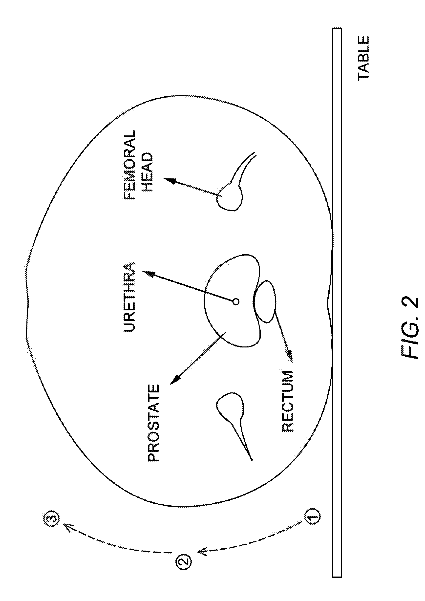
[0037]There has not been any prior art methods or devices described that can safely deliver high doses of radiation to the target(s) in the human pelvis while sparing a critical structure completely surrounded by cancerous tissue (example: the urethra in the prostate gland).

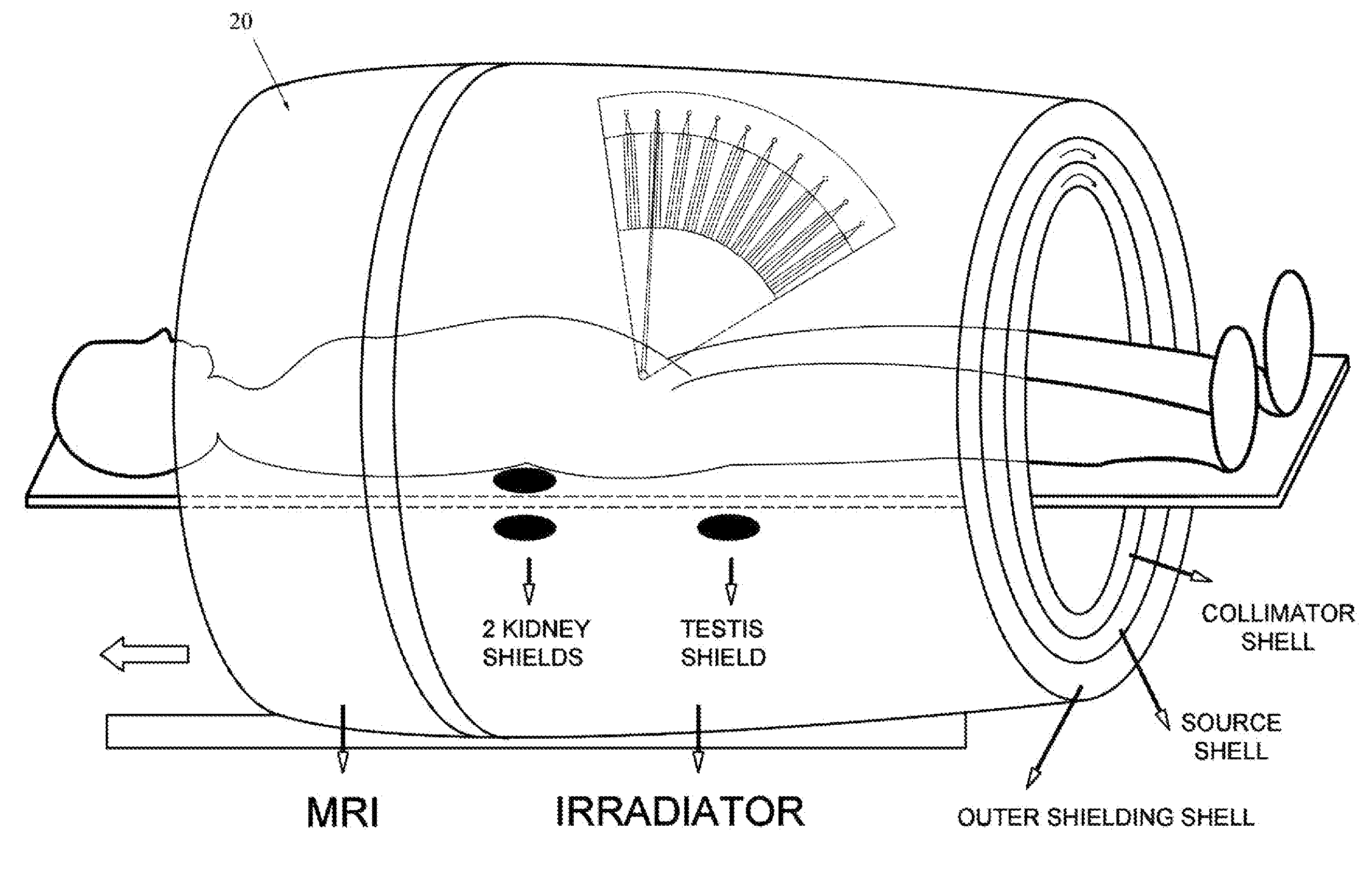
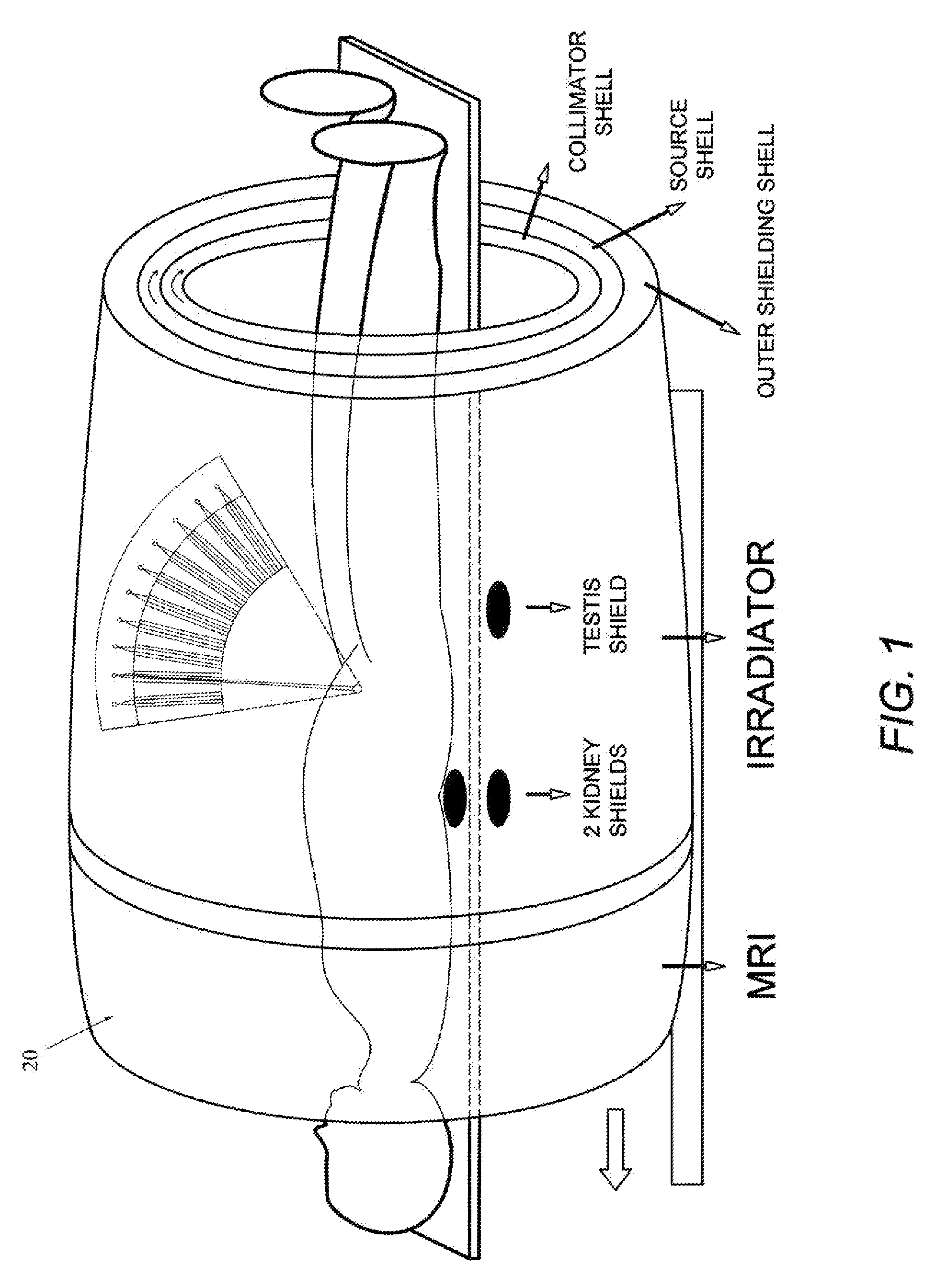
[0038]The present invention is a new device and method for delivering radiation to the human pelvis, such as the prostate gland, while avoiding irradiation of internal and external normal tissues. The device includes: 1) a unique fan geometry of radiation sources; 2) a special collimation method and apparatus to sculpt the radiation borders; 3) an integrated three-dimensional imager and a special tissue interface imaging system to locate and track critical boundaries in real-time; 4) a dynamic patient support system, which is shared by the said imager and the irradiation system; and 5) motorized custom shielding filters to further protect neighboring normal tissues such as the kidneys and femoral heads.

[0039]A sc...


PUM


Abstract

A method and device for image guided dynamic radiation treatment of prostate cancer and other pelvic lesions including: 1) a unique fan geometry of radiation sources; 2) a special collimation method and apparatus to sculpt the radiation borders; 3) an integrated three-dimensional imager and a special tissue interface imaging system to locate and track critical boundaries in real-time; 4) a dynamic patient support system, which is shared by the said imager and the irradiation system; and 5) motorized custom shielding filters to further protect neighboring normal tissues such as the kidneys and femoral heads. The fan geometry utilizes a plural number of radiation sources arranged specifically for irradiating tumors in the human pelvis while not harming critical structures, and the collimation sculpts the radiation borders using motorized shields for different sensitive structures. This allows high doses of radiation to be delivered to lesions within the human pelvis, such as the prostate, while sparing its surrounding structures, such as the rectum and bladder, as well as the urethra that is contained inside the prostate.

Description

CROSS-REFERENCE TO RELATED APPLICATION(S) [0001]The present application derives priority from U.S. provisional patent application Ser. No. 61 / 210,766 filed 23 Mar. 2009.BACKGROUND OF THE INVENTION [0002](1) Field of the Invention[0003]The present invention relates to a method and system for delivering radiation treatments to cancerous tissues within the human pelvis, such as the prostate gland and the cervix, while avoiding tissues that would otherwise be damaged by aggressive radiation treatment.[0004](2) Description of Prior Art[0005]Radiation therapy and radiosurgery are established methods of treating patients with certain malignant and benign diseases. Radiotherapy is typically given over many episodes of treatment and generally involves treating larger volumes of tissue that often include normal structures that may be affected adversely by the radiation treatment. The strategy of spreading the therapy over many episodes separated in time is chosen to enable recovery of normal ...

Claims


Application Information

Patent Timeline
IPC IPC(8): A61N5/00A61B5/055
CPCA61N5/1049A61N2005/1061A61N5/1084A61N5/1067
Inventor WANG, CHAO
Owner XCISION MEDICAL SYST
Why Eureka
Try Eureka