System and method for treating tumors

a tumor and system technology, applied in the field of tumor system and method, can solve the problem that the field may not pass into the tumor effectively

Inactive Publication Date: 2010-09-23
PULSE BIOSCI INC
View PDF21 Cites 66 Cited by
  • Summary
  • Abstract
  • Description
  • Claims
  • Application Information

AI Technical Summary

Benefits of technology

[0009]In delivering nanosecond pulsed electric fields (nsPEF) to a region of tissue, such as a tumor, it is possible to precisely control the number of pulses delivered as well as the frequency of those pulses to deliver electrotherapy via an electrode assembly designed to draw tissue into a recessed cavity in o...

Problems solved by technology

When nsPEF is applied to a tissue region such as a tumor, if a large resistance between the electrode and the tumor res...

Method used


Image

  • System and method for treating tumors
  • System and method for treating tumors
  • System and method for treating tumors

Examples


example 1

[0084]In optimizing the device, multiple experiments have shown that tumors, such as melanoma tumors, may be eliminated utilizing nsPEF when exposed to 100 ns long pulses having a 15 ns rise time where the minimum number of pulses range from, e.g. 1500 to 2000 pulses, as illustrated in the chart of FIG. 10 which shows the Optimum Pulse Number where a majority of tumors were successfully treated after a single treatment when pulsed with at least 1500 to 2000 pulses. Accordingly, as shown in the graph of FIG. 11, the Percent Efficacy after one treatment is shown to increase from 1500 pulses and higher.

[0085]As also indicated in the chart of FIG. 12 which illustrates Optimum Amplitude of the pulses, the number of tumors successfully treated after a single treatment begins to rise at higher amplitudes, e.g., from 25 kV / cm. Thus, the minimum pulse amplitude observed is 30 kV / cm in this example while the optimum pulse amplitude is 40 kV / cm or greater in this example for effectively treati...

example 2

[0087]In this particular example, Murine B16-F10 melanoma cells transfected with enhanced green fluorescent protein (eGFP) were obtained and stored in liquid nitrogen until use. These cells were cultured and injected into 4-6 week old female Nu / Nu mice (immunodeficient, hairless, albino) using standard procedures at four injection sites each. Tumors were detected visually by the bulges they produced and by GFP detection under fluorescent microscopy.

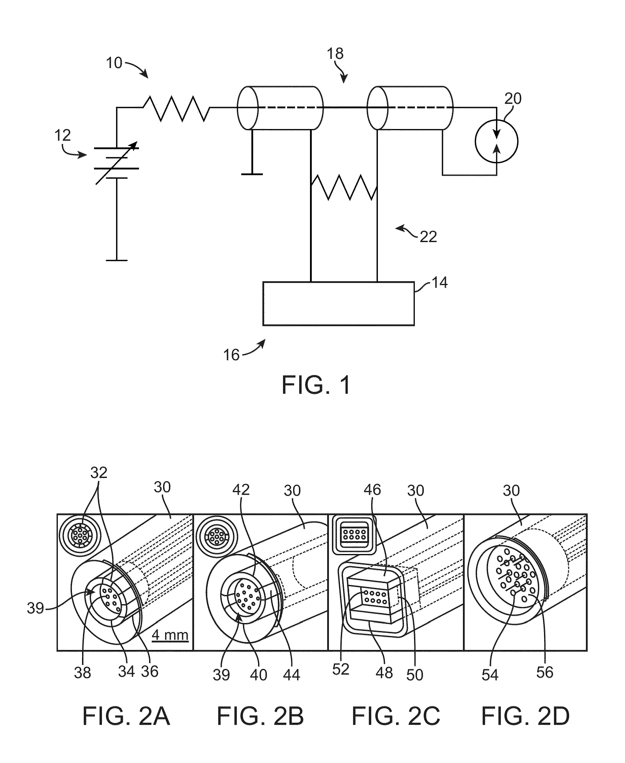
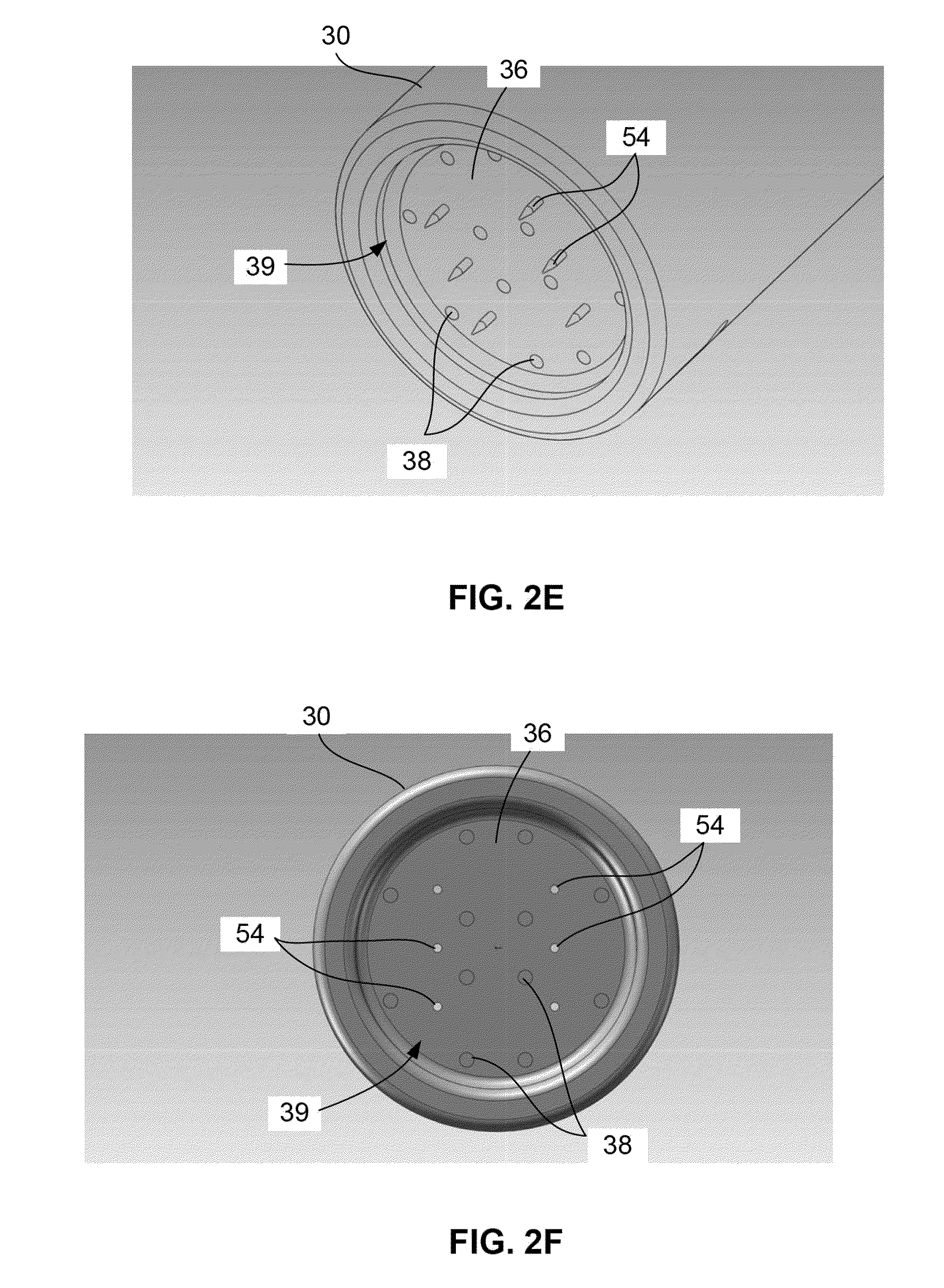
[0088]Various suction electrode assemblies, shown in FIG. 13, were used where electrode assemblies 110, 112, and 114 each had a recessed cavity with an inner diameter of about 4 mm and a depth of about 2 mm while the electrode assembly 116 utilized an array of needles positioned within the recessed cavity where a distance between the center needle and each of the outer needles was about 2 mm. In each of the assemblies, one or more electrodes 118 were used to discharge the energy into the treated tissue while the remaining electrodes funct...

example 3

[0095]Typical melanoma responses to nsPEF therapy in the 10-25 kV / cm range were recorded where four melanomas on one mouse were treated with either 10, 15, 20 or 25 kV / cm nsPEF (2000 pulses, 100 ns, 7 Hz). The GFP fluorescence at each respective pulse amplitude over a period of 0, 1, 6, and 8 days were recorded, as shown in FIG. 21, as were the trans-illumination, as shown in FIG. 22, and reflected light images, as shown in FIG. 23. The temperature increase inside a tumor over a period of several minutes during nsPEF application were also recorded, as shown in FIG. 24.

[0096]A pulse amplitude of 30 kV / cm with 100 ns long pulses were applied beginning at the indicated frequency 120 with frequency of 1 Hz and 5 Hz. Pulsing was stopped at the indicated frequency 122 for 5 Hz and at 124 for 1 Hz. The 1 Hz pulse application increased tumor temperature by 2° C. while the 5 Hz pulse application increased the temperature by 7° C.

[0097]The appearance of nsPEF-treated skin on the indicated day...


PUM


Abstract

Systems and methods for treating tumors on or within internal organs of mammals that have been imaged with endoscopic ultrasound are described. The system uses an expandable bipolar electrode assembly that can be imaged by ultrasound and can penetrate, e.g., the stomach, intestine or bowel wall, etc. and be positioned in or around the tumor on an internal organ while being guided by an operator who visualizes its position with ultrasound imaging. It utilizes an electrode assembly that extends down an internal cavity in the endoscope to allow the operator to spread the electrodes for pulse delivery of a nanosecond pulsed electric field (nsPEF) to the tumor.

Description

CROSS-REFERENCE TO RELATED APPLICATIONS[0001]This application claims the benefit of priority to U.S. Prov. 61 / 161,043 filed Mar. 17, 2009 and 61 / 186,798 filed Jun. 12, 2009, and is also a continuation-in-part of U.S. Pat. App. 12 / 722,441 filed Mar. 11, 2010, each of which is incorporated herein by reference in its entirety.FIELD OF THE INVENTION[0002]This application is directed to systems and methods for treating tumors on internal organs that have been identified using endoscopic ultrasound by precisely positioning a pulsed field delivery device on or in the tumor guided by ultrasound imaging.BACKGROUND OF THE INVENTION[0003]Endoscopic ultrasound (EUS) combines endoscopy and ultrasound in order to obtain images and information about the digestive tract and the surrounding tissue and organs. Endoscopy refers to the procedure of inserting a long flexible tube via the mouth or the rectum to visualize the digestive tract, whereas ultrasound uses high-frequency sound waves to produce i...

Claims


Application Information

Patent Timeline
IPC IPC(8): A61B18/14A61B8/00
CPCA61B8/12A61B18/1477A61B2019/5278A61B2018/1425A61B2018/143A61B18/1492A61B2090/3782
Inventor NUCCITELLI, RICHARD LEENUCCITELLI, PAMELASHEIKH, SALEHTRAN, KEVINATHOS, BRIANKREIS, MARK
Owner PULSE BIOSCI INC
Why Eureka
Try Eureka