Method for diagnosing lung cancers using gene expression profiles in peripheral blood mononuclear cells

Active Publication Date: 2010-10-07
THE WISTAR INST OF ANATOMY & BIOLOGY
View PDF16 Cites 19 Cited by
  • Summary
  • Abstract
  • Description
  • Claims
  • Application Information

AI Technical Summary

Benefits of technology

[0020]In another aspect, a novel method for selecting significant genes in comparative gene expression studies is provided. This novel method, i.e., SVM-RCE, combines K-means and Support Vector Machines (SVMs) to identify and score (rank) those gene clusters for the purpose of classification by (i) initially using K-means to group genes into clusters; and (ii) using recursive cluster elimination (RCE) to iteratively remove those clusters of genes that contribute the least to the classification performance.
[0021]In yet a further aspect, a composition for diagnosing or evaluating a lung cancer in a mammalian subject is provided. This composition includes (a) three or more polynucleotides or oligonucleotides, wherein each polynucleotide or oligonucleotide hybridizes to a different gene, gene fragment, gene transcript or expression product from mammalian peripheral blood mononuclear cells (PBMC), or (b) three or more ligands, wherein each ligand binds to a different gene expression product from mammalian peripheral blood mononuclear cells (PBMC). The gene, gene fragment, gene transcript or expression product is selected from among (i) the genes of Table V; (ii) the genes of Table VI; (iii) the genes of Table VII, or (iv) genes from a combination of these Tables.
[0022]In one embodiment, the composition includes polynucleotides or oligonucleotides that hybridize to, or ligands that bind the expression products of, the first 29 genes of Table V (hereinafter referred to as “the 29 gene classifier”) or a subset thereof. This embodiment is particularly useful for

Problems solved by technology

The use of serial CT in this group of patients runs the risk of delaying potential curable therapy, along with the costs of repeat scans, the not-insignificant radiation doses, and the anxiety of the patient.
Although each of these approaches has its own merits, none has yet passed the exploratory stage in the effort to detect patients with early stage lung cancer, even in high-risk groups, or patients which have a preliminary diagnosis based on radiological and other clinical factors12.
While the effect

Method used


Image

  • Method for diagnosing lung cancers using gene expression profiles in peripheral blood mononuclear cells
  • Method for diagnosing lung cancers using gene expression profiles in peripheral blood mononuclear cells
  • Method for diagnosing lung cancers using gene expression profiles in peripheral blood mononuclear cells

Examples


example 1

Patient Subject and Control Subjects for PBMC Samples

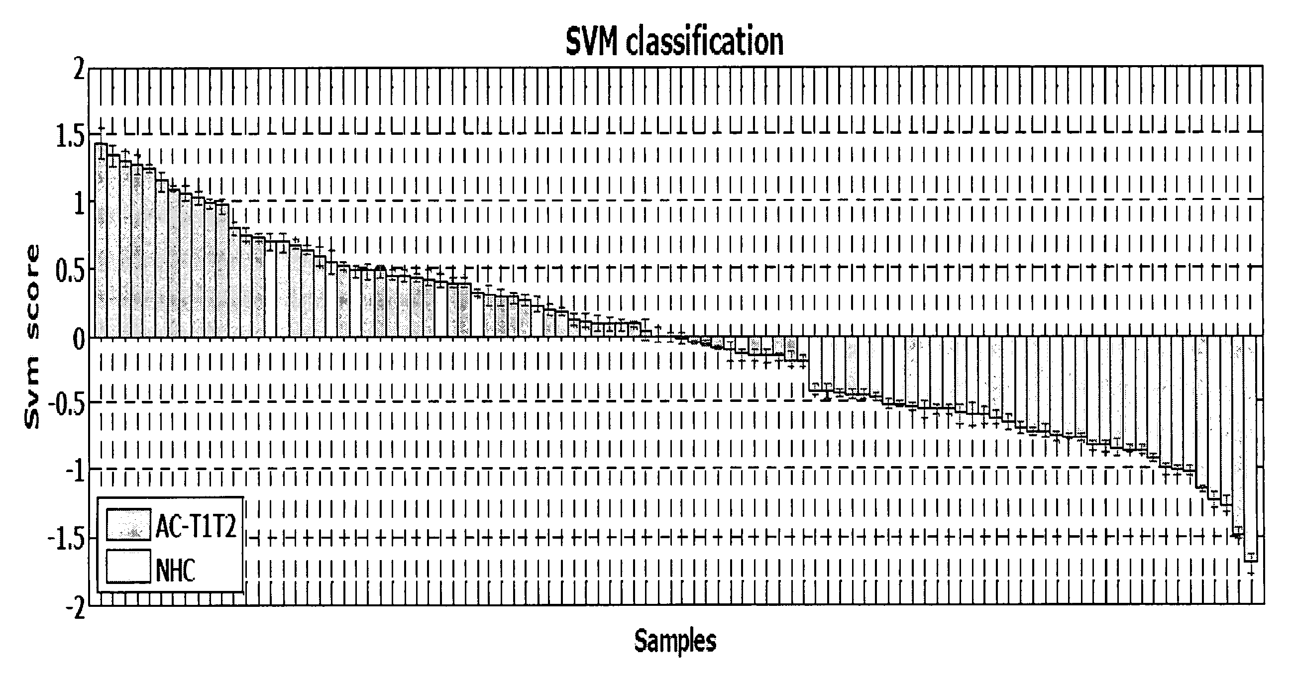
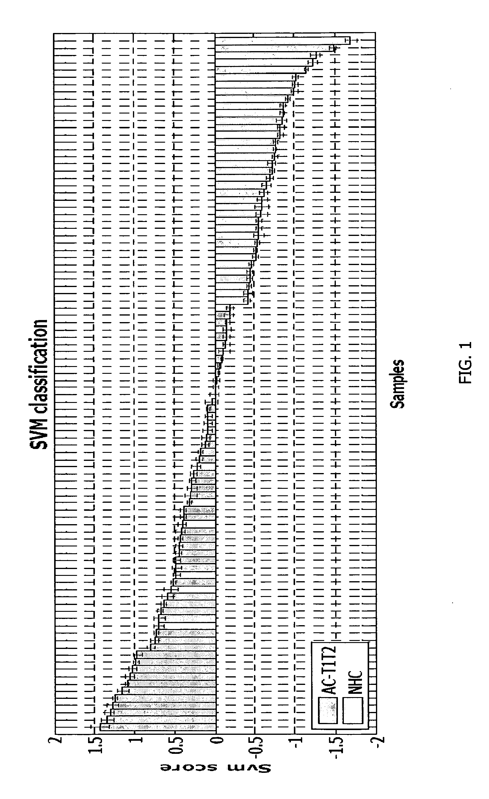
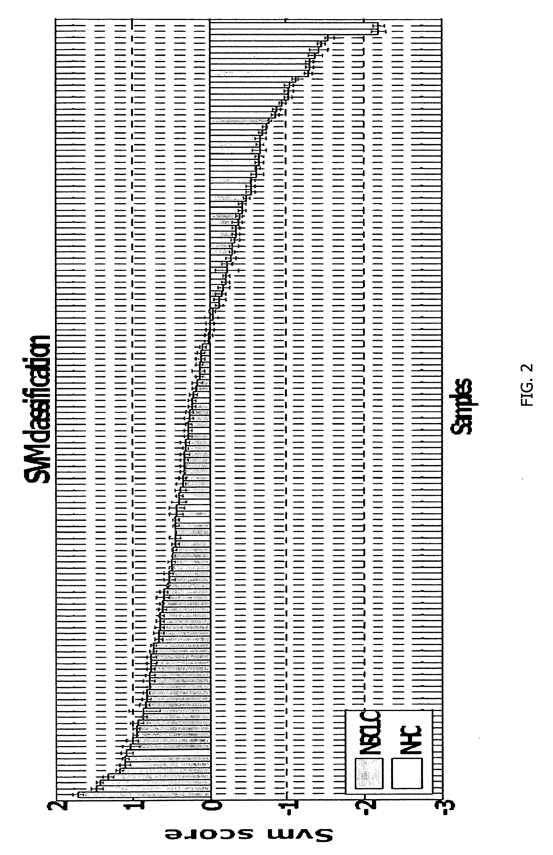
[0135]PBMC samples and clinical information were collected from 300 lung cancer patients and 150 controls, including samples from 16 patients collected pre- and post-surgery. Patient subjects and control subjects both have the key risk factor for lung cancer, i.e., smoking, and many of the patient subjects and non-healthy controls (NHCs) have smoking-related diseases such as COPD. The major difference between the 2 classes is the presence of a malignant nodule in the patient class.

[0136]A. Patient Subjects

[0137]Patient populations useful in providing data for the development of the gene expression profiles described herein include newly diagnosed male and female patients with early stage lung cancer. Inclusion criteria for selection of these patients were patients a representative number of African-American patients (about 15%), Hispanics (5%), and no Pacific Islanders. The age range of the patients was from 50-80 years. They were...

example 2

Sample Collection Protocols and Processing

[0141]Blood samples were collected in the clinic by the tissue acquisition technician. Blood is collected in two CPT® tubes (Becton-Dickenson). CPT tubes were evacuated blood collection tubes containing FICOLL reagent below a gel insert and an anti-coagulant above the gel. This is a very efficient and easy way to directly isolate PBMC. Blood is collected from the same patients during their 2-6 month follow-up visit in the clinic after surgery. Blood samples were collected in PAXgene tubes from a subset of patients and control subjects. All coded samples, including tissue blocks and blood components (PBMC, serum, and plasma) were stored based on subject identification in marked freezer storage boxes at −80 C.°. Collected samples were processed through a variety of routine steps that have been highly standardized. Samples were processed as batches (usually 20-50 samples) of both cases and controls rather than as individual samples were collect...

example 3

Methods of Processing Data for Gene Expression Profiling

[0144]The ILLUMINA BeadChip is a relatively new method of performing multiplex gene analysis. The essential element of BeadChip technology is the attachment of oligonucleotides to silica beads. The beads were then randomly deposited into wells on a substrate (for example, a glass slide). The resultant array was decoded to determine which oligonucleotide-bead combination is in which well. The decoded arrays may be used for a number of applications, including gene expression analysis. These arrays have the same gene coverage as Affymetrix arrays (47,000 probes for 27,000 genes including splice variants) but use 50-mer oligonucleotides rather than 25-mers and thus provide greater specificity.

[0145]The data analysis pipeline procedures using Matlab functions, coded PDA and SVM with RFE and SVM-RCE, were routinely and successfully used as evidenced by previous publications and as described herein.

[0146]A. Data Pre-Processing and Arr...


PUM

PropertyMeasurementUnit
Compositionaaaaaaaaaa
Gene expression profileaaaaaaaaaa
Orderaaaaaaaaaa

Abstract

Methods and compositions are provided for diagnosing lung cancer in a mammalian subject by use of three or more selected genes, e.g., a gene expression profile, from the peripheral blood mononuclear cells (PBMC) of the subject which is characteristic of disease, a stage of the disease, or enables prognosis of recurrence of disease. The gene expression profile includes three or more genes of Table I, Table II, Table III, Table IV, Table V, Table VI or Table VII herein. Detection of changes in expression in the selected genes forming the gene expression profile from that of a reference gene expression profile are correlated with non-small cell lung cancer (NSCLC). One composition for use in such diagnosis includes three or more PCR primer-probe sets, wherein each primer-probe set amplifies a different polynucleotide sequence from the gene expression profile. Another composition for similar use contains a plurality of polynucleotides immobilized on a substrate, which probes hybridize to three or more gene expression products from genes in the gene expression profile. Still another composition involves detection of the protein expression products of genes from the gene expression profile.

Description

STATEMENT REGARDING FEDERALLY SPONSORED RESEARCH OR DEVELOPMENT[0001]This invention was made with government support under Grant No. R01 CA125749 awarded by the National Institutes of Health. The government has certain rights in this invention.BACKGROUND OF THE INVENTION[0002]Lung cancer is the most common worldwide cause of cancer mortality. In the United States, lung cancer is the second most prevalent cancer in both men and women and will account for more than 174,000 new cases per year and more than 162,000 cancer deaths. In fact, lung cancer accounts for more deaths each year than from breast, prostate and colorectal cancers combined2.[0003]The high mortality (80-85% in five years), which has shown little or no improvement in the past 30 years, emphasizes the fact that new and effective tools to facilitate early diagnoses prior to metastasis to regional nodes or beyond the lung are needed6.[0004]High risk populations include smokers, former smokers, and individuals with markers...

Claims


Application Information

Patent Timeline
IPC IPC(8): C12Q1/68C40B40/06C12M1/34
CPCC12Q1/6886C12Q2600/16C12Q2600/112C12Q2600/118C12Q2600/158G01N33/57423
Inventor SHOWE, MICHAELSHOWE, LOUISEYOUSEF, MALIKALBELDA, STEVEN M.VACHANI, ANILKOSSENKOV, ANDREI V.
Owner THE WISTAR INST OF ANATOMY & BIOLOGY
Why Eureka
Try Eureka