Two-dimensional photonic bandgap structures for ultrahigh-sensitivity biosensing
a biosensing and photonic crystal technology, applied in the field of photonic crystal (phc) arrays, can solve the problems of large sensing area, large detection limit, and relatively large sensing area
- Summary
- Abstract
- Description
- Claims
- Application Information
AI Technical Summary
Benefits of technology
Problems solved by technology
Method used
Image
Examples
example 1
Fabrication of Two-Dimensional Photonic Crystal and Microcavity Biosensor Setup
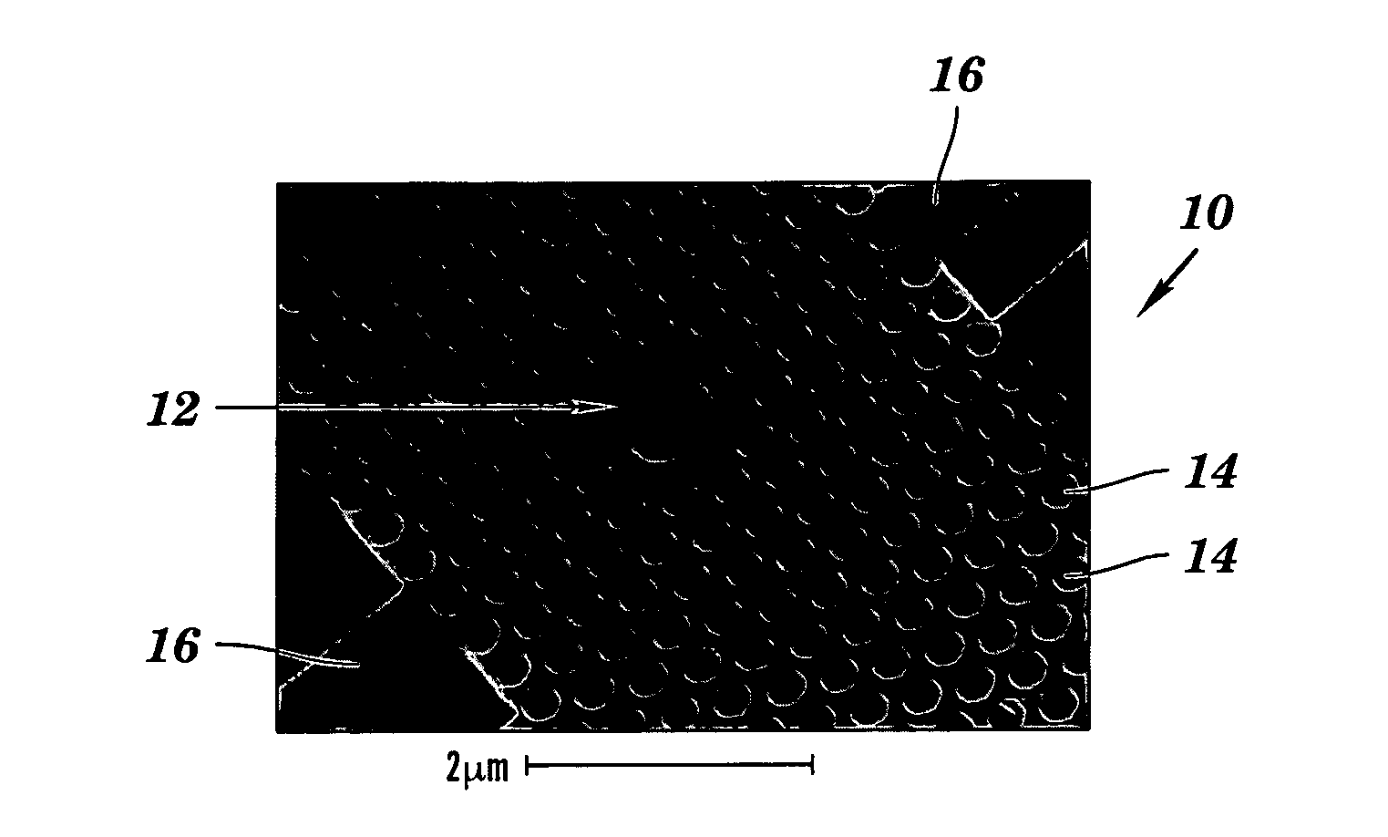
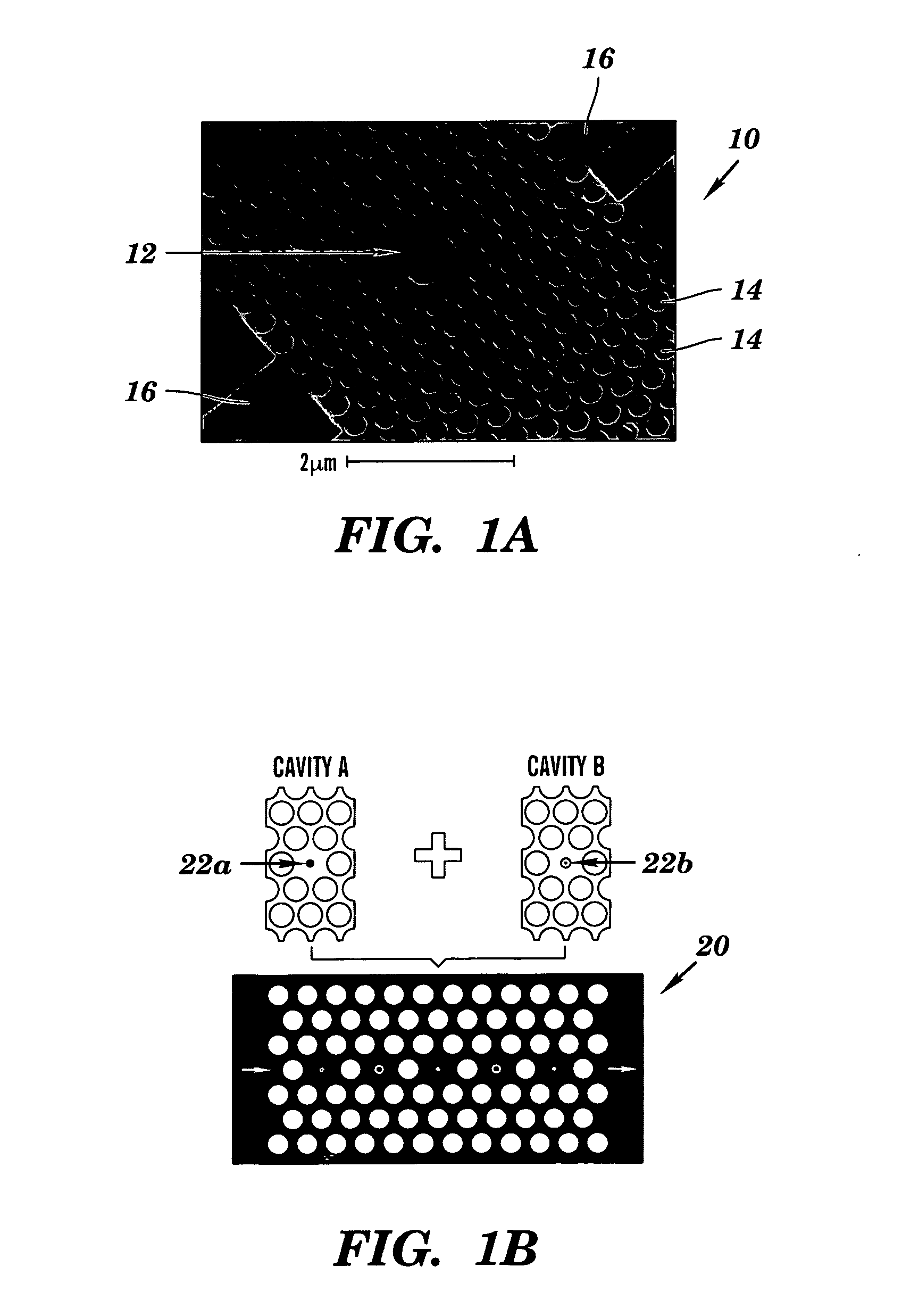
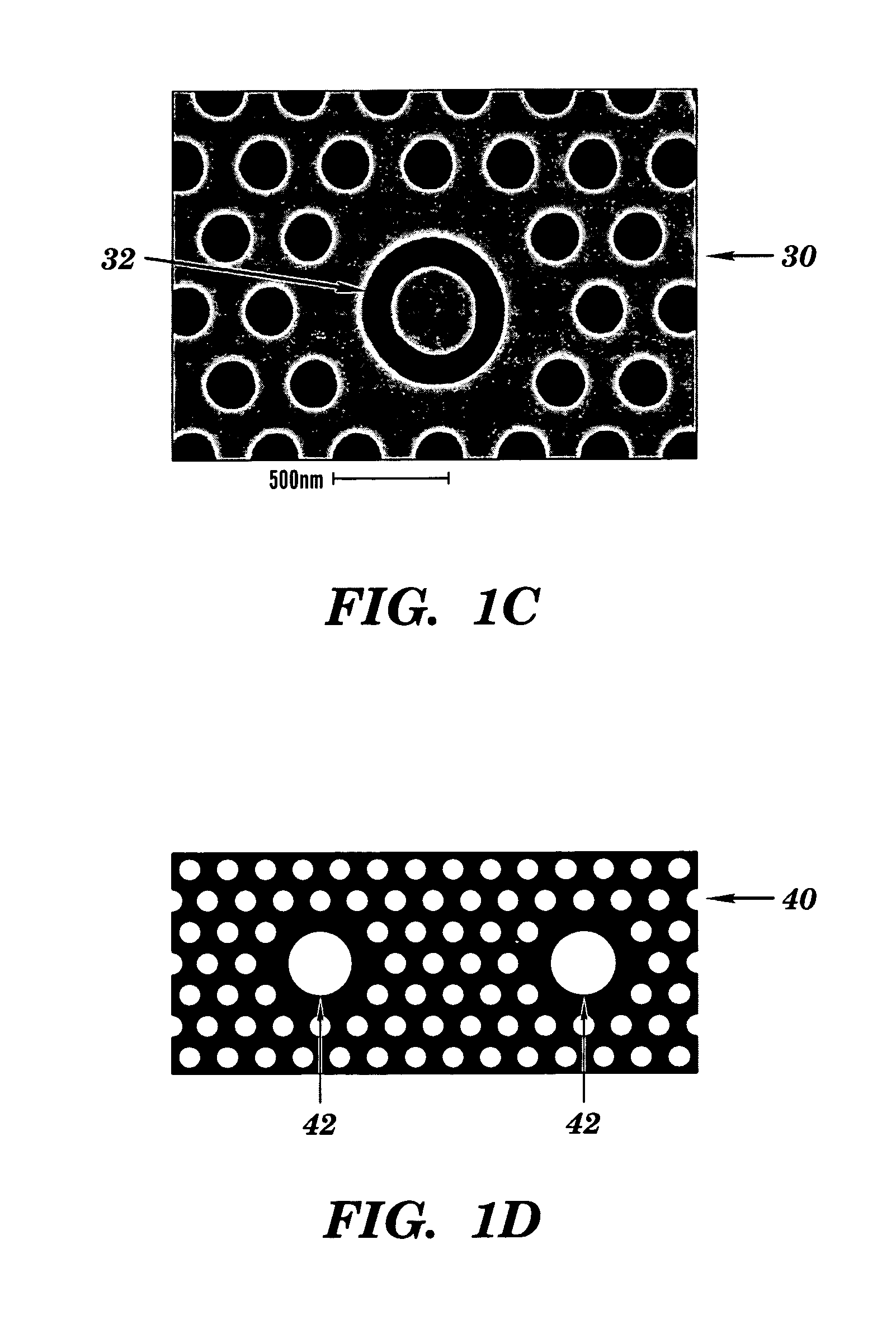
[0103]The structure depicted in FIG. 8 contains a hexagonal array of cylindrical air pores in a 400 nm-thick silicon (Si) slab separated from the Si substrate by 1 μm of SiO2 to provide a good vertical confinement for the propagation modes. The photonic crystal has a lattice constant a of 465 nm and a pore diameter d of 270 nm. The defect was introduced by reducing the center pore diameter to 140 nm. Such a configuration gives rise to a resonance in the bandgap close to 1.58 μm for even (TE-like) modes. Here, TE-like mode was studied because there is no bandgap for TM-like modes beneath the light cone. Two tapered ridge waveguides were used to couple light in and out of the microcavity. They were tapered from 2 mm down to ˜0.7 μm to match the mode of the microcavity. Light is coupled along the Γ-M direction, because the resonance mode in-plane leakage is mainly in the Γ-M direction and, hence, the couplin...
example 2
Sensor Performance Characterization for Binding of Bovine Serum Albumin Using Glutaraldehyde Probe
[0106]To characterize the sensor performance, glutaraldehyde-bovine serum albumin (BSA) binding was used as the model system because glutaraldehyde has a strong affinity for BSA. The pore size of the device is ˜30 times larger than the protein hydrodynamic diameter (Kuntz et al., “Hydration of Proteins and Polypeptides,”Adv. Protein Chem. 28:239-345 (1974); Squire et al., “Hydrodynamic Properties of Bovine Serum Albumin Monomer and Dimer,”J. Biochem. 7:4261-4272 (1968), each of which is hereby incorporated by reference in its entirety), which guarantees a high infiltration efficiency of the proteins into the device and facilitates the uniform formation of a monolayer-thick coating on the pore walls.
[0107]To prepare the surface for the capture of BSA proteins, the device was first thermally oxidized at 800 to form a silica-like internal surface. The sensor was then treated with 2% amino-...
example 3
Sensor Sensitivity for Binding of Streptavidin Using Biotin Probe
[0114]Two important benchmarks for a biosensor are sensitivity and selectivity. Previous experiments demonstrated the capability of detecting the dehydrated protein layer thickness as thin as 1 Å. However, glutaraldehyde-BSA binding is a non-specific binding process; thus, it only shows the presence of bio-molecules inside the microcavity without specifying the type of proteins.
[0115]To demonstrate the selectivity of this device, biotin-streptavidin coupling was used as a model system. First the device was functionalized with the probe molecule (Sulfo-NHS-LC-LC-Biotin), which has an extremely high binding affinity for the target molecule (Streptavidin). Each streptavidin molecule has four equivalent sites for biotin which makes it an excellent molecular linker in many assays. As shown in FIG. 12A, the target molecules were immobilized on the pore walls in the presence of the probe molecules. The experimental results sh...
PUM
 Abstract
Description
Claims
Application Information
 - R&D
- Intellectual Property
- Life Sciences
- Materials
- Tech Scout
- Unparalleled Data Quality
- Higher Quality Content
- 60% Fewer Hallucinations
Browse by: Latest US Patents, China's latest patents, Technical Efficacy Thesaurus, Application Domain, Technology Topic, Popular Technical Reports.
© 2025 PatSnap. All rights reserved.Legal|Privacy policy|Modern Slavery Act Transparency Statement|Sitemap|About US| Contact US: help@patsnap.com


