
Markers of matrix gene expression and cellular differentiation in chondrocytes

a chondrocyte and matrix gene technology, applied in the field of tissue engineering, can solve the problems of limited knowledge of the molecular level processes involved in the pathogenesis of oa, influence the phenotypic stability of the cell culture, and compromise the chondrogenic capacity of the said cells,

Inactive Publication Date: 2010-12-30
UNIV GENT
View PDF0 Cites 5 Cited by
  • Summary
  • Abstract
  • Description
  • Claims
  • Application Information

AI Technical Summary

Benefits of technology

[0024]Thus, in an even further embodiment, the phenotypic stability of the cell culture of isolated pluripotent cells and / or chondrocytes, is further characterised in the capability of said cells to retain COL2A1 and aggrecan expression upon reconstitution in their endogenous environment.
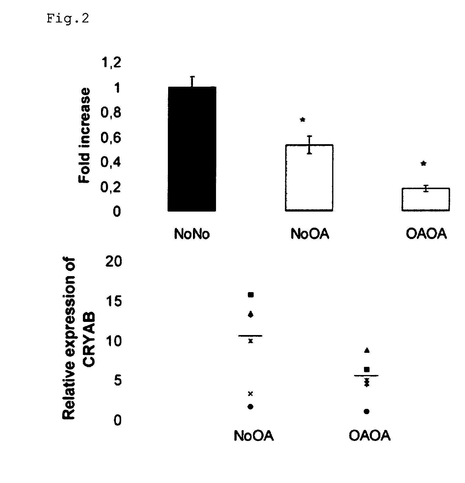
[0029]In a second objective, the present invention provides the use of the methods according to the invention, to identify cell culture conditions or treatments (compounds) that enhance the phenotypic stability of a cell culture. It is known for instance, that extensive cell expansion of isolated pluripotent skeletal cells, compromises the chondrogenic capacity of said cells. Some treatments, such as for example addition of growth factors / reagents, or cell culture conditions, such as for example non-adherent growth conditions, may influence the phenotypic stability of the cell culture. Monitoring the expression of CRYAB, optionally with HSP27, the present invention provides the tools to identify treatments and / or cell culture conditions that prevent or delay the dedifferentiation of the isolated pluripotent skeletal cells, and thus enhance the chondrogenic capacity of the in vitro culture of cells.

Problems solved by technology

Until now, the knowledge of the processes involved in the pathogenesis of OA at the molecular level is limited.

Method used


Image

  • Markers of matrix gene expression and cellular differentiation in chondrocytes
  • Markers of matrix gene expression and cellular differentiation in chondrocytes
  • Markers of matrix gene expression and cellular differentiation in chondrocytes

Examples


example 1

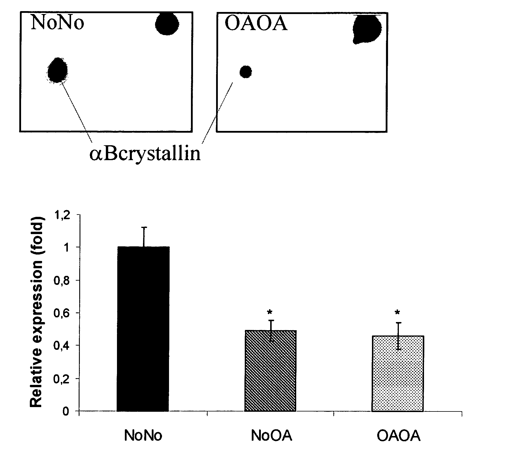
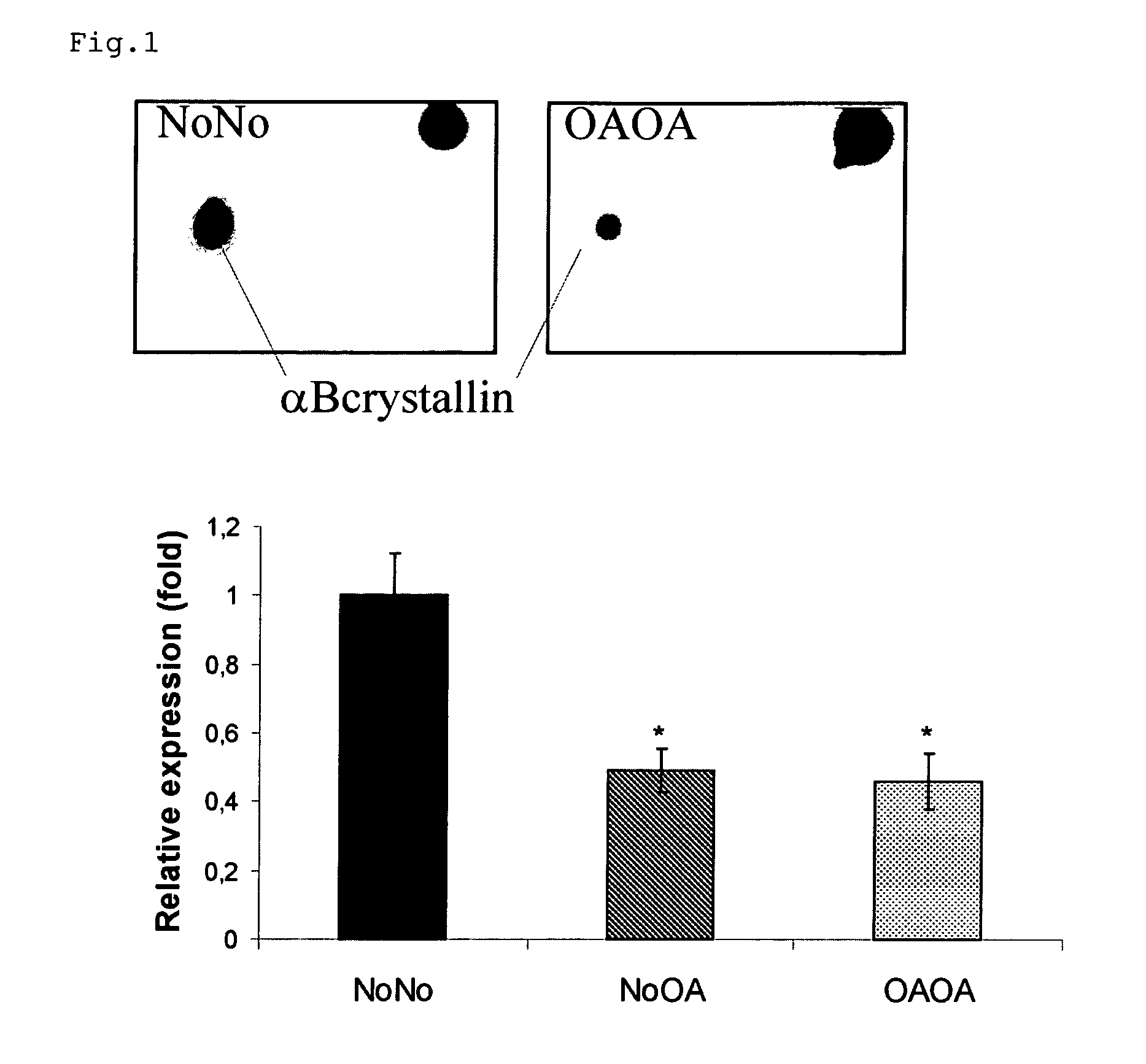
αBcrystallin, a Potential Mediator of Matrix Gene Expression in Chondrocytes During the Development of Osteoarthritis

1.1 Materials and Methods

2-DE Analysis

[0103]In a preliminary study, protein extracts of chondrocytes (6 NoNo, 7 NoOA and 7 OAOA samples) were analyzed by a 2-DE approach, as described in [8]. Briefly, soluble and hydrophobic fractions of protein extracts of chondrocytes were separated by 2-DE. Sypro Ruby stained gels were scanned and analyzed using PDQuest V 7.1. Statistically significant differentially expressed spots (p<0.05; Mann-Whitney U-test) were excised and subjected to tandem mass spectrometry for identification.

Isolation of Chondrocytes

[0104]Human articular chondrocytes were isolated as previously described. Articular knee cartilage from donors without arthropathy (NoNo) (3 male, 2 female, mean age: 44±25 years) was obtained within 24 h post-mortem. All donors had died as a result of trauma or a brief illness and none of them had been receiving corticosteroi...

example 2

HSP27 in OA-Affected Chondrocytes

2.1 Materials and Methods

Isolation and Culture of Chondrocytes

[0120]Human articular knee cartilage was obtained from 4 donors (one male, three female, median age ±SD: 70±10.9 years) after total knee arthroplasty. The study protocol was approved by the local Ethics Committee. The cartilage was diced into small fragments and chondrocytes were isolated by sequential enzymatic digestion (hyaluronidase, pronase, collagenase (Sigma-Aldrich, Steinheim, Germany)) as described in detail elsewhere [9]. Trypan blue exclusion revealed that >95% of the cells were viable after isolation.

[0121]Chondrocyte cultures in alginate beads were prepared and maintained as described previously [8]. For each sample, 15-17.5×10⁶ chondrocytes were lysed and treated as described below.

Sample Preparation and Protein Digestion

[0122]Total cell lysate—Isolated chondrocytes were resuspended and lysed in a denaturing buffer containing 8 M urea. After reduction and alkylation, proteins...

example 3

Further Characterization of the End Point Marking the Phenotypic Change

3.1. Materials and Methods

Real-Time PCR

[0129]After the culture period, Trizol (Invitrogen) was added to the isolated cells, and RNA was extracted according to the manufacturer's instructions, followed by an additional purification step (RNeasy mini-kit (Qiagen)). This step included the digestion of DNA by deoxyribonuclease I (Invitrogen). cDNA was synthesized with oligo(dT) primers using the Superscript kit (Invitrogen).

[0130]Real-time PCR was performed using the ABI 7000 Sequence Detection System (Applied Biosystems). Each reaction utilized 20 μl of iTaq Supermix with Rox (Bio-Rad, Hercules, Calif.) and 5 μl of cDNA and was performed in triplicate. The thermocycler conditions were 2 min at 50° C., followed by 2 min at 95° C. and 45 cycles, each at 95° C. for 15 s and 60° C. for 1 min. Expression levels were normalized to those of human GAPDH and PPIA. Relative quantitation was calculated using the 2−ΔΔCt method....


PUM

PropertyMeasurementUnit
Fractionaaaaaaaaaa
Fractionaaaaaaaaaa
Fractionaaaaaaaaaa

Abstract

The invention relates generally to the field of tissue engineering. More particularly, the present invention relates to methods for identifying a population of cells suitable for the repair of connective tissue, including cartilage. The invention further provides methods and compositions related to the generation of a population of cells suitable for the repair of cartilage, in particular in the repair of cartilage degeneration associated with osteoarthritis. Methods of using said cells thus identified or thus generated, in methods to extend the period of cell manipulation, i.e. to increase the yield of cells suitable in the aforementioned methods, are also provided by the present invention.

Description

FIELD OF THE INVENTION[0001]The invention relates generally to the field of tissue engineering. More particularly, the present invention relates to methods for identifying a population of cells suitable for the repair of connective tissue, including cartilage.[0002]The invention further provides methods and compositions related to the generation of a population of cells suitable for the repair of cartilage, in particular in the repair of cartilage degeneration associated with osteoarthritis.[0003]Methods of using said cells thus identified or thus generated, in methods to extend the period of cell manipulation, i.e. to increase the yield of cells suitable in the aforementioned methods, are also provided by the present invention.BACKGROUND TO THE INVENTION[0004]Osteoarthritis (OA) has become one of the major health problems in the western world [1]. The key hallmark of this disease is a slow progressive degeneration of the articular cartilage [2]. Articular cartilage forms a speciali...

Claims


Application Information

Patent Timeline
IPC IPC(8): A61K35/12C12Q1/02C12N5/071C12Q1/68A61P19/02
CPCG01N33/5023G01N2800/105G01N33/56966G01N33/5073A61P19/02
Inventor LAMBRECHT, STIJNDEFORCE, DIETERELEWAUT, DIRKVERBRUGGEN, AUGUST
Owner UNIV GENT
Learn More