
Home healthcare management system and hardware

a technology for managing systems and home healthcare, applied in the field of home healthcare management systems, can solve problems such as cumbersome translation of medical knowledge into a series of questions that can lead the patient into a diagnosis, high testing costs, and inability to meet patient needs, so as to reduce insurance costs

Active Publication Date: 2011-01-20
THE CLEAR LAKE MEDICAL FOUND PATENTS
View PDF98 Cites 21 Cited by
  • Summary
  • Abstract
  • Description
  • Claims
  • Application Information

AI Technical Summary

Benefits of technology

[0037]The present disclosure provides a very low cost system for at-home healthcare management, and an optional Internet linked professional and administrating supporting structure. Families unable to afford insurance will be able to run laboratory tests and address their own primary health care needs without the additional cost of monthly insurance premiums. It is contemplated that the home healthcare personal computer (HHPC) and tests could be purchased at a local drug store.
[0040]More specifically, and in relation to digital imaging, the present invention solves the problem of having to: 1) use large format cameras for the extra overlapping pixels; 2) use encoders for fine positioning; 3) contend with encoder induced image position distortion; 4) rely on a slide-based coordinate system; 5) employ computationally expensive algorithms for pixel alignment and pixel striping; 6) have prior knowledge of magnification and resolution for positioning, display, and internet transfer; and 7) having to discard images due to corrupted header files. The invention uses motion and velocity vectoring methods for capturing and tiling images using a digital microscope. Motion and velocity vectoring methods are fast and typically have better than half-pixel resolutions thereby fulfilling Nyquist requirements and mitigating error accumulation found in prior art pixel alignment methods.
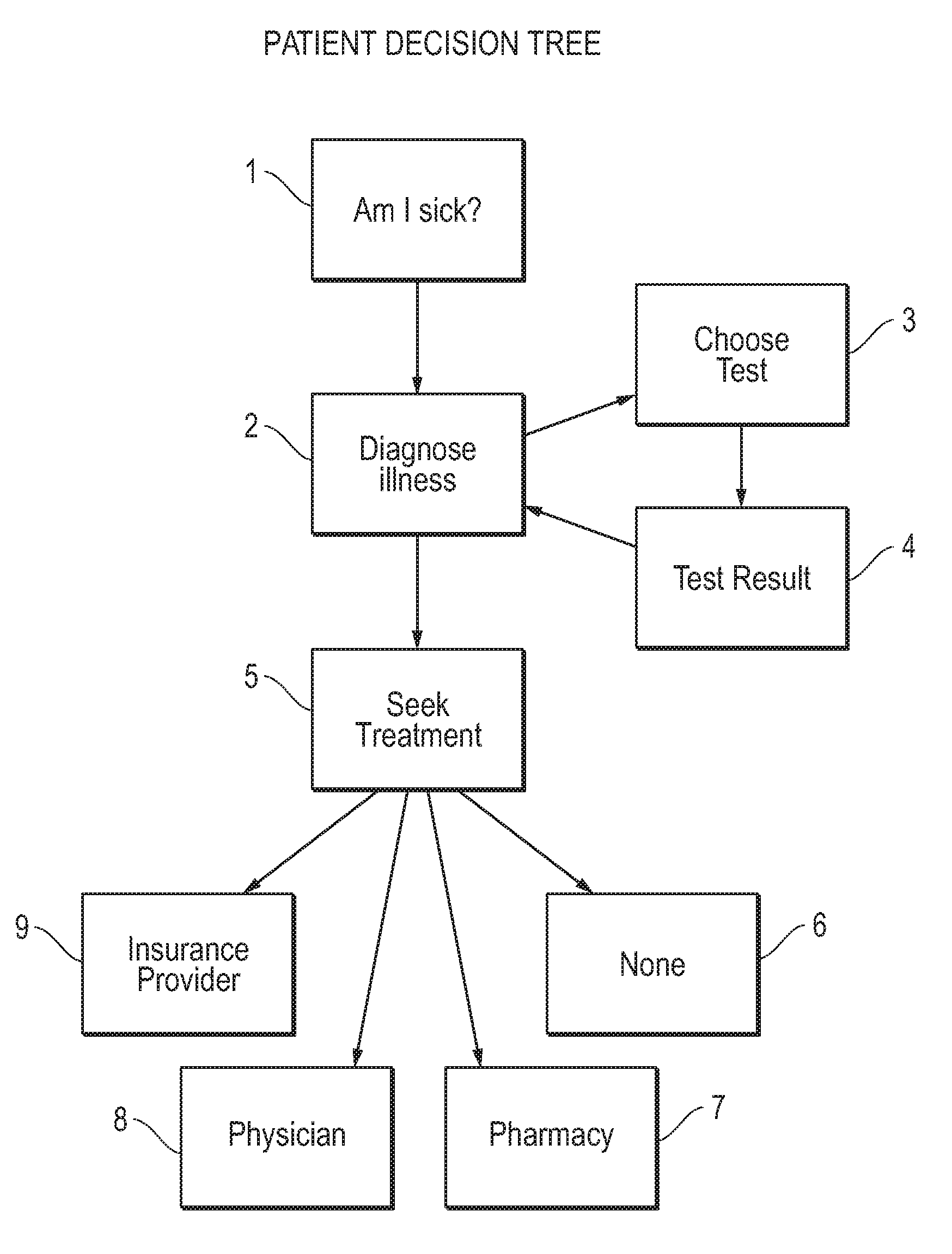
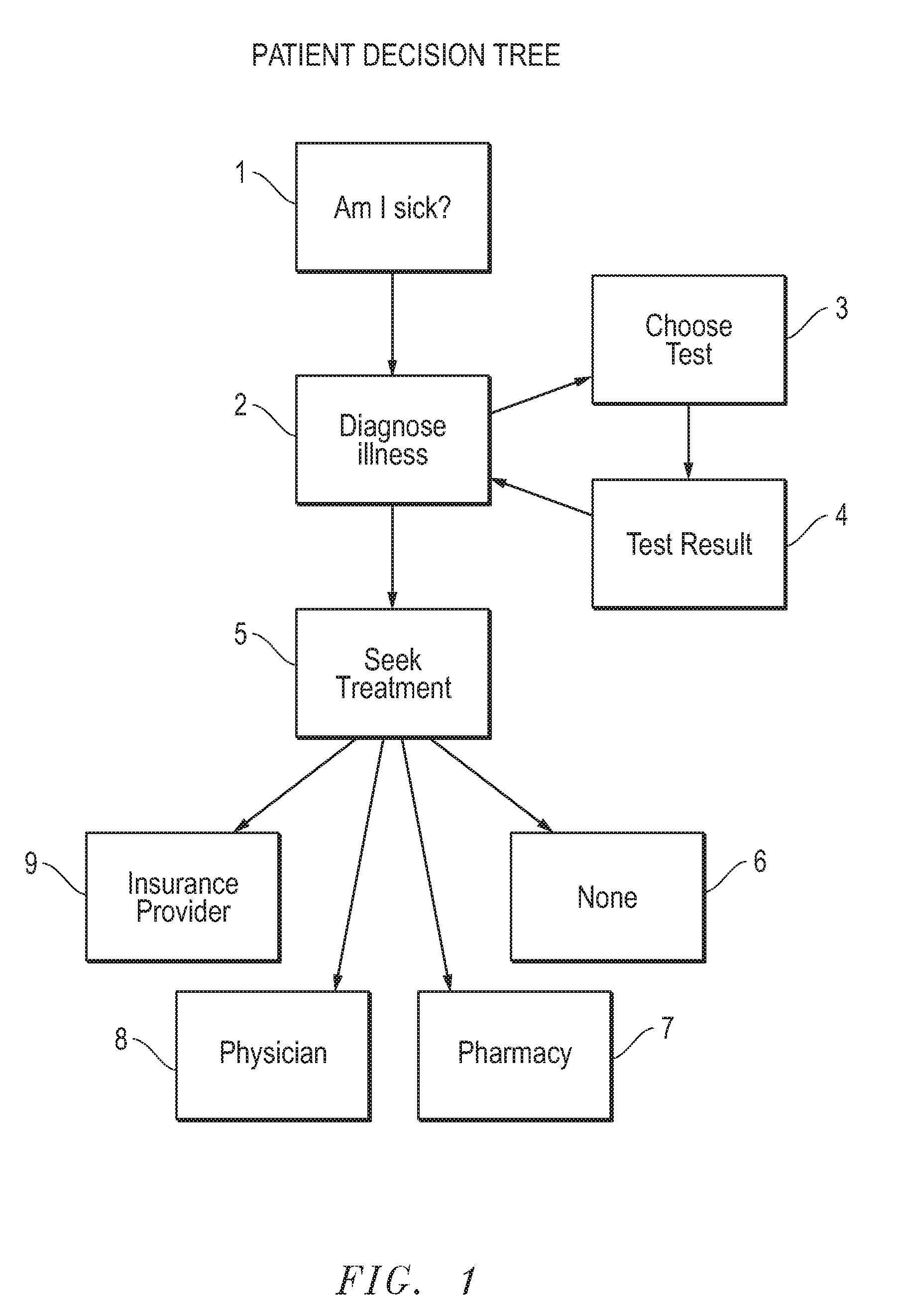
[0042]In one embodiment and example of the system, and illustrating the implementation of the optional insurance provider support, Health Care Management (HCM) insurers would provide patients with an at-home instrument for laboratory and physiological testing, an Internet link to professional and administrative support, and credit for five disposable tests per year per patient. When a patient feels ill, for an example a sore throat, the patient conducts the basic physiological tests, e.g., blood pressure and temperature, then runs a Strep Test (streptococcal) thereby using one of the five allocated tests per year. If symptoms are mild the patient may not wish to proceed any further, having assured themselves that they do not have strep throat. However, if symptoms or concerns are more severe the patient may run a Mono Spot test (mononucleosis). If the Strep Test was positive, the patient may opt to connect through the Internet to their insurer and have an antibiotic prescription sent to the nearest pharmacy (or have the pharmacy deliver the prescription to their home). The insurer provides other services over the internet as well; they may provide guidance and recommend additional tests, they may visually link the patient to a physician for a visual assessment or to answer questions, or they may recommend additional evaluation at a nearby medical facility, providing directions and making the appointment, or providing services to backup medical records and monitor progress. In my practice, most patients knew what was wrong and were seeking a confirmatory test and a prescription for relief. Moving primary care into the hands of the patients is more efficient and will greatly reduce insurance costs while improving health care. Today, patients are better educated, have access to vast amounts of medical information through the internet, and are capable of diagnosing most common illnesses, and have general knowledge of the therapeutics required having had or known others with similar illnesses.

Problems solved by technology

This was in part due to the limited availability of education, and due to the limited access to medical knowledge bases.
Prior to the general availability of the Internet, patients had to rely on limited medical resources.
The translation of medical knowledge into a series of questions that can lead the patient into a diagnosis is cumbersome, impractical and fraught with error.
Often the listener is uncertain that the press 1 service is exactly what they are looking for, and may try another, ending up at an incorrect end-point.
Testing can be expensive because it includes the cost of the facilities to house the test and personnel, the cost of the instrument that conducts the tests, the government certification and compliant costs, the personnel costs to draw blood, personnel to maintain instruments, personnel to certify compliance, the cost of the test itself, and the administrative costs (e.g., computers, personnel, accounting, health care insurance, etc).
In addition to the issue of non-medical personnel reviewing records and photos, there is an issue of security where servers are broken into or files, taken home on laptops, are by third parties.
However, the number of tests is limited due to the special instrumentation and computer processing required.
Health care costs continue to soar in spite of newer technologies purporting to reduce cost; primary care providers cannot support the 300 million people in the United States, and the spill-over is disrupting emergency care facilities.
The medical paradigm of physician centric primary care is unable to sustain the quality of patient care administered two decades ago.
Digital microscopes are used to enlarge an object, for example a red blood cell in a whole blood smear, but are not designed for colorimetric or fluorometric laboratory assays such as glucose testing, antigen testing and the like.
The tradeoff is a considerably larger objective size, more optical elements in the objective, greater difficulty in assembling and aligning the optical elements, and the need to extend the optical path beyond the objective, i.e., where an additional set of optical elements are required for image formation.
However, when a slide is removed from a digital microscope and later placed back into the same position in the same digital microscope, the original encoder coordinates are no longer valid.
Relying on the fixed coordinate approach to scan and tile slides is costly, cumbersome, and fraught with error.
Encoders are expensive and add complexity and points of failure.
Moreover, others have confused encoder resolution with accuracy.
Moreover, the absolute accuracy is only valid in a specific environment at a precise temperature.
Relying on encoders for abutting image tiles can lead to erroneous image results due to the absolute positional inaccuracy.
For example, if a specific lot of encoders is used for deriving a coordinate system, and that lot is 3,000 nm longer than it should be, images collected and assembled based on that coordinate system will be distorted; this could possibly misdirect a clinical decision.
Encoders, in general, add cost to the system, increase the degree of electrical and software complexity increase assembly and maintenance costs, and add another possible failure point.
Not only is this costly in time, but a typical pathology laboratory would generate terabytes of useless data that federal law mandates must be properly stored for many years, backed up, and security maintained.
Superfluous images of virtual microscope slides, as is seen in the prior art, unnecessarily displaces important information.
There is significant programming and file overhead with the prior art coordinate based image tiling.
A header file is needed for coordinate translation, and if lost or corrupted, image formation can fail.
Additionally, image display is slow—when a file is downloaded from the hard drive it must first go through a coordinate translation algorithm that is unique to coordinate based image tiling to derive the image display coordinates.
Consequently, rounding errors and offset error inaccuracies accumulate with every captured image.
The accumulated error distorts both column and row alignment of tiled images.
This method requires a more expensive camera because of the extra pixels required to overlap images.
Segmentation algorithms for calculating mal-alignment are inaccurate, slow, and computationally intensive.
And, preprocessing images to strip out unused pixels in overlapping area around the image periphery requires significant computation time thereby further slowing down storage.
However, such detection methods require a specific sequence of preparation that is beyond the training of the average person.
Automation comes at a substantial price: capital equipment costs, federal regulatory certification costs, training costs, maintenance costs, and facilities costs.
And, the automation equipment is not disposable.
Consequently, many diagnostic tests cannot be run in small laboratories, physician offices, or at patient homes.
This is especially true of PCR assays, where processing errors are poorly tolerated and the processing methods and temperatures are more complex.
Certain materials related to Delran and high molecular weight polypropylenes may reduce adherence but none eliminate adherence.
A washing step limits the use of many assays to skilled personnel.
Many assays require thermal management—especially PCR assays where thermal management is complex.
They can be heavy and costly, and they are not disposable.
This is one of the technical difficulties that has impeded the general introduction of genetic testing.
Size, durability, and reliability are focus issues for first responder emergencies e.g., anthrax release, bioterrorism, avian flu pandemic, where assays need to be run immediately without prior training, or in difficult terrain such as in a cave, or in a situation where diverting attention to the assay could prove deadly.

Method used


Image

  • Home healthcare management system and hardware
  • Home healthcare management system and hardware
  • Home healthcare management system and hardware

Examples


Embodiment Construction

[0062]The present disclosure provides a patient centric system wherein the patient is responsible for diagnosis, testing, management and medical records handling within their home, and wherein their insurer offers optional ancillary services through an Internet connection. Those services include, but are not limited to, test evaluations, consultations with professional health care providers, prescription services, medical records maintenance, and administrative services. A person of ordinary skill in the art will recognize that other services can also be provided with the disclosed system.

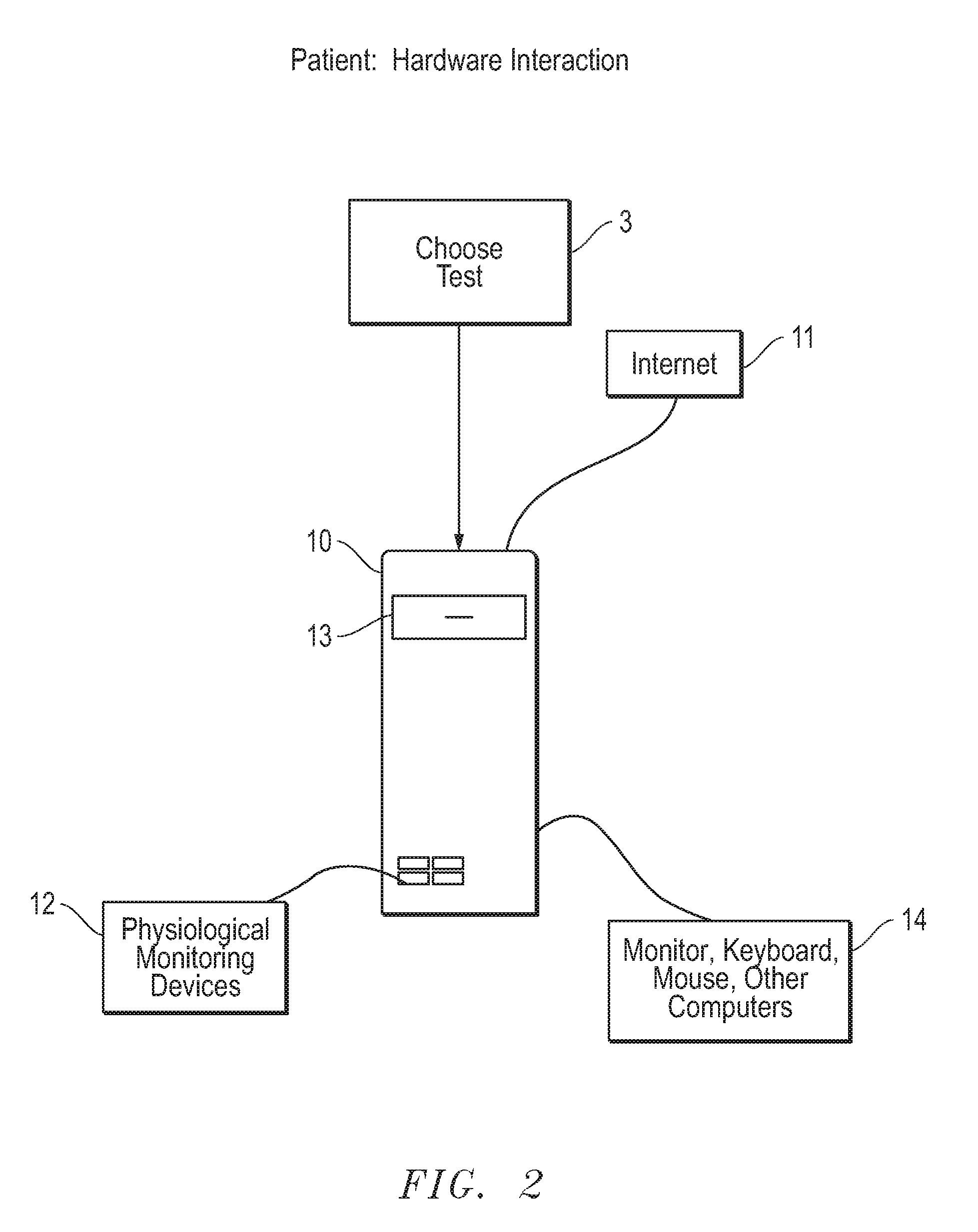
[0063]The system comprises a computer with interfaces for physiological testing, a digital microscope for evaluating diagnostic tests, a connection to the Internet, and an optional insurer computer Internet connection that supports a network for data base and data storage, for connections to video and audio based consultations, and for administrative support.

[0064]The process of adapting a PC to re...


PUM


Abstract

A home healthcare management system wherein a patient conducts self diagnoses and self testing, and manages their own medical records at home. A digital microscope is utilized as part of the system that is smaller, of lower cost, faster, of a higher dynamic range, and has a higher resolution than conventional microscopes. The microscope consists of an illumination source, a spatial sub-sampling device and a detector device. The digital microscope provides a vectored method of collecting images from a digital microscope that is independent of the optical resolution, and a slide based coordinate system, and a method of displaying images and communicating such images over the Internet in a file format that does not require a header or prior knowledge of magnification, coordinate system, or tiling structure. The system further includes an interface for physiological monitoring devices and a connection to the Internet for more comprehensive services.

Description

CROSS-REFERENCE TO RELATED APPLICATIONS[0001]This application claims the benefit of U.S. Provisional Application Ser. No. 61 / 225,693, filed on Jul. 15, 2009, entitled: HOME HEALTHCARE MANAGEMENT SYSTEM AND HARDWARE, by inventor Glenn F. Spaulding.STATEMENT REGARDING FEDERALLY SPONSORED RESEARCH OR DEVELOPMENT[0002]Not Applicable.REFERENCE TO A “MICROFICHE APPENDIX”[0003]Not Applicable.BACKGROUND OF THE INVENTION[0004]1. Field of the Invention[0005]The present invention relates to a system for providing home healthcare management. More specifically, the present invention relates to a home healthcare system for providing home diagnoses, testing, and management of the information derived therefrom.[0006]2. Description of the Related Art[0007]Medical Diagnosis & Diagnostics[0008]Throughout recorded time health care has increasingly relied upon trained healthcare personnel. This was in part due to the limited availability of education, and due to the limited access to medical knowledge b...

Claims


Application Information

Patent Timeline
IPC IPC(8): A61B5/00H04N7/18H04N5/225G06K9/00G01J3/46G06Q50/00G06Q10/00G06Q40/00G16H20/10G16H40/63G16H40/67
CPCA61B5/0006A61B5/0008G06Q50/22G02B21/0004G06F19/3418A61B5/02055G16H40/67G16H20/10G16H40/63
Inventor SPAULDING, GLENN F.
Owner THE CLEAR LAKE MEDICAL FOUND PATENTS
Learn More