Bacteriophage/Quantum-Dot (Phage-QD) Nanocomplex to Detect Biological Targets in Clinical and Environmental Isolates

a technology of phage-qd and nanocomplex, which is applied in the field of bacteria/quantumdot (phage-qd) nanocomplex to detect biological targets in clinical and environmental isolates, can solve the problems of low signal-to-noise ratio due to detection pathogenic bacteria, low sensitivity and rapidity, and low photo-stability such as fast photobleaching

Inactive Publication Date: 2011-04-14
GOVERNMENT OF THE UNITED STATES OF AMERICA AS REPRESENTED BY THE SEC OF COMMERCE THE NAT INST OF STANDARDS & TEHCNOLOGY +1
View PDF0 Cites 26 Cited by
  • Summary
  • Abstract
  • Description
  • Claims
  • Application Information

AI Technical Summary

Problems solved by technology
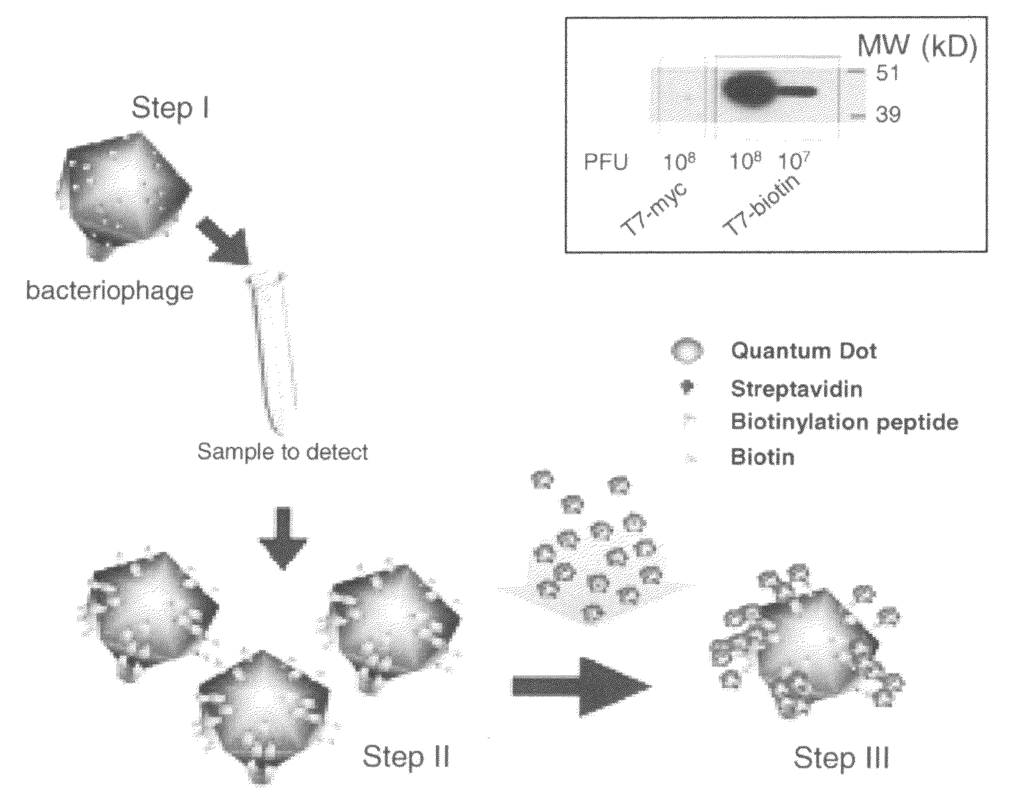
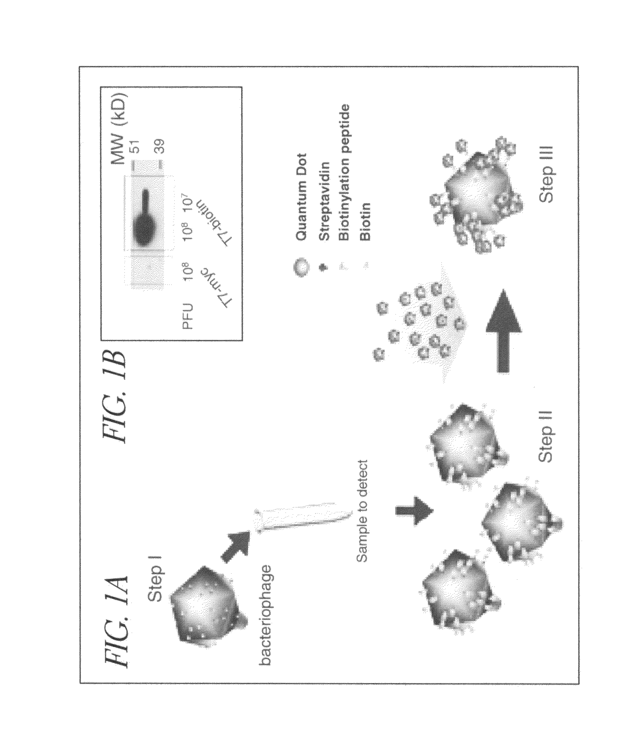
Two limiting features when detecting pathogenic bacteria are sensitivity and rapidity.
Common fluorophores (e.g., green fluorescence protein and luciferase) used as reporters have two major disadvantages: low signal-to-noise ratio due to auto-fluorescence of clinical samples and of bacterial cells and low photo-stability such as fast photobleaching.

Method used


Image

  • Bacteriophage/Quantum-Dot (Phage-QD) Nanocomplex to Detect Biological Targets in Clinical and Environmental Isolates
  • Bacteriophage/Quantum-Dot (Phage-QD) Nanocomplex to Detect Biological Targets in Clinical and Environmental Isolates
  • Bacteriophage/Quantum-Dot (Phage-QD) Nanocomplex to Detect Biological Targets in Clinical and Environmental Isolates

Examples


example 1

Engineering the T7-Bio and T7-Myc Phages

[0093]We used the T7Select System (Novagen) for engineering and packaging of DNA into T7 phage particles. For the T7-bio we used two phosphorylated primers: 3′L6bio and 5′L6bio, containing overhang sequences for ligation with HindIII and EcoRI digested phage arms DNA, respectively (upper case), a 6 amino acid linker coding sequence (underlined), followed by the biotinylation peptide coding DNA (lower case) and a stop codon (bold):

3′L6bio:(SEQ ID NO: 6)5′-AGCTTttagtgccattcgattttctgagcttcgaagatgtcgttcaggcctgaaccacgcggccgcaacG-3′5′L6bio:(SEQ ID NO: 7)5′-AATTCgttgcggccgcgtggttcaggcctgaacgacatcttcgaagctcagaaaatcgaatggcactaaA-3′.

[0094]The primers were annealed to each other by heating at 95° C. for 5 min in ligation buffer and cooling at room temperature, ligation to T7 arms was done as recommended by the manufacturer. For the engineering of the T7-myc phage we used the primers MYC1: 5′-AATTCtggtggcagcggatctgagcagaagctgatcagcgaggaagatcttaattaaA-3′ (...

example 2

Engineered Phage Containing birA Gene and Biotinylation Domain

[0099]The birA gene was engineered into phage along with a biotinylation domain to allow more of the displayed peptides to be biotinylated, thereby increasing the detection sensitivity. The BPL of E. coli, birA gene, was clone as a transcriptional fusion with the phage Capsid-bio under the phage promoter.

[0100]This engineered phage (capsid-bio-birA) had about a 100 fold higher level of biotinylation than the Capsid-bio engineered phage as judged by western blot analysis with streptavidin-HRP. This new engineered phage overcomes potential limitation of the endogenous BirA protein such that most of the displayed biotinylation domain becomes biotinylated.

example 3

Phage-QD Complexes and Analysis of Quantized Levels of QDs in Complexes Binding to Bacteria

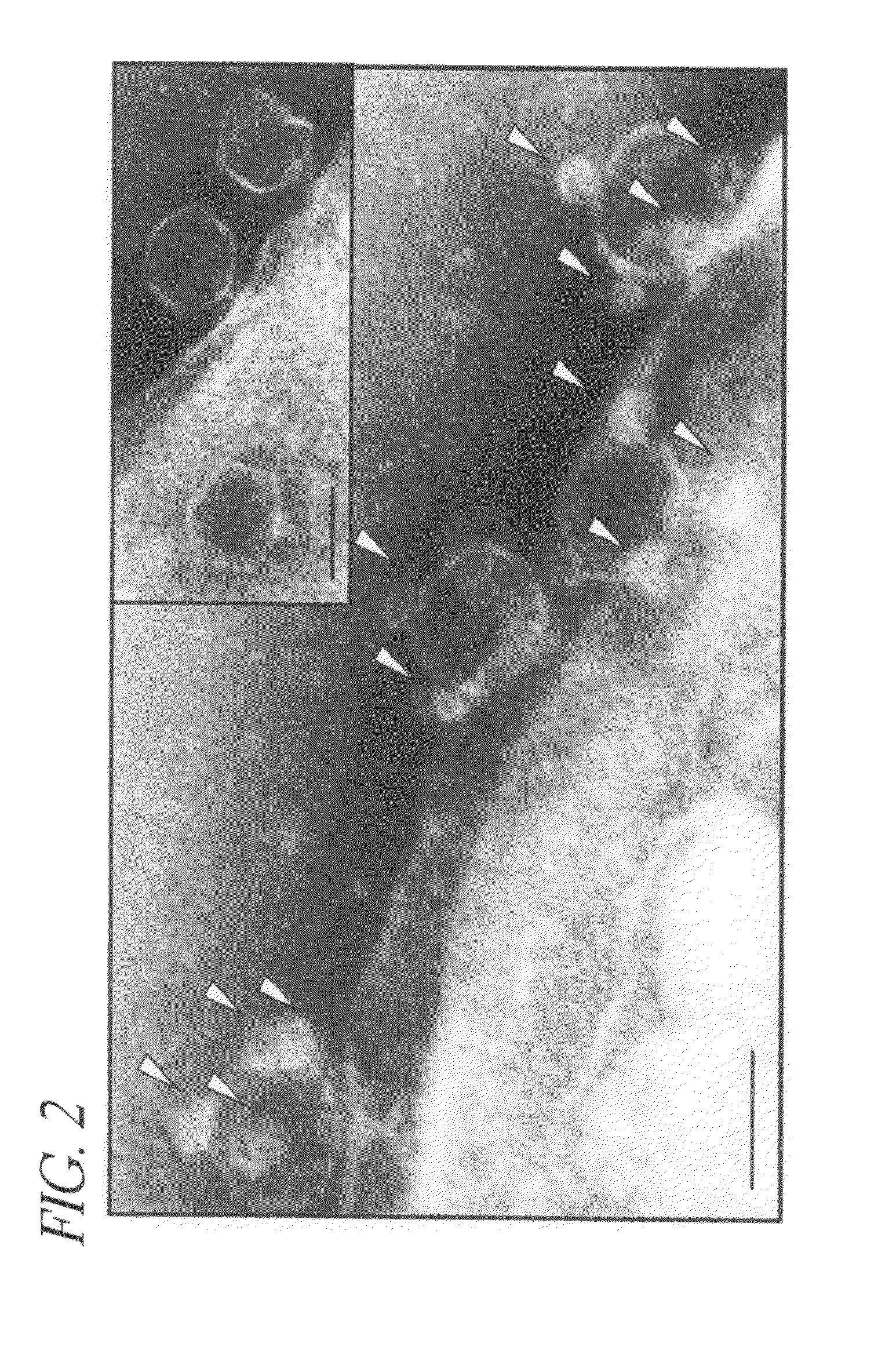
[0101]A fluorescence image of phage-QD complexes' spread on a glass coverslip is shown in FIG. 18A (top). Each bright spot in the image exhibits fluorescence signal from one or two of QDs attached onto different phage. The image was time-averaged from 500 movie frames taken at the rate of 100 ms per frame. In FIG. 18A (bottom), a time-transient intensity along the line of (a-b) shows that the fluorescent spot near “a” shows a single level quantized blinking indicative of one QD, while the other fluorescent spot near “b” shows two-levels of quantized blinking from two QDs. m1 and m2 in the intensity scale bar correspond to two local maxima of the (occurrence vs. intensity) histogram calculated from the intensity fluctuation of the 2 QD spot.

[0102]Time-averaged bright field and fluorescence imaging of bacteria cells was done after an attempt to bind maximum number of phage-QD complexes by adding...


PUM

PropertyMeasurementUnit
timeaaaaaaaaaa
full width half maximumaaaaaaaaaa
massaaaaaaaaaa

Abstract

The invention is related to a non-biotinylated bacteriophage that comprises a nucleic acid sequence encoding a biotinylation domain, a complex that comprises a biotinylated bacteriophage and a biotin-specific ligand conjugated bioconjugate, and a method of detecting a bacterial cell in a sample comprising contacting the sample with a non-biotinylated bacteriophage that comprises a nucleic acid sequence encoding a biotinylation domain, wherein the bacteriophage is specific to the bacterial cell.

Description

RELATED APPLICATIONS[0001]This application claims the benefit of U.S. Provisional Application No. 60 / 654,784 filed Feb. 18, 2005, which is hereby incorporated by reference in its entirety.FIELD OF THE INVENTION[0002]The invention pertains to combining quantum dots (QDs) with engineered phage for the specific identification of biological targets including bacterial strain(s) or cells from clinical or environmental isolates.BACKGROUND OF THE INVENTION[0003]The number and diversity of bacteriophages in the environment provide a promising natural pool of specific detection tools for pathogenic bacteria. Currently there are several methods for detection of pathogenic bacteria that exploit phage (McKinstry, M. & Edgar, R. in Phages: Their Role in Bacterial Pathogenesis and Biotechnology, eds. Matthew, K.-W., Friedman, D.-I., & Adhya, S.-L. (ASM Press, Washington, D.C. 2005), pp. 430-440): a plaque assay for detection of Mycobacterium tuberculosis (McNerney, R. et al. 2004 J Clin Microbiol...

Claims


Application Information

Patent Timeline
Patent Type & Authority Applications(United States)
IPC IPC(8): C12Q1/70C12N7/00A61K35/13
CPCA61K35/13B82Y15/00G01N33/588G01N33/56911C12N2810/10
Inventor HWANG, JEESEONGEDGAR, ROTEMMCKINSTRY, MICHAELGIULIAN, GARYMERRIL, CARLADHYA, SANKAR
Owner GOVERNMENT OF THE UNITED STATES OF AMERICA AS REPRESENTED BY THE SEC OF COMMERCE THE NAT INST OF STANDARDS & TEHCNOLOGY
Why Eureka
Try Eureka