Methods to increase permeability of corneal epithelium and destabilize stromal collagen fibril network

a stromal collagen and network technology, applied in the field of corneal epithelium permeability increase and destabilization of stromal collagen network, can solve the problems of corneoplasty induced astigmatism or double vision, wear of retainer lenses, and difficulty in accurately placing contact lenses with respect to the axis of vision to control the reshaping of corneal tissue, etc., to facilitate the stabilization of the reshaping corneal curvature and enhance the ocular delivery molecul

Inactive Publication Date: 2011-04-14
EUCLID SYST CORP
View PDF14 Cites 15 Cited by
  • Summary
  • Abstract
  • Description
  • Claims
  • Application Information

AI Technical Summary

Benefits of technology

[0024]The disclosure describes methods of treating the cornea with agents that disrupt epithelial cell junctures. These methods can be used to enhance the ocular delivery of any molecule of interest, such as the ocular drugs used in the treatment of glaucoma and the stabilizing agents used

Problems solved by technology

A limitation of orthokeratology is that reshaped corneal tissue keeps a memory of its original curvature, and tends to relax and return to the original curvature after the lenses are removed.
A disadvantage of such a modality is that it requires the wearing of retainer lenses every night in order to keep the cornea from regressing to its former shape.
However, it has been found that it is difficult to accurately place the shaping contact lens with respect to the axis of vision to control the reshaping of the corneal tissue.
In some unsuccessful applications, corneoplasty has induced astigmatism or double vision due to errors caused by misplacing the shaping contact lens.
In addition, because all three steps are performed in one visit, the patient lacks an opportunity to react to the result of reshaped corneal tissue.
The patient cannot “try and see” or guide the clinician to help achieve a better outcome during the process.
However, the penetration of these extracellular matrix molecules is limited due to fact that the intrinsic conjunctional epithelial tissue layer f

Method used


Image

  • Methods to increase permeability of corneal epithelium and destabilize stromal collagen fibril network
  • Methods to increase permeability of corneal epithelium and destabilize stromal collagen fibril network
  • Methods to increase permeability of corneal epithelium and destabilize stromal collagen fibril network

Examples


example 1

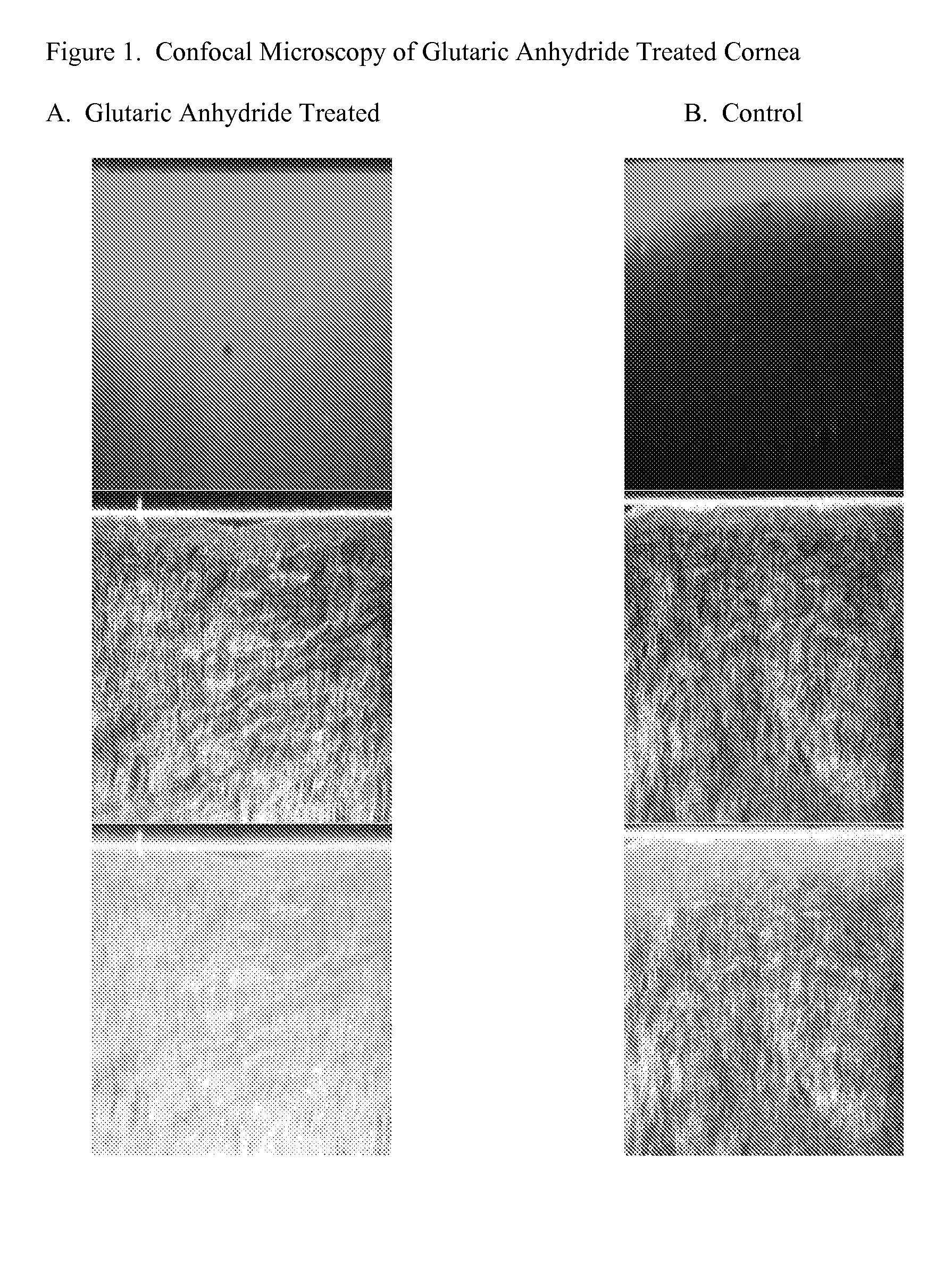
Confocal Microscopy to Evaluate Decorin Penetration into Human Donor Corneal Tissue

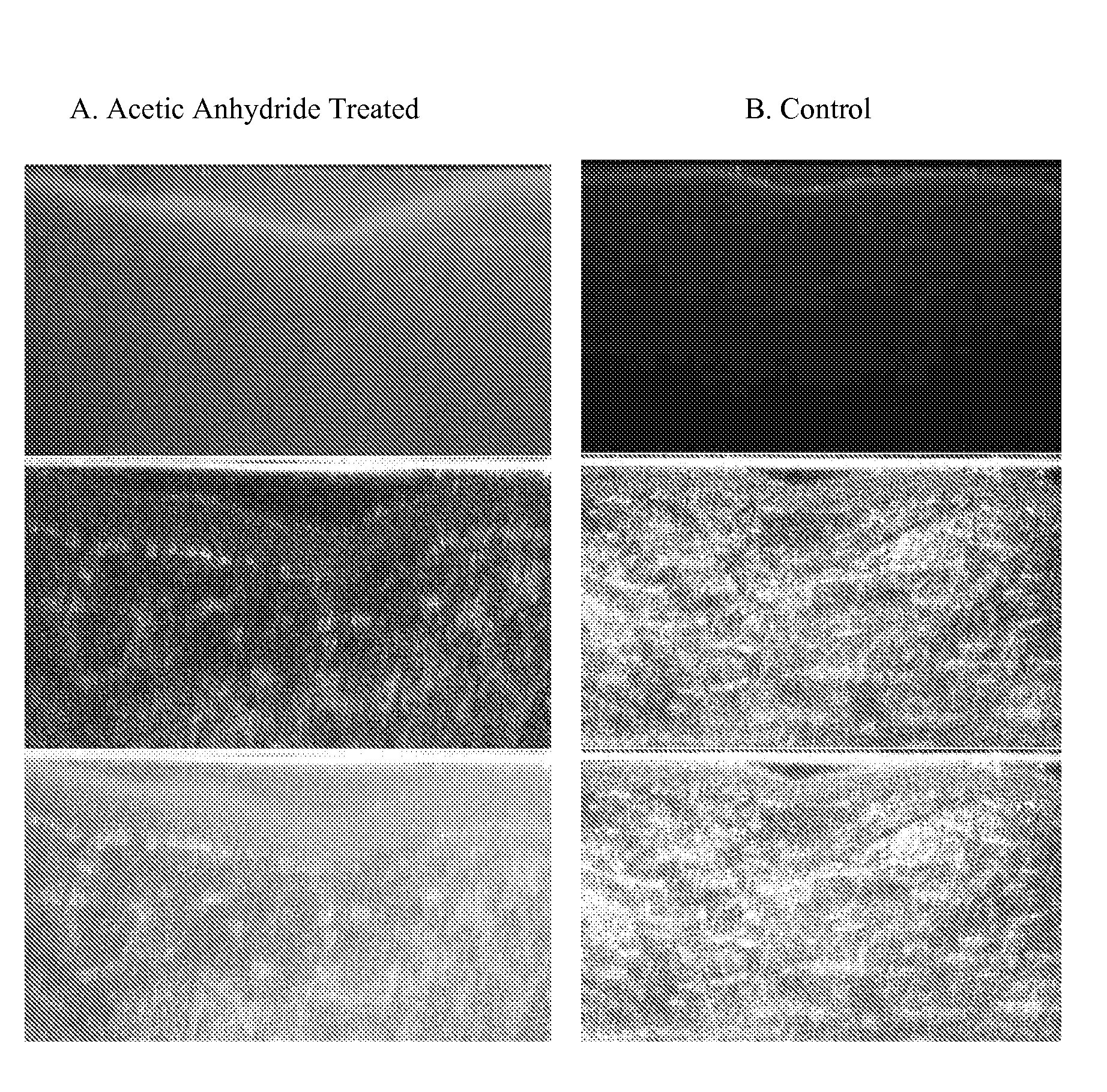
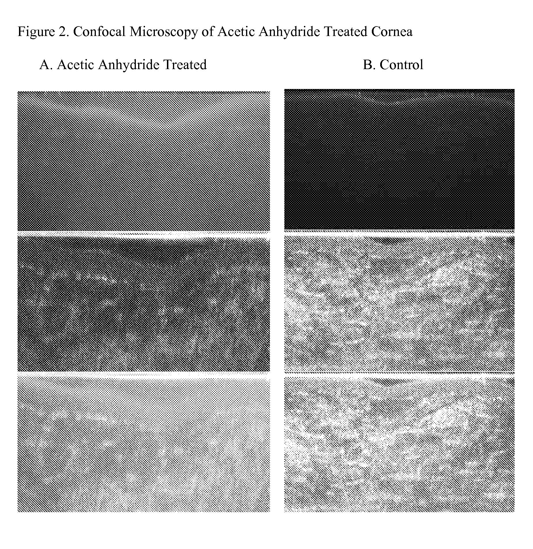
[0082]Confocal Microscopy of glutaric anhydride treated cornea. Four donor corneas were obtained from Insight Biomed (Minneapolis, Minn.). All cornea tested negative for viral contamination. Corneas were rejected for transplantation due to storage expiration, low endothelial cell counts or other factors not related to epithelial cell integrity and stromal structure. All corneas were examined by slit-lamp microscopy for epithelial cell integrity. Corneas were stored in Optisol (Bausch & Lomb) for storage. Confocal microscopy was conducted at the Department of Surgical Research, Dartmouth-Hitchcock Medical Center, Lebanon, N.H. Two corneas were untreated controls and two were treated with glutaric anhydride followed by application of fluorescent-tagged human recombinant decorin. Human recombinant Decorin was prepared from CHO—S cells (Cardinal Health) and exhibited a concentration of 3.7 mg / mL in 10 mM ...

example 2

Transmission Electron Microscopy to Evaluate Decorin Binding to Collagen Stromal Fibers in Human Donor Corneal Tissue

[0087]The following study was conducted at the Department of Surgical Research, Dartmouth-Hitchcock Medical Center, Lebanon, N.H. Five adult female cats were included in the study. All animals procured from Liberty Laboratories and were identified by ear tattoo. Ocular toxicity was determined by slit-lamp examination and measurement of endothelial cell structure. Decorin penetration into the corneal stroma was determined utilizing transmission electron microscopy. Cornea treated with decorin were reacted with Quinolinic Blue Stain (Cupromeronic blue) in buffered formalin. This reagent stains small proteoglycan structures such as decorin.

[0088]Cats were placed in three treatment groups. One eye from each group was treated with decorin. Three eyes were controls. Treated eyes were exposed to 50 μg of decorin for 1 day, 3 days or 5 days. Decorin solution was administered ...

example 3

Effects of Acylation Treatments in Corneal Hysteresis in the Feline Model

[0094]Measurements of Corneal Hysteresis (CH) in the animals treated in Example 2. Corneal Hysteresis is a measure of the biomechanical strength of the cornea and is measured using the Reichert Ocular Response Analyzer. The Reichert Ocular Response Analyzer utilizes a dynamic bi-directional applanation process to measure the biomechanical properties of the cornea and the Intraocular Pressure of the eye. The basic output of the measurement process is a Goldmann-correlated pressure measurement (IOPG), and a new measure of corneal tissue properties called Corneal Hysteresis (CH). CH values are shown in Table 1.

TABLE 1CH values for Feline Study #1Corneal Hysteresis of Feline Cornea followingAcylation and Decorin TreatmentsAnimal IBT4Animal QKU8Animal QNO6TreatmentControlTreatedTreatedTreatedControlTreatedInitialND4.06.47.0ND6.3Post-ND7.08.07.5ND7.0ProparacainePost-GAND3.35.36.4ND6.4Post-AA #1ND3.27.05.7ND5.7Post-AA...


PUM

PropertyMeasurementUnit
Shapeaaaaaaaaaa
Permeabilityaaaaaaaaaa
Hydrophilicityaaaaaaaaaa

Abstract

Methods of increasing the permeability of corneal epithelium to facilitate the diffusion of agents into the collagen fibrillar network of the stroma are provided. Used in combination, these methods open the epithelium to facilitate diffusion of stabilization molecules into the stroma and dissociate bridging molecules from stromal collagen fibers, thereby priming the collagen fibrillar network for restabilization by stabilization molecules. These methods can be used to increase the effectiveness and longevity of non-invasive corneal reshaping, such as orthokeratology, for correcting myopia, hyperopia and astigmatism.

Description

[0001]This application claims priority to U.S. Provisional Application Nos. 61 / 064,730 filed Mar. 24, 2008, and 61 / 064,731 filed Mar. 24, 2008, the contents of which are all incorporated herein by reference.FIELD OF THE INVENTION[0002]The present disclosure relates to methods of increasing the permeability of corneal epithelium to allow diffusion of molecules, such as collagen binding molecules, into the corneal stroma and to methods of temporarily destabilizing the collagen fibrillar network of the stroma. The treatments described herein (1) open the epithelium to enhance the diffusion of molecules into the stroma and (2) dissociate bridging molecules from stromal collagen fibers, thereby priming the collagen fibrillar network for restabilization by stabilization molecules. Such treatments are important to improve the effectiveness and longevity of non-invasive corneal reshaping to correct myopia, hyperopia and astigmatism.BACKGROUND[0003]Orthokeratology procedure: Orthokeratology ...

Claims


Application Information

Patent Timeline
IPC IPC(8): A61K38/39A61K31/365A61K31/194A61P27/02A61K38/17
CPCA61K31/185A61K31/341A61K38/1709A61K38/39A61K39/39A61K45/06A61K2300/00A61P27/02A61P43/00A61F9/0008A61M35/003A61M2005/2093
Inventor DEWOOLFSON, BRUCEDEVORE, DALE
Owner EUCLID SYST CORP
Why Eureka
Try Eureka