Human omental mesothelial cells, methods of isolation and uses thereof

a technology of omental mesothelial cells and methods, applied in the field of human omental mesothelial cells, methods of isolation and uses thereof, can solve the problems of peritoneal dialysis failure, fibrin deposition, peritoneal adhesion, etc., and achieve the effect of preventing peritoneal dialysis failure, preventing peritoneal dialysis, and preventing peritoneal dialysis

Inactive Publication Date: 2011-05-05
ZENBIO
View PDF2 Cites 22 Cited by
  • Summary
  • Abstract
  • Description
  • Claims
  • Application Information

AI Technical Summary

Benefits of technology

[0022]Another embodiment of the present invention comprises a method of providing a source of omentum, myocardial, liver, lung, renal, peritoneal, intestinal or pancreatic mesothelial cells comprising providing a substantially pure isolated population of human omentum, myocardial, liver, lung, renal, peritoneal, intestinal or pancreatic mesothelial cells and using the cells to co-culture with another selected cell and then observing the effect on the selected cell.
[0023]Another embodiment of the present invention comprises a method of providing a source of omentum mesothelial cells comprising providing a substantially pure isolated population of human omentum mesothelial cells, and isolation of the conditioned medium from the substantially pure population of omentum mesothelial cells, and using the conditioned medium to treat another selected cell, and then observing the effect on the selected cell.

Problems solved by technology

Damage to serosal surfaces by infection or surgery can lead to a disruption in the mesothelial layer, fibrin deposition, and peritoneal adhesions.
Asbestos fibers interact directly with mesothelial cells, where they are ingested and lead to DNA damage from reactive oxygen species.
Damage to the mesothelial cells lining the peritoneal cavity reduces the effectiveness of solute transfer and ultimately results in failure of peritoneal dialysis.
Damage to mesothelial cells occurs because they are in direct contact with nonphysiologic dialysis fluid during long dwell times. Osmotic agents, high glucose levels, and low pH contribute to injuring the peritoneum and denuding the surface of mesothelial cells.
This increases fibrosis within the mesothelium, disrupting the ultrastructure, and decreasing the effectiveness of peritoneal dialysis.
However, long term peritoneal dialysis continues to result in mesothelial cell injury and ultimate failure of the treatment.
This process degrades the tight junctions between cells, causes a morphology change to fibroblast-like cells, and increases their migratory and invasive properties.
Overall, EMT decreases the integrity of the peritoneal lining greatly increasing transport rates and rendering peritoneal dialysis ineffective.

Method used


Image

  • Human omental mesothelial cells, methods of isolation and uses thereof
  • Human omental mesothelial cells, methods of isolation and uses thereof
  • Human omental mesothelial cells, methods of isolation and uses thereof

Examples


example 1

[0072]A sample approximately 10 grams of omental tissue is removed from the greater or lesser omentum of a subject. The tissue piece is transferred to a cryovial and placed in a liquid nitrogen freezer for long term storage or used immediately for mesothelial cell isolation.

[0073]The thawed or freshly isolated tissue sample is tested for pathogens and the test sample precisely weighed. The tissue sample is then mixed with an equal volume of a solution containing of 0.25% trypsin and incubated undisturbed for 20 minutes at 23-25° C. The tissue fragments are then removed from the solution and the trypsin solution containing the mesothelial cell suspension is centrifuged at 1200 rpm for 5 minutes at 20° C. The resulting pellet containing mesothelial cells is resuspended in DMEM-F12 medium containing fetal bovine serum (5-10%) and epidermal growth factor (5 ng / ml), and cells are plated in tissue cultureware. Mesothelial cells can then be expanded in Media 199 containing 5% fetal bovine ...

example 2

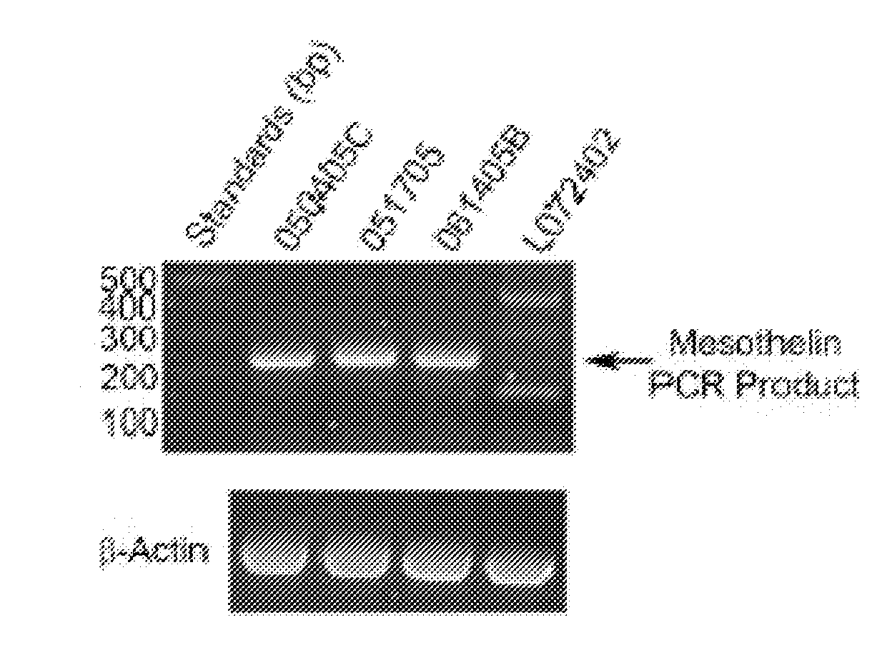
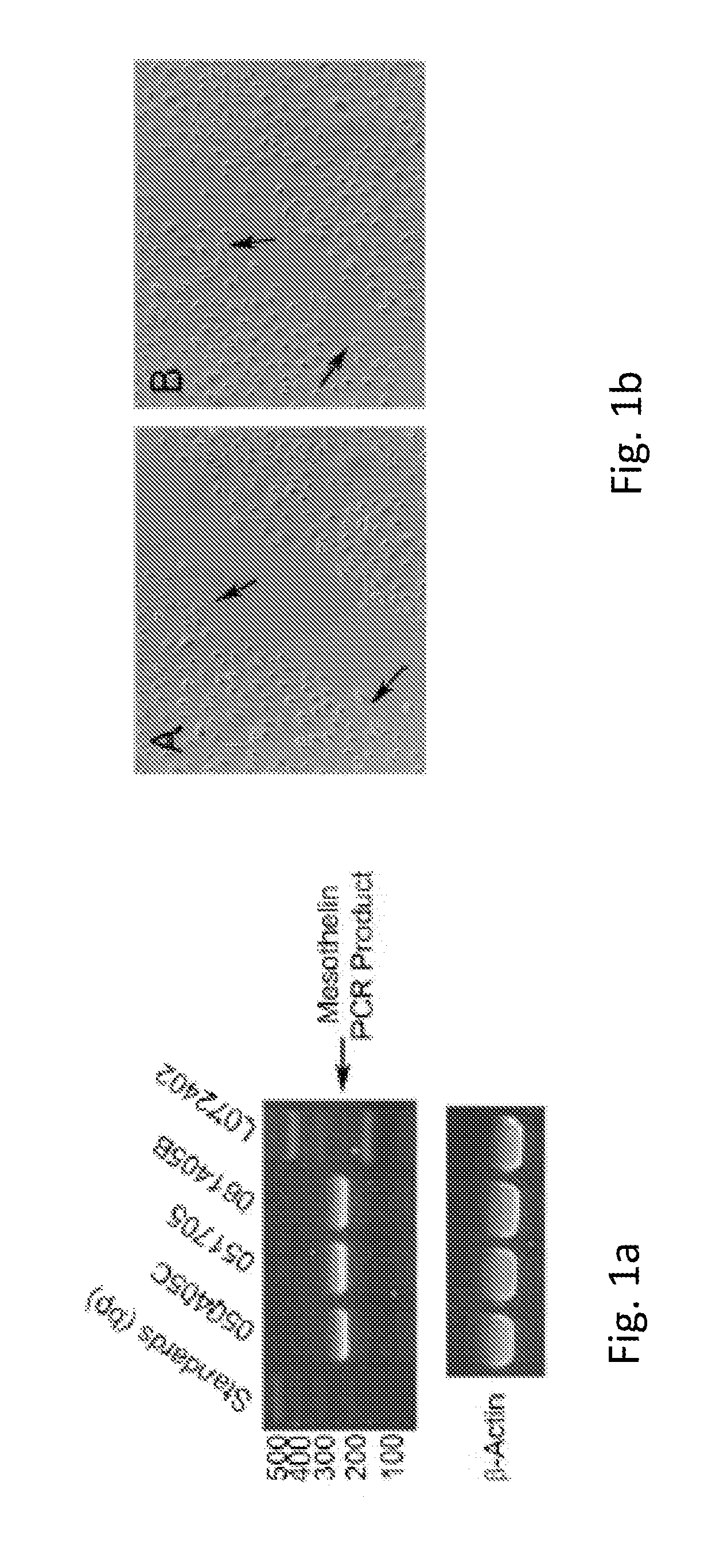
[0074]Mesothelin is expressed in the purified omental mesothelial cells. This was shown by reverse-transcription polymerase chain reaction (RT-PCR) assay, using RNA isolated from 3 unique donor lots of omental mesothelial cells. FIG. 1a shows mesothelin expression analyzed by RT-PCR. 1 microgram of total RNA from three mesothelial lots and one preadipocytes lot (L072402) was used as a template for RT-PCR reaction. Primers for mesothelin or Beta-actin were added to assess expression of each gene.

example 3

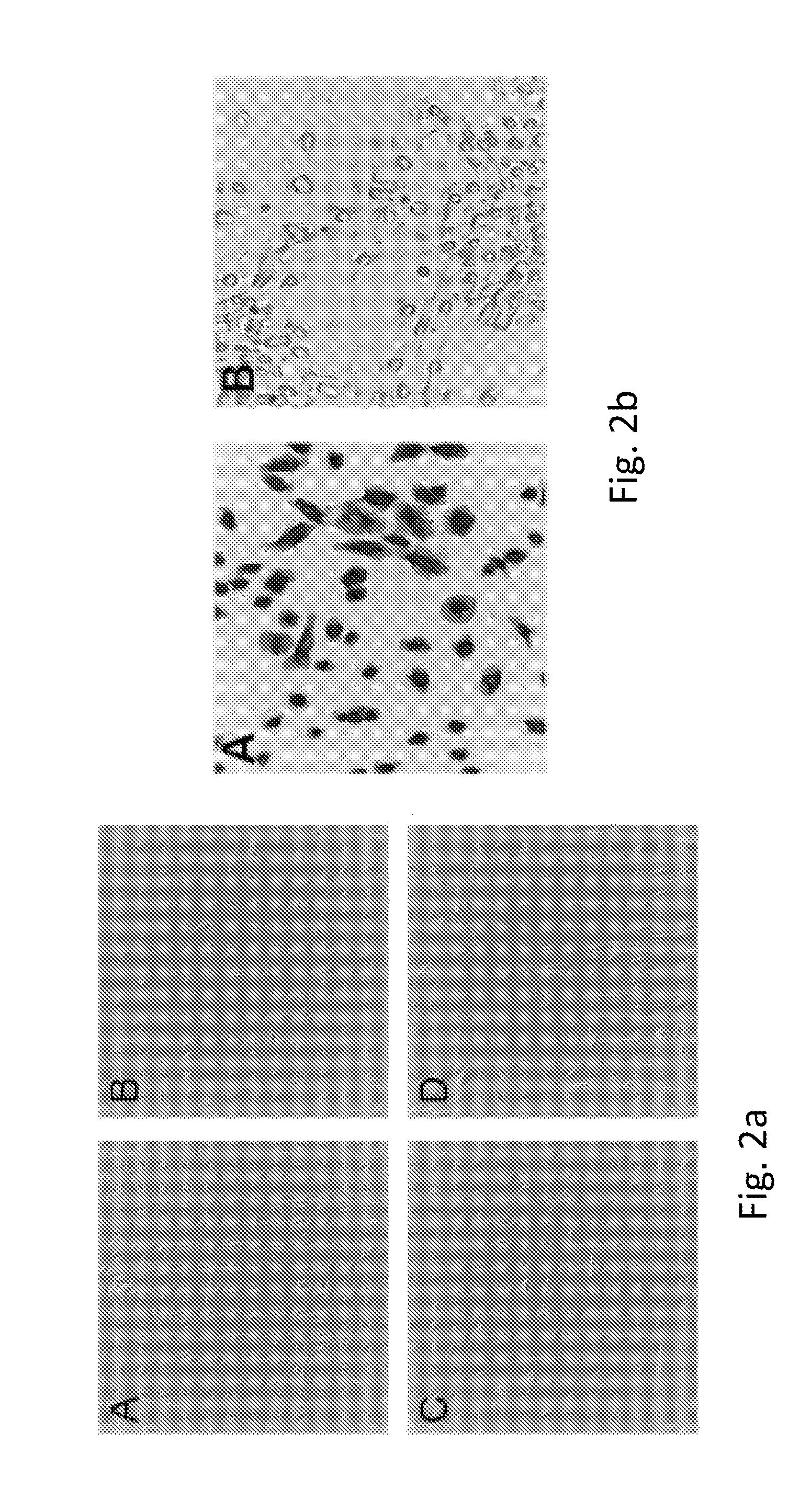
[0075]Mesothelial cells are known to be involved in wound healing. As shown in FIG. 1b, mesothelial cells were grown on collagen coated plates and the cell monolayer scrapped to mimic wounding. Fibroblastic mesothelial cells were recruited to the wound site after 3 hours (A) and 18 hours (B). Fibroblast mesothelial cells are indicated by arrows.


PUM

PropertyMeasurementUnit
survival timeaaaaaaaaaa
temperatureaaaaaaaaaa
physicalaaaaaaaaaa

Abstract

The present invention discloses novel methods and omental, myocardial, liver, lung, renal, peritoneal, intestinal and pancreatic mesothelial cells which are useful for a number of procedures including drug discovery, co-culturing, cell therapy and bioassay. The invention provides a method for isolating these cells that improves upon the methods previously used and provides cells isolated in quantity. The present invention provides a list of secreted proteins from omentum mesothelial cells that can be utilized in the described cell based assays.

Description

[0001]This application claims priority of U.S. provisional application No. 61 / 256,770 filed on Nov. 1, 2009 and U.S. provisional application No. 61 / 259,316 filed on Nov. 9, 2009 included herein in their entirety by reference.GOVERNMENT SPONSORED STATEMENT[0002]This invention was made with government support under R44RR024302-02 awarded by National Institutes of Health. The government has certain rights in the invention.COPYRIGHT NOTICE[0003]A portion of the disclosure of this patent contains material that is subject to copyright protection. The copyright owner has no objection to the reproduction by anyone of the patent document or the patent disclosure as it appears in the Patent and Trademark Office patent files or records, but otherwise reserves all copyright rights whatsoever.BACKGROUND OF THE INVENTION[0004]1. Field of the Invention[0005]The present invention relates to the isolation and use of substantially pure cultures of human omental mesothelial cells. Specifically, the is...

Claims


Application Information

Patent Timeline
Patent Type & Authority Applications(United States)
IPC IPC(8): C12Q1/02C12N5/071G01N33/53
CPCC12N2503/02C12N5/0653
Inventor BUEHRER, BENJAMIN MARCUSCHEATHAM, RICHARD BENTLEYNICOLL, JAMES BRADFORDPIERACCINI, PETER EARL
Owner ZENBIO
Why Eureka
Try Eureka