Methods and compositions for topoisomerase i modulated tumor suppression

Inactive Publication Date: 2011-06-09
RG BIOPHARMA
View PDF1 Cites 0 Cited by
  • Summary
  • Abstract
  • Description
  • Claims
  • Application Information

AI Technical Summary

Benefits of technology

[0009]The invention is based upon the discovery that cells resistant to topoisomerase inhibitors frequently have a deficiency in topoisomerase I serine phosphorylation, rendering them less sensitive (or insensitive) to the cytotoxic effect of topoisomerase I inhibitors. The deficiency in topoisomerase I phosphorylation reduces the ability of topoisomerase I to bind p14ARF (ARF), an activator protein. Thus, cancer cells can be assessed for their sensitivity to topoisomerase I inhibitors, prior to initiating therapy, by measuring the level of serine phosphorylation of topoisomerase I, its activity, and / or its ability to bind ARF. Likewise, cells can be sensitized to the effects of topoisomerase I inhibitors by increasing the amount of serine phosphorylation of topoisomerase I, or by increasing ARF-topoisomerase I complex formation by increasing, for example, the amount of ARF available for complexation with serine phosphorylated topoisomerase I.
[0011]Accordingly, in one aspect, the invention provides a method for increasing the sensitivity of a cell to a topoisomerase I inhibitor by contacting the cell with an agent that increases the level of topoisomerase I serine phosphorylation.
[0017]In another aspect, the invention provides, for a cell expressing increased phosphorylation of topoisomerase I, a method for increasing the sensitivity of a said cell to a topoisomerase I inhibitor by contacting the cell with an agent that increases the ARF-topoisomerase I complex formation.
[0042]In another aspect, the invention provides a method for inducing apoptosis, cell killing, and / or growth arrest in a cell by contacting the cell with an agent that inhibits the binding of ARF to topoisomerase I. The binding may be inhibited by an antibody or other binding agent (e.g. a peptide, an aptamer, or a peptidomimetic) which disrupts the interaction between ARF to topoisomerase I. The agent may bind directly to ARF or to topoisomerase I and may competitively or non-competitively inhibit the ARF-topoisomerase I binding interaction. Suitable antibodies include, for example, ARF-specific antibodies and topoisomerase I-specific antibodies. Alternatively, a phosphatase that dephosphorylates topoisomerase I may be used to reduce ARF binding to topoisomerase I. A CK2 inhibitor such as TBB (4,5,6,7-tetrabromobenzotriazole) could also be used to reduce CK2-mediated phosphorylation of topoisomerase I. In preferred embodiments, the method disrupts existing ARF-topoisomerase I complexes. In other embodiments, ARF binding to HDM2 is increased. In other embodiments, p53 biological activity is increased.
[0053]In another aspect, the invention provides methods of increasing the sensitivity of a cancer patient to a topoisomerase inhibitor, comprising administering a CKII activator to the cancer patient in an amount sufficient to decrease ratio of unphosphorylated topoisomerase I to phosphorylated topoisomerase I. In some embodiments, the CKII activator is 1-ethyl-4,5-dicarbamoylimidazole.

Problems solved by technology

However, some cancers are not sensitive to such topoisomerase I inhibitors.

Method used


Image

  • Methods and compositions for topoisomerase i modulated tumor suppression
  • Methods and compositions for topoisomerase i modulated tumor suppression
  • Methods and compositions for topoisomerase i modulated tumor suppression

Examples


example 1

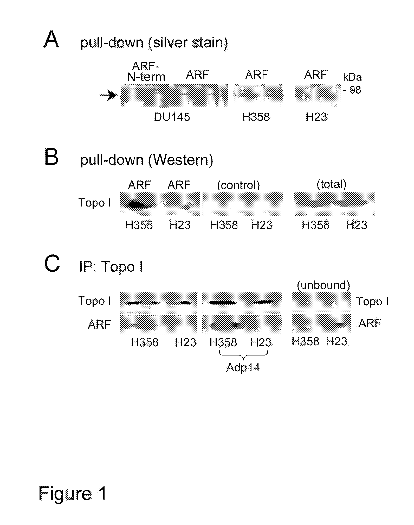
Defective ARF / Topoisomerase I Complex Formation in H23 Cells

[0117]FIG. 1A shows a silver stained gel following a pull-down assay in which immobilized human ARF-thioredoxin fusion protein (or the N-terminal domain (1-64) of ARF) was used to compare ARF-binding proteins from DU145 (prostate cancer), H358, and H23 (non-small cell lung carcinoma) cell RIPA lysates.

[0118]Topoisomerase I bound to full-length ARF (ARF, FIG. 1A) but not the ARF N-terminal domain (ARF-N-term, amino acid residues 1-64, FIG. 1A) encoded by ARF's first exon (exon 1β). This is consistent with previous reports that topoisomerase I binds to ARF through the ARF C-terminal, exon 2-encoded domain (Ayrault, et al., Oncogene 2006; 25(19):2827 (correction); Olivier, et al., Oncogene 2003; 22(13):1945-54). H23 cells appeared to have significantly less topoisomerase I activity compared to that measured in H358 cells (FIG. 1A, far right lane).

[0119]Western blot analysis confirmed that the level of topoisomerase I was reduc...

example 2

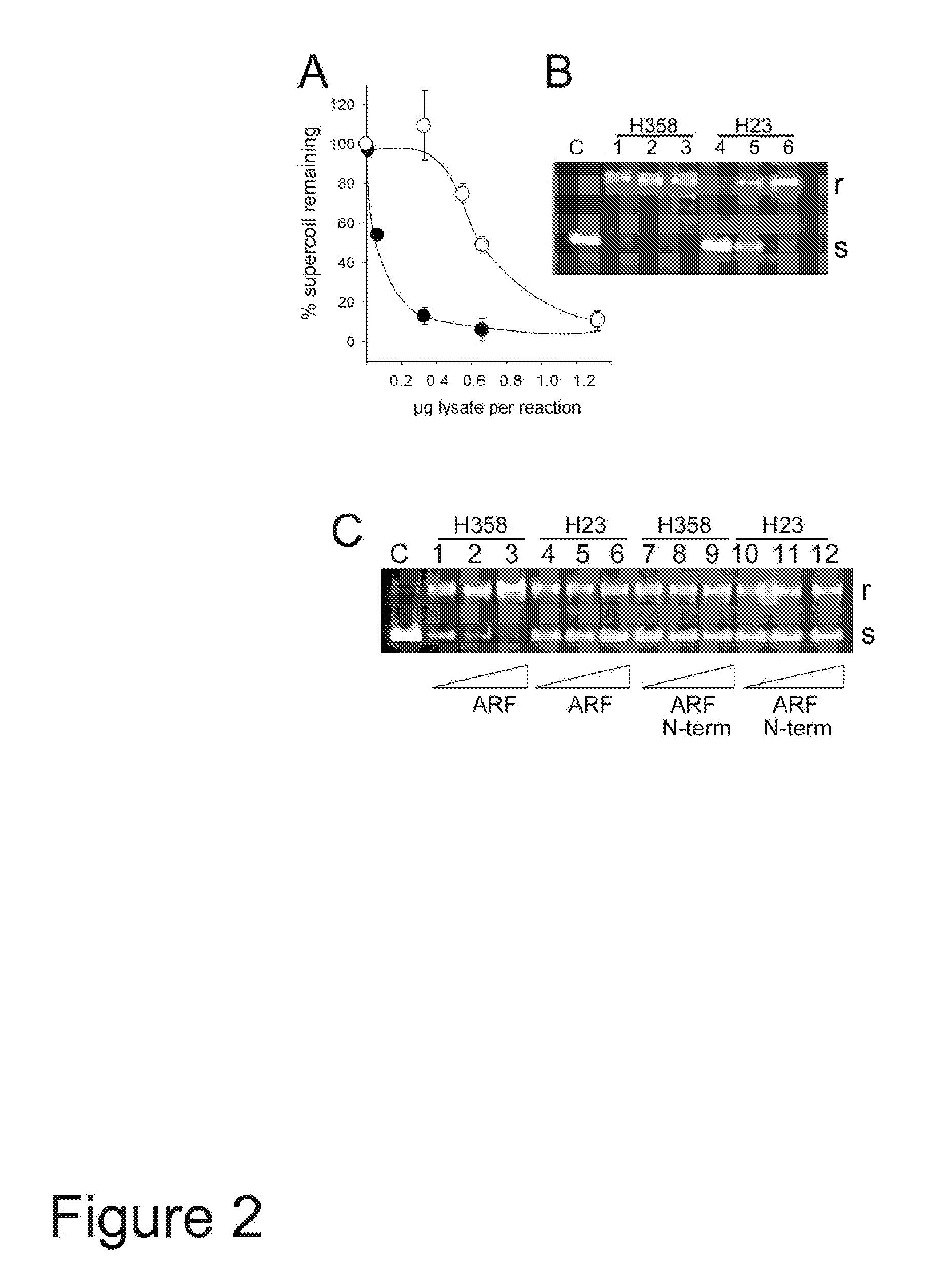
H23 Nuclear Extracts have Reduced Topoisomerase I Activity which Cannot be Stimulated by ARF

[0126]H23 and H358 nuclear extracts were compared for topoisomerase I activity in vitro, and investigated whether the activities could be stimulated by the addition of recombinant thioredoxin-ARF. As shown in FIG. 2A, H358 topoisomerase I was found to be more effective at relaxing supercoiled plasmid DNA than was H23 topoisomerase, achieving 50% relaxation at about 0.06 μg nuclear extract per reaction, some 10-fold lower than the amount of H23 extract needed to achieve the same level of relaxation (0.6 μg extract per reaction). A typical electrophoretic profile of the reaction products with increasing amounts of nuclear extract is shown in FIG. 2B in which 0.32, 0.65, or 1.3 μg of H358 cell extract (lanes 1-3) or H23 (lanes 4-6) were added in each reaction. “r” is the relaxed (non-supercoiled) plasmid and “s” is the supercoiled form.

[0127]Similar assays were carried out using the amount of ea...

example 3

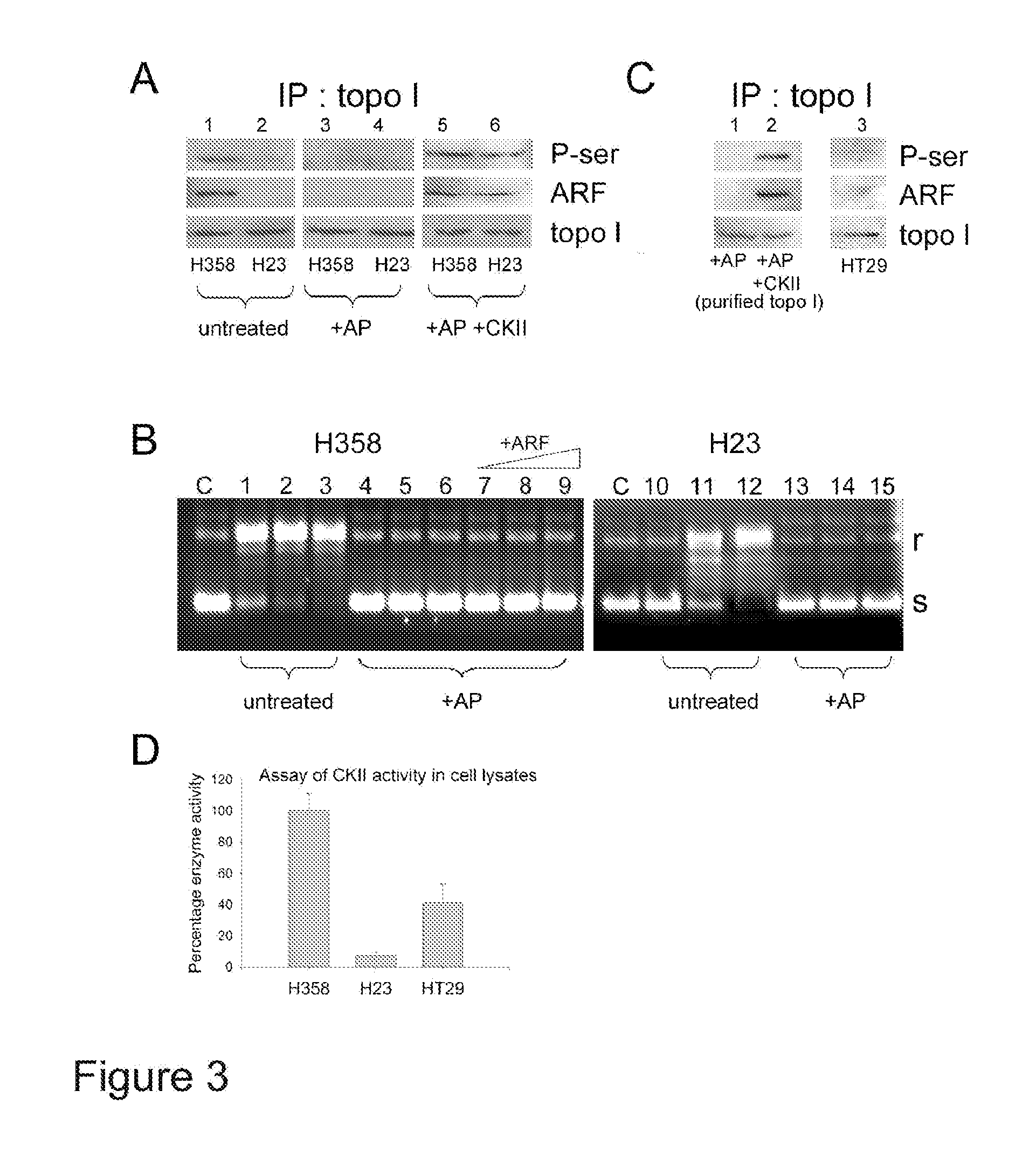
Topoisomerase I is Activated by Both Phosphorylation and ARF Binding

[0129]A topoisomerase I immunoprecipitation analysis followed by Western detection of phosphoserine revealed that H358 cells expressed a serine-phosphorylated topoisomerase I (FIG. 3A, lane 1, top row). A similar analysis of phosphotyrosine revealed no evidence for tyrosine phosphorylation (data not shown). Similar results were found in PC-3 cells (data not shown). In contrast, serine-phosphorylated topoisomerase I was only weakly detectable in H23 cells (FIG. 3A, lane 2, top row).

[0130]Treatment of both H358 and H23 nuclear extracts with alkaline phosphatase (AP) eliminated serine phosphorylation (FIG. 3A, lanes 3, 4, top row) and abolished their topoisomerase I activity in vitro (FIG. 3B, lanes 4-6 and lanes 13-15). The dephosphorylated topoisomerase I from H358 cells could no longer be activated by addition of increasing amounts of ARF fusion protein (FIG. 3B, lanes 7-9). Furthermore, while topoisomerase I co-imm...


PUM

PropertyMeasurementUnit
Ratioaaaaaaaaaa
Sensitivityaaaaaaaaaa

Abstract

Disclosed herein are methods and compositions for determining the sensitivity or enhancing the sensitivity of cells to the effects of topoisomerase I inhibitors. Also disclosed are methods and compositions for inducing cell death, apoptosis and / or growth arrest which may be used for tumor suppression.

Description

CROSS-REFERENCE TO RELATED APPLICATIONS[0001]This application is a continuation in part of application Ser. No. 12 / 377,498, filed Feb. 13, 2009, which is a 35 USC 371 National Phase Entry Application from PCT / US07 / 018,387, filed Aug. 16, 2007, and designating the U.S., which claims the priority of U.S. Provisional Application No. 60 / 822,774 filed Aug. 18, 2006, the disclosures of which are incorporated herein by reference in their entireties.RIGHTS IN THE INVENTION[0002]This invention was supported in part by grants from the NIH / NCI (CA 111868, CA 135369) and from the California Tobacco-Related Disease Research Program (11RT-0074). The Government may have certain rights in this invention.FIELD OF INVENTION[0003]This invention relates to the field of cancer therapy and diagnostics.BACKGROUND OF INVENTION[0004]The following discussion of the background of the invention is merely provided to aid the reader in understanding the invention and is not admitted to describe or constitute pri...

Claims


Application Information

Patent Timeline
IPC IPC(8): A61K31/4375A61K31/4164A61P35/00C12Q1/02G01N33/68C12Q1/68A61P35/02
CPCA61K31/4164A61K31/4375C12Q1/6886C12Q1/533G01N33/574G01N2333/99G01N2800/52C12Q2600/106A61P35/00A61P35/02
Inventor GJERSET, RUTH A.BANDYOPADHYAY, KEYA
Owner RG BIOPHARMA
Why Eureka
Try Eureka