
Methods and dressings for sealing internal injuries

a technology for internal injuries and dressings, applied in the direction of metabolism disorders, extracellular fluid disorders, peptide/protein ingredients, etc., can solve the problems of increased expense, significant morbidity, blood vessel injuries, etc., and achieve the effect of reducing the flow of fluid

Inactive Publication Date: 2011-08-11
HDG
View PDF90 Cites 12 Cited by
  • Summary
  • Abstract
  • Description
  • Claims
  • Application Information

AI Technical Summary

Benefits of technology

"The present invention provides solid dressings that can treat wounded internal mammalian tissue, particularly human tissue. The method involves applying to wounded internal tissue at least one haemostatic material consisting essentially of a fibrinogen component and a fibrinogen activator for a time sufficient to reduce the flow of fluid from the wounded tissue. The haemostatic material can be cast or formed from a single aqueous solution containing the fibrinogen component and the fibrinogen activator or formed as a single piece. The various solid and frozen haemostatic materials useful in the inventive methods are also described."

Problems solved by technology

There are a large number of medical procedures that result in injuries to blood vessels.
Current sealing products and devices have one or more deficiencies, usually due to their inadequate performance, or their reliance upon non-natural components that interfere with normal healing.
These complications can lead to significant morbidity, increased expense, a requirement for additional procedures and / or devices, extended time in the medical facility and conversion of outpatients to inpatients.
Nevertheless, despite this poor performance, even these devices are currently used since the costs and consequences of procedure-induced complications is so high (See Resnic et al: Am J. Cardiol. 2007 Mar. 15; 99(6):766-70).
These additional complications may lead to prolonged closure procedures, hospitalization, the requirement for surgical repair, and even tissue loss or death.
These devices are convenient and gaining in popularity, but their overall safety appears over estimated.
Indeed, far from being risk free, these devices may be associated with unique levels of hemorrhagic and cardiac risks including myocardial infarction, stroke and death (See Rao, S.
Significant risks have been reported to be associated with all classes of vascular closure devices.
Most seriously, the severity and the difficulty in treating complications are generally greater when vascular closure devices are used (See Nehler et al.
The use of such devices is even associated with higher risks among patients having complications of pseudoaneurysms, failure to successfully treat such pseudoaneurysms, blood loss, transfusions, extensive operations to correct the problems and arterial infections (See Sprouse et al.
Moreover, some of these complications can be deadly, particularly in patients with diabetes, obesity and previously implanted devices (all conditions commonly found in patients in whom such closure devices are frequently used) (See Hollis and Retiring.
Major complications for most hernia repair procedures include pain and the requirement to re-do the repair (See American College of Surgeons.
These agents have met with varying degrees of success when used in animal models of traumatic injuries and / or in the field, and with limited success in the sealing of therapeutic vascular injuries.
As liquids, however, these fibrin sealants have not proved useful outside certain specific procedures.
While these fibrinogen-thrombin dressings do not require the pre-mixing needed by liquid fibrin sealants, their utility for field applications is limited by a requirement for storage at 4° C. and the necessity for pre-wetting with saline solution prior to application to the wound.
These dressings are also not effective against high pressure, high volume bleeding.
While this dressing has shown great success in several animal models of hemorrhage, the bandage is fragile, inflexible, and has a tendency to break apart when handled.
To date, none of these dressings have been approved for use or are available commercially.
To date, however, all of these remain little-used in therapy, most likely due to the difficult and time consuming preparation requirements for two-component liquid fibrin sealant compositions.
However, the time and efforts associated with preparing such sealants make them less than ideal for daily clinical use
While the procedure described in U.S. Pat. No. 6,762,336 has been shown to be useful for evaluating haemostatic dressings, it failed to replicate faithfully the requirements for success in vivo.

Method used


Image

  • Methods and dressings for sealing internal injuries
  • Methods and dressings for sealing internal injuries

Examples


example 1

In order to apply the haemostatic test articles to the surface of an injured artery surrounded by a tissue stimulant, the test articles were housed in cylindrical molds made of 10 or 3 mL polypropylene syringes (Becton Dickinson) with the luer-lock end removed. The plungers were withdrawn to the 6 mL and 2 mL mark respectively. For dressings utilizing a backing, the support material was cut and placed into each mold and pushed down until it was adjacent to the plunger. Once prepared the molds were placed upright and surrounded by dry ice, leaving the opening exposed at the top. 1 ml of fibrinogen and 0.15 mL of thrombin (with or without backing material dispersed within) were dispensed into the 10 mL molds and 1 ml of fibrinogen and 0.15 mL of thrombin (with or without support material dispersed within) were dispensed into the 3 mL molds, which were allowed to freeze for 5 minutes. The molds were then placed into the −80° C. freezer for at least two hours before being placed into a ...

example 2

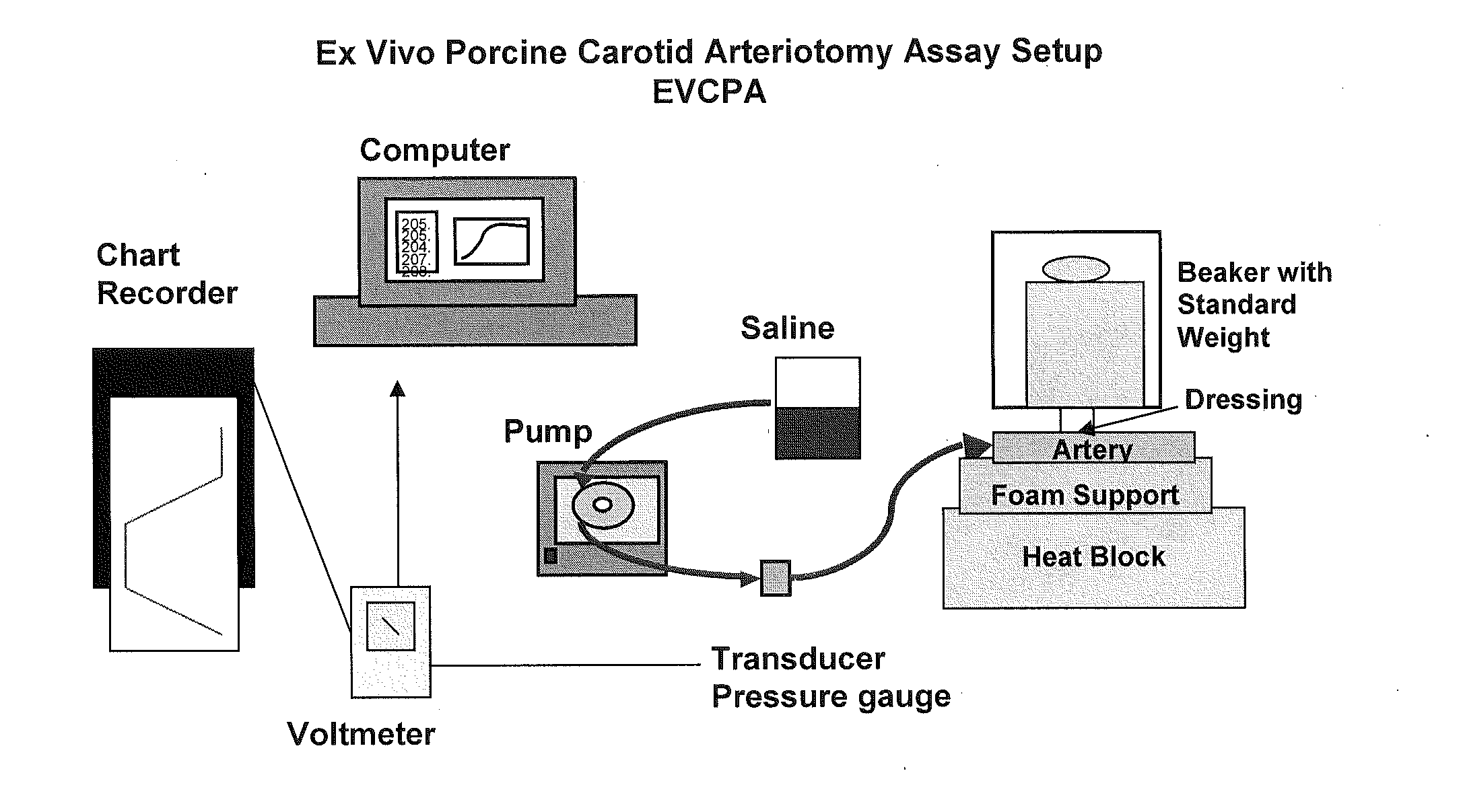
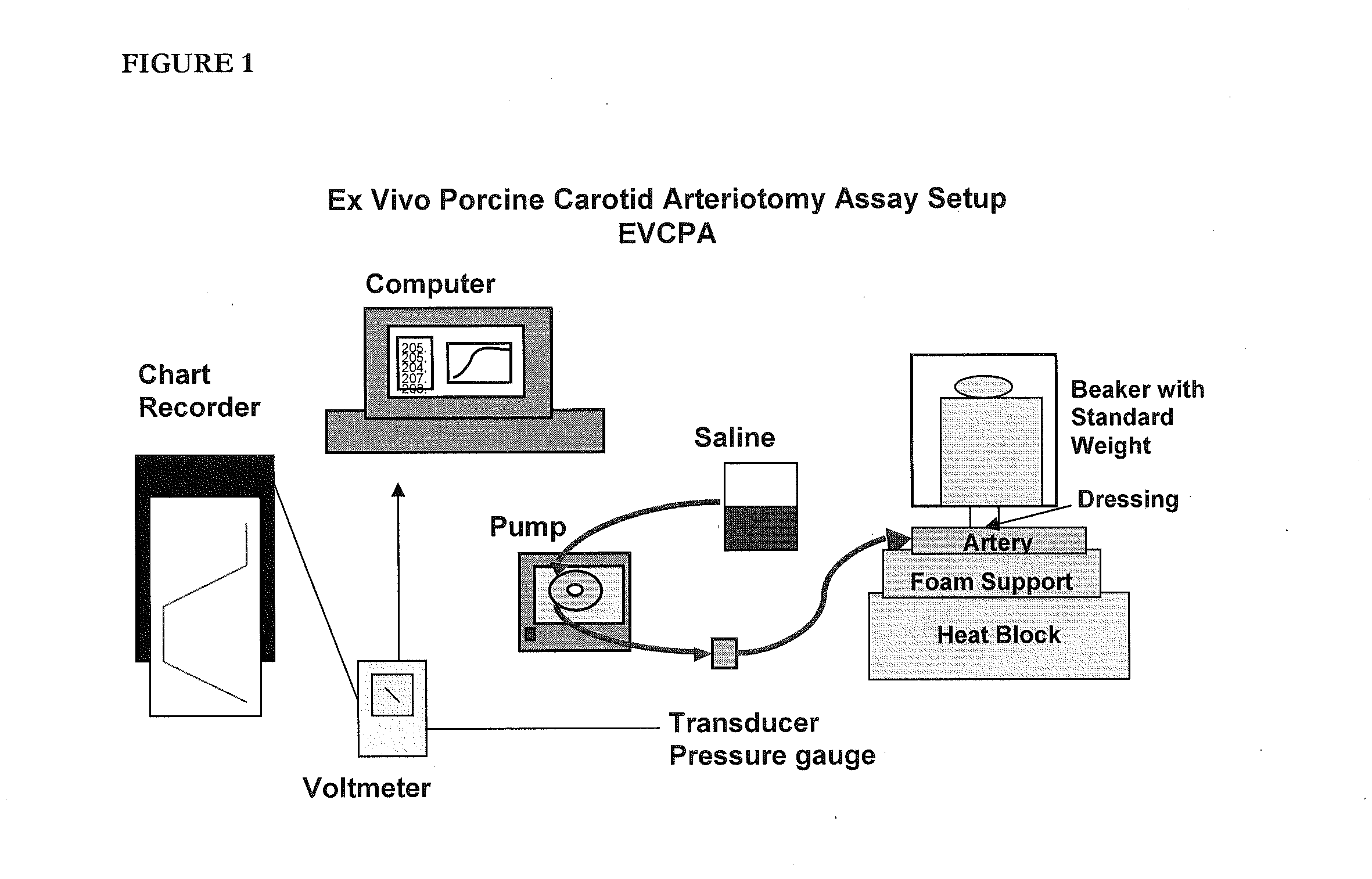
Similar to the need to evaluate a test article in the context of sealing and injury deep within surrounding tissue, there was also a need to test products that can seal injured tissue where the injured vessels are smaller and thinner-walled than an aorta. The following assay accomplishes this goal.

According to this modification, the porcine carotid artery is attached to a barbed female connector using cotton thread with the connective tissue side exposed. This is in contrast to the standard EVPA where the internal side is exposed. As the carotid arteries used in the VA model are more elastic and friable than the aorta, it is more difficult to treat or abrade the surface without damaging and compromising the artery. To ensure that no tears have occurred during the removal of the bulk of the connective tissue, the artery is connected to the barbed connector and solution is pumped into it. If the artery is intact, a 1.5 mm hole is punched into the artery using a biopsy punch.

After the ...

example 3

For all dressings, ERL fibrinogen lot 3130 was formulated in CFB. The final pH of the fibrinogen was 7.4±0.1. The fibrinogen concentration was adjusted to 37.5 mg / ml. Once prepared the fibrinogen was placed on ice until use. Thrombin was formulated in CTB. The final pH of the thrombin was 7.4±0.1. The thrombin was adjusted to deliver 0.1 units / mg of Fibrinogen or 25 Units / ml thrombin. For the group with shredded support material dispersed within, it was cut into approximately 1 mm×1 mm pieces and dispersed within the thrombin solution prior to filling the molds. Once prepared the thrombin was placed on ice until use. The temperature of the fibrinogen and thrombin prior to dispensing was 4° C.±2° C. Cylindrical molds made of 10 or 3 mL polypropylene syringes (Becton Dickinson) with the luer-lock end removed were used. The plungers were withdrawn to the 6 mL and 2 mL mark respectively. For dressings utilizing a support material, the support material was cut and placed into each mold a...


PUM

PropertyMeasurementUnit
temperaturesaaaaaaaaaa
pressuresaaaaaaaaaa
temperaturesaaaaaaaaaa

Abstract

Disclosed are solid and frozen haemostatic materials and dressings consisting essentially of a fibrinogen component and a fibrinogen activator. Also disclosed are methods of treating internal wounded tissue in a mammal by applying one or more of these haemostatic materials and dressings.

Description

FIELD OF THE INVENTIONThe present invention relates to dressings for treating internal injured tissue in a mammalian patient, such as a human and methods of using the same.BACKGROUND OF THE INVENTIONThere are a large number of medical procedures that result in injuries to blood vessels. Similarly, there are numerous examples of bleeding caused by traumatic injuries, hematological disorders, and from unknown causes. When the site of bleeding is not readily accessible, such as an injured vessel located deep within the flesh, or inside a body cavity, a simple and effective method of hemorrhage control that can access the site within the body and seal the injured vessel is needed. Similarly, tissue may be divided by either traumatic injury or surgical procedure, and require sealing to approximate the edges of the injury in order to restore function. Current sealing products and devices have one or more deficiencies, usually due to their inadequate performance, or their reliance upon non...

Claims


Application Information

Patent Timeline
Patent Type & Authority Applications(United States)
IPC IPC(8): A61B17/03
CPCA61B17/00491A61B17/0057A61B2017/0065A61L26/009A61L15/32A61L15/64A61L26/0042A61F2013/00472A61K38/363A61K38/4833C12Y304/21005A61P17/02A61P29/00A61P3/02A61P31/00A61P31/10A61P31/12A61P33/00A61P35/00A61P7/02A61P9/00A61L15/44A61L2300/254A61L2300/252A61L2400/04A61L15/28A61L2300/23
Inventor MACPHEE, MARTINKANELLOS, JERRYWILMER, BELINDABEALL, DAWSON
Owner HDG
Learn More