Method for formation of renal tubules

a technology of renal tubules and tubules, which is applied in the direction of kidney/kidney cells, artificial cell constructs, biochemical apparatus and processes, etc., can solve the problems of inability to clear nanoparticles above inability to address in vivo models, and inability to clear nanoparticles over a certain size limit, etc., to achieve easy image, high resolution, and facilitate microscopic monitoring of the transport process in the present tubules

Inactive Publication Date: 2011-09-29
ZINK DANIELE +3
View PDF3 Cites 4 Cited by
  • Summary
  • Abstract
  • Description
  • Claims
  • Application Information

AI Technical Summary

Benefits of technology

[0016]The tubules provided by the present methods may be used as an in vitro model for studying tubular functions and transport processes occurring in tubular cells. Microscopic monitoring of the transport processes in the present tubules can be greatly facilitated due to the fact that the tubules may be easily imaged with high resolution using, for example, confocal microscopy, since the tubules are not formed within a gel matrix. As well, transcellular transport of fluorescent substrates may be more easily detected by fluorescent imaging of such tubules than by the use of current two-compartment systems that involve cell growth on porous membranes and measurement of substrate concentration in the different compartments. Transcellular transport can be easily detected by increases in the cellular and luminal levels of fluorescence and can be monitored online by using live cell microscopy.
[0017]The tubules may also be used as models for pathological processes affecting renal tubules, such as for example tubular necrosis. Tubular necrosis is frequently associated with kidney disease. The processes leading to tubular necrosis are not well understood, and can be difficult to address using in vivo models. The tubules of the present invention provide a convenient in vitro model not provided by monolayer cultures, and alterations in the tubule structure can more easily be examined than with tubules embedded in 3D gels.

Problems solved by technology

The processes leading to tubular necrosis are not well understood, and can be difficult to address using in vivo models.
However, nanoparticles above a certain size limit cannot be cleared by glomerular filtration.

Method used


Image

  • Method for formation of renal tubules
  • Method for formation of renal tubules
  • Method for formation of renal tubules

Examples


example 1

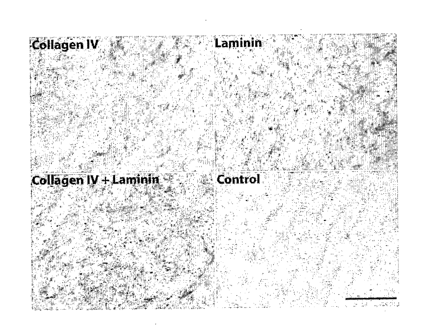
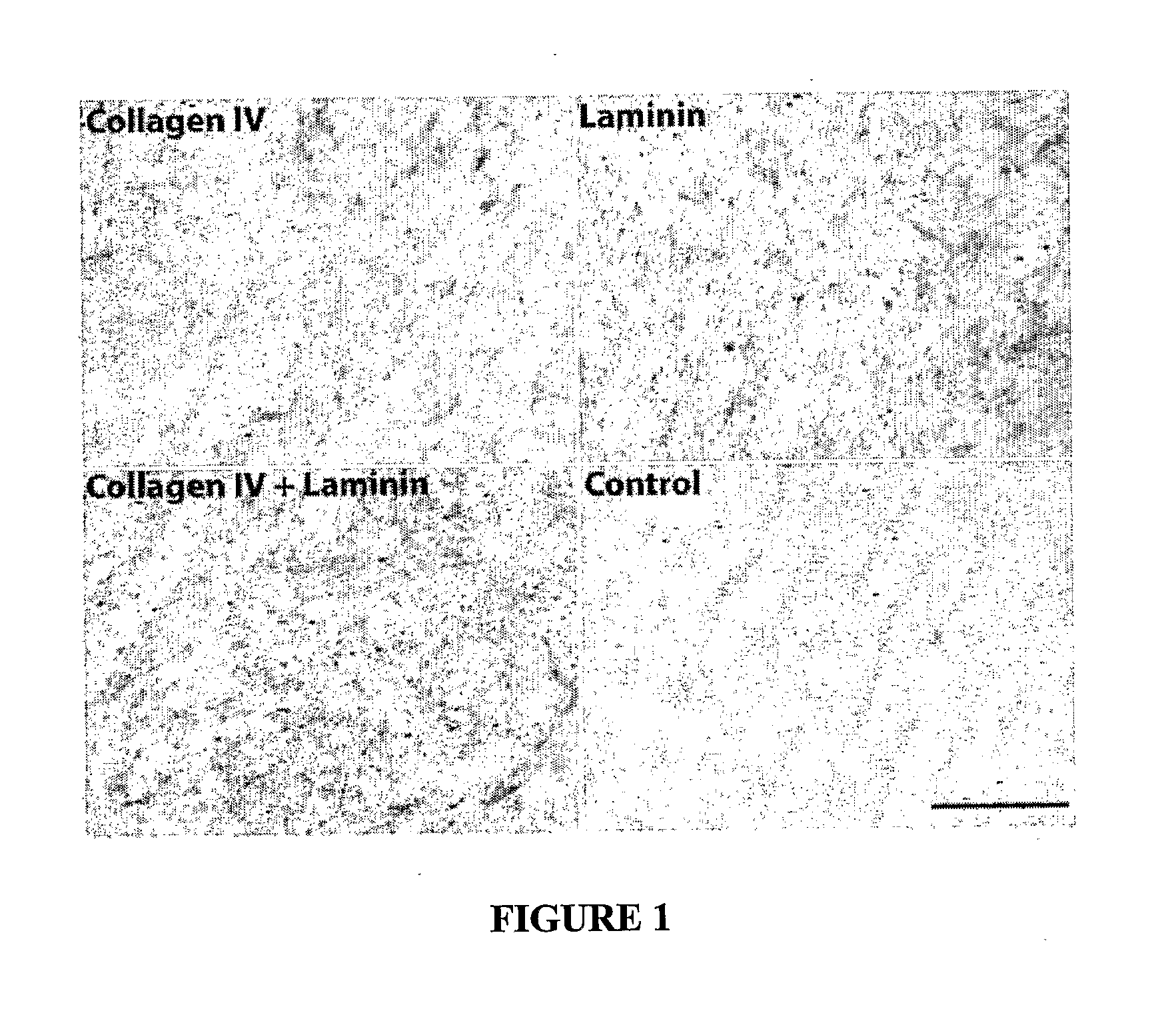
[0126]ECM coatings were tested and the performance of primary human renal proximal tubule cells was monitored during extended periods of several weeks. During this process, it was observed that the formation of differentiated monolayers by human primary renal proximal tubule cells (HPTCs) was closely coupled to tubulogenesis, which occurred on solid surfaces. It was found that tubule formation is faster on ECMs consisting of collagen IV+laminin. The longer the monolayer is maintained, the later tubule formation occurs. This example relates to the experiments leading to tubule formation on solid surfaces.

[0127]Materials and Methods

[0128]Cell culture assay: All cell culture media used were also supplemented with 1% penicillin / streptomycin solution (ScienCell Research Laboratories, Carlsbad, Calif., USA), and all cells were cultivated at 37° C. in a 5% CO2 atmosphere. HPTCs were obtained from ScienCell Research Laboratories and were cultivated in basal epithelial cell medium supplement...

example 2

[0177]The four panels in FIG. 5 show different parts of a 2D tubule consisting of HPTCs (Scale bars: 100 μm).

[0178]First, histidine-coated quantum dots (QDs, green fluorescence) were added to culture medium. (DAPI counterstaining of cell nuclei: blue). 20 hours later the specimens were fixed and the 2D tubules were examined by fluorescence microscopy. The images show that the QDs were uptaken by HPTCs. There was no evidence for transport of the QDs into the tubular lumen (i.e. no enrichment of QDs in the lumen of the 2D tubule).

[0179]Next, transcellular transport of the QDs was examined using TRANSWELL™ plates where a monolayer of HPTCs grew on the lower side of a porous membrane separating two compartments (apical sides of cells facing the bottom compartment). QDs were added to the upper compartment. Occurrence of QDs in the bottom compartment of the TRANSWELL™ system (corresponding to the tubular lumen) could indicate transcellular transport. However, occurrence of QDs in the basa...

example 3

Materials and Methods

[0184]Cell culture: Different batches of HPTCs were obtained from ScienCell Research Laboratories (Carlsbad, Calif., USA). Cells were cultivated in basal epithelial cell medium supplemented with 2% fetal bovine serum (FBS) and 1% epithelial cell growth supplement (ScienCell Research Laboratories). In some experiments, TGF-β1 (R&D Systems, Minneapolis, Minn., USA) was added at a concentration of 10 ng / ml after monolayer formation.

[0185]Cells were cultivated on uncoated multi-well plates (Nunc, Naperville, Ill., USA) or plates coated with human laminin or other ECMs as described in Zhang et al. Biomaterials 2009 30: 2899-2911. The seeding density was 5×10⁴ cells / cm2, unless otherwise indicated.

[0186]The wells of diagnostic printed slides have a diameter of 2 mm and a glass bottom. No ECM coating was applied in experiments using the printed slides.

[0187]Cells were seeded at a density of 2.65×10⁵ cells / cm2 into glass capillaries (inner diameter=0.58 mm; Sutter Instr...


PUM

PropertyMeasurementUnit
lengthaaaaaaaaaa
diameteraaaaaaaaaa
penetration depthaaaaaaaaaa

Abstract

There is provided a method of making a renal tubule. The method comprises seeding renal tubule cells onto a solid surface; culturing the renal tubule cells in a liquid growth medium to form a monolayer on the solid surface; and continuing culturing the renal tubule cells to form a tubule.

Description

CROSS-REFERENCE TO RELATED APPLICATION[0001]This application claims benefit of, and priority from, U.S. provisional patent application No. 61 / 193,467, filed Dec. 2, 2008, the contents of which are incorporated herein by reference.FIELD OF THE INVENTION[0002]The present invention relates generally to methods for forming renal tubules in gel-free in vitro systems.BACKGROUND OF THE INVENTION[0003]Many drugs and xenobiotics (e.g. antibiotics, p-aminohippurate) cannot be cleared efficiently in the kidney by glomerular filtration. Such substances are then actively transported by the cells of the proximal tubule from the bloodstream into the glomerular filtrate flowing in the lumen of the renal tubule. Organic anion transporters, organic cation transporters and the p-glycoprotein coded by the multi drug resistance locus play a major role in such transcellular transport processes. Due to their functions in drug transport, proximal tubule cells are a major target for toxic drug effects in th...

Claims


Application Information

Patent Timeline
Patent Type & Authority Applications(United States)
IPC IPC(8): A01N1/00C12Q1/02C12N5/071
CPCC12N5/0686C12N2501/385C12N2501/15C12N2501/11
Inventor ZINK, DANIELEYING, JACKIE Y.ZHANG, HUISHITASNIM, FARAH
Owner ZINK DANIELE
Why Eureka
Try Eureka