
Methylation detection

a technology of methylation and detection, applied in the field of methylation detection, can solve the problems of high sensitiveness and accuracy, and achieve the effect of improving the success ra

Active Publication Date: 2011-11-24
ONCOMETHYLOME SCI
View PDF8 Cites 0 Cited by
  • Summary
  • Abstract
  • Description
  • Claims
  • Application Information

AI Technical Summary

Benefits of technology

"The present invention provides an improved method for detecting the presence and amount of methylated or unmethylated genes in a DNA-containing sample. This method involves selectively modifying unmethylated cytosine residues in the DNA with a reagent that produces detectable residues, but does not modify methylated cytosine residues. The method uses a real-time detection technique that allows for the quantitative analysis of the methylated or unmethylated gene of interest as the reaction progresses. The method can be used for detecting the methylation status of any gene of interest and has applications in diagnostics and research. The methylation status of certain genes, such as MGMT, WRN, BRCA1, PTEN, and NDRG4, can be reliably determined using the method."

Problems solved by technology

This methodology is highly sensitive and accurate, but has drawbacks for routine clinical use.

Method used


Image

  • Methylation detection
  • Methylation detection
  • Methylation detection

Examples


example 2

Amplifluor Experiments: Testing Different Primer Sequences and Reading Temperatures

[0233]Amplifluor is a primer-based methodology. It lacks the third oligo nucleotide “probe” that typifies TaqMan and Molecular Beacon technologies. Without this third layer of specificity, amplifluor primers can fluoresce in response to non-specific amplification such as primer-dimers. Therefore it is important to carefully select primer sequences to overcome this behavior.

[0234]Initial Results

[0235]Initial MGMT amplifluor results were obtained using the primer set shown in Table 3. The final primer concentrations in the Amplifluor reaction mix were 100 nM for both forward primer / detector and reverse primer. 12.5 μl of iTaq™ Supermix with Rox (BioRad, 2× buffer) were used per PCR reaction. The total volume per reaction, including 5 μl of no-template control (NTC: water control with no DNA present), was 25 μl. The following thermal profile was used on the ABI 7900 HT SDS instrument: Stage1: 50° C. for ...

example 3

Additional Genes Tested Through Amplifluor

[0253]We developed a direct real-time fluorescence based methylation-specific PCR assay (real-time MSP assay) to define the methylation status of the MGMT promoter. Briefly, genomic DNA is deaminated using sodium bisulphite after isolation. 5-Methyl Cytosine is refractory to this chemical modification. Unmethylated Cytosine quantitatively turns into Uracil during this process. After amplification of the DNA sequences using methylation specific primers, the detection of the amplified DNA sequences is carried out using the amplifluor technology. The modification for the amplifluor primers is 5′ FAM internal Dabcyl.

[0254]The quantitation process depends on fluorescent light which is emitted only when the detector is bound to its complementary sequence.

[0255]Analyte quantitations for several markers additional to MGMT were successfully performed using this technology. These consisted of parallel amplification / quantification processes using speci...

example 4

Reproducibility of the MGMT Amplifluor Assay

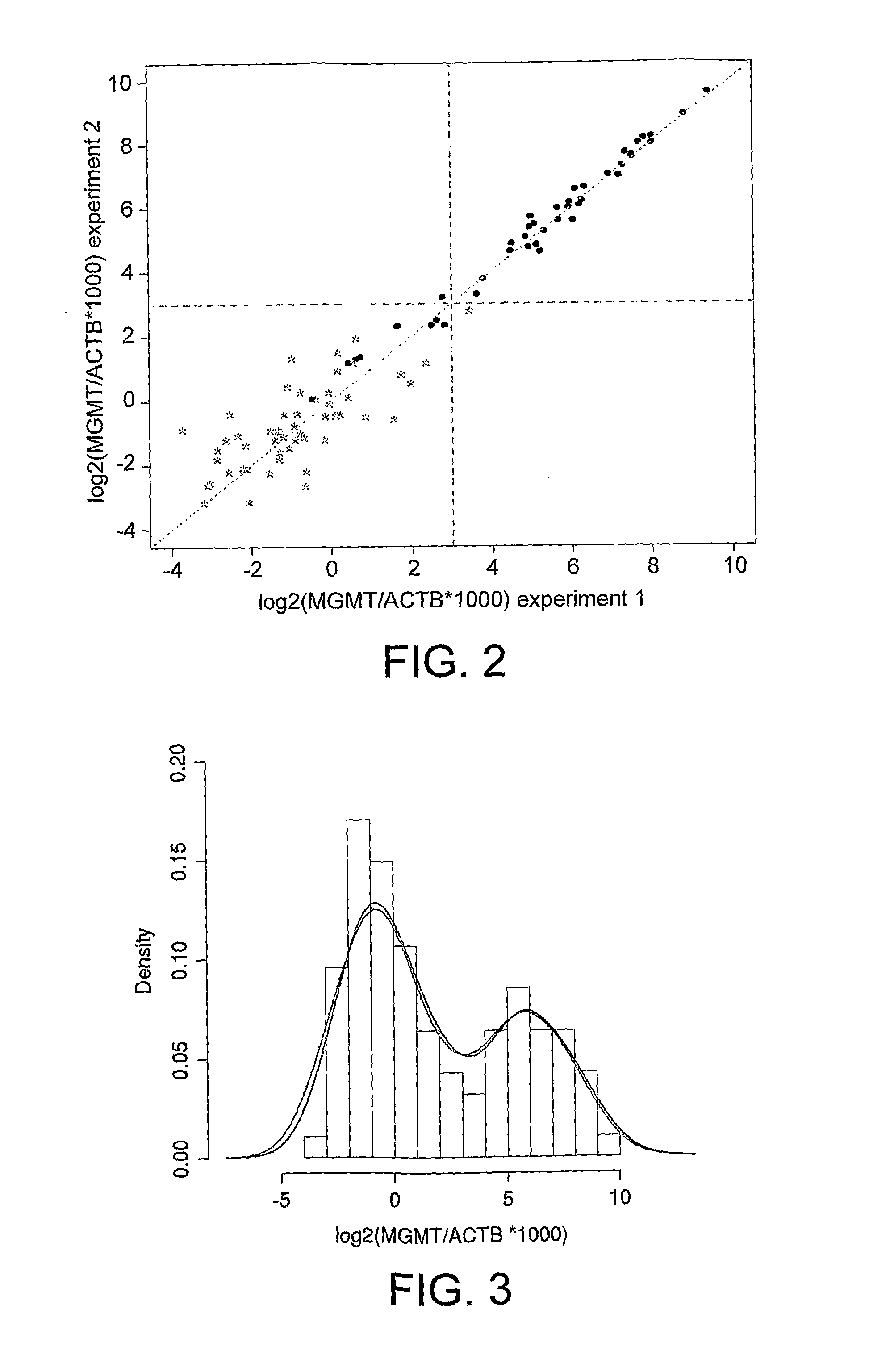
[0302]To test the reproducibility of the MGMT amplifluor assay as described in example 1, 75 available clinical samples were processed twice through real-time MSP: once at OncoMethylome Sciences (ONCO) and once at Laboratory Corporation of America@ Holdings (LabCorp®). Both laboratories obtained comparable results.

[0303]Materials and Methods

[0304]Sample Preparation

[0305]75 formalin-fixed, paraffin-embedded (FFPE) glioma tissue samples were available for testing through the MGMT amplifluor real-time MSP. For each glioma tumor sample, 4 10 μm consecutive sections were prepared on glass slides. The prepared sample sections were then divided between the 2 sites and processed in parallel (including sample preparation) according to each laboratory's respective protocols (both real-time MSP). LabCorp® was blinded to the results obtained by ONCO.

[0306]Real-Time MSP Protocol Followed by ONCO

[0307]DNA isolation, modification and analyte quantitatio...


PUM

PropertyMeasurementUnit
total reaction volumeaaaaaaaaaa
wavelengthaaaaaaaaaa

Abstract

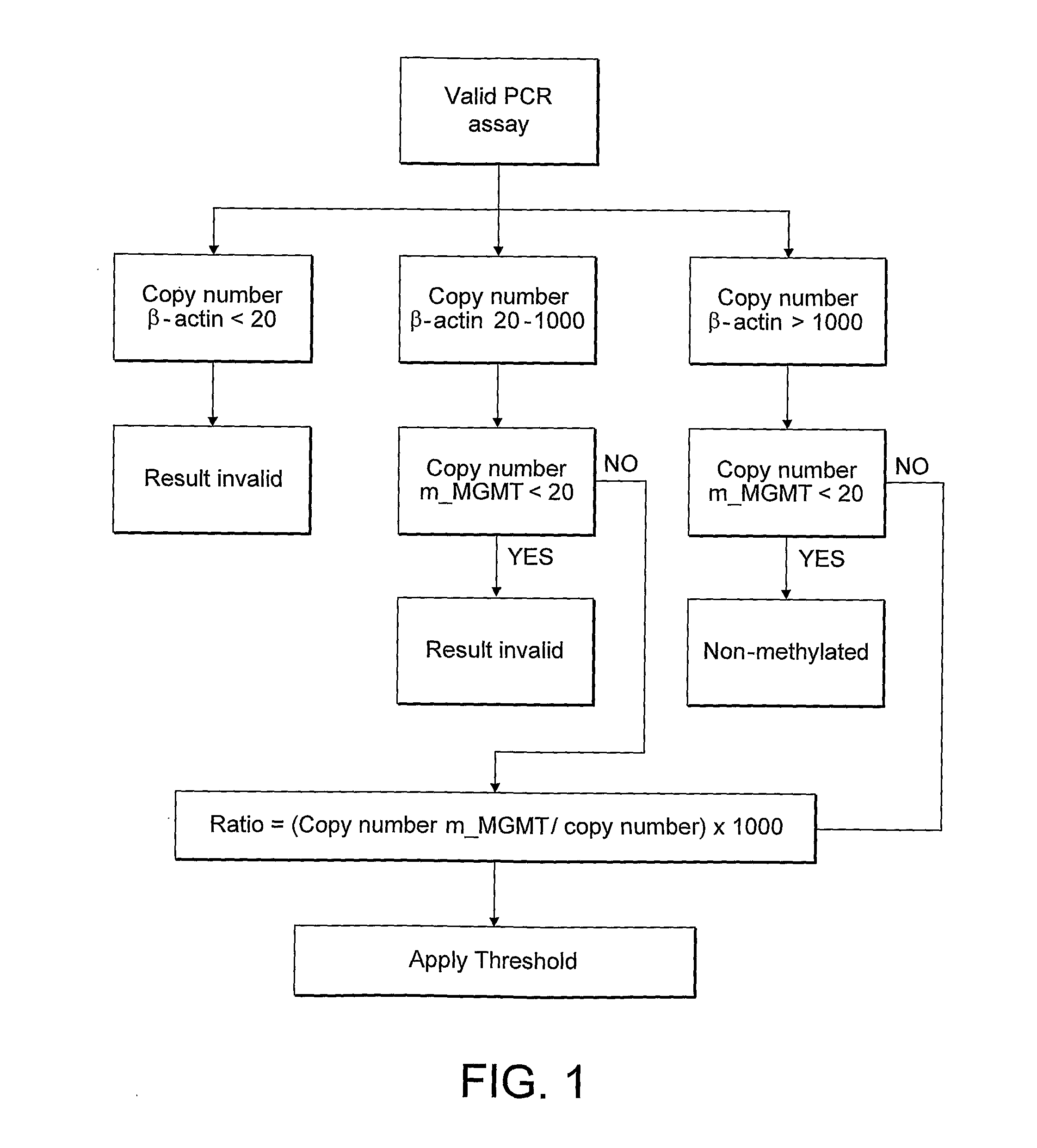
A real-time method of detecting the presence and / or amount of a methylated or unmethylated gene of interest in a DNA-containing sample, comprises the steps of: (a) contacting the DNA-containing sample with a reagent which selectively modifies unmethylated cytosine residues in the DNA to produce detectable modified residues but which does not modify methylated cytosine residues (b) amplifying at least a portion of the methylated or unmethylated gene of interest using at least one primer pair, at least one primer of which is designed to bind only to the sequence of methylated or unmethylated DNA following treatment with the reagent, wherein at least one primer in the primer pair is a primer containing a step loop structure carrying a donor and an acceptor moiety of a molecular energy transfer pair arranged such that in the absence of amplification, the acceptor moiety quenches fluorescence emitted by the donor moiety upon excitation and during amplification, the stem loop structure is disrupted so as to separate the donor and acceptor moieties sufficiently to produce a detectable fluorescence signal which is detected in real-time to provide an indication of the gene copy number of the methylated or unmethylated gene of interest (c) quantifying the results of the real-time detection against a standard curve for the methylated or unmethylated gene of interest to produce an output of gene copy number. The method may be characterized in that the amplification is considered valid where the cycle threshold value is less than (40). Alternatively the method, which may be carried out in real-time or at end point, is characterized in that the portion of the gene which is amplified is between approximately (50) and 250 bp, especially for plasma and serum samples.

Description

FIELD OF THE INVENTION[0001]The present invention is concerned with detection of epigenetic modifications in nucleic acids. More specifically, the invention relates to methods of detecting methylated or unmethylated forms of a gene of interest. The methods of the invention involve amplification techniques, in particular fluorescence based real-time and end-point PCR methods.BACKGROUND TO THE INVENTION[0002]Gene methylation is an important regulator of gene expression. In particular, methylation at cytosine residues found in CpG di-nucleotide pairs in the promoter region of specific genes can contribute to many disease conditions through down regulation of gene expression. For example, aberrant methylation of tumour suppressor genes can lead to down regulation of these genes and is thus associated with the presence and development of many cancers. Patterns of aberrant gene methylation are often specific to the tissue of origin. Accordingly, detection of the methylation status of spec...

Claims


Application Information

Patent Timeline
Patent Type & Authority Applications(United States)
IPC IPC(8): C12Q1/68C07H21/04A61K38/08
CPCA61K38/08A61K38/10C12Q1/6844C12Q1/6886A61K39/0011C12Q2600/16C12Q2600/158C12Q2600/154C12Q2525/301C12Q2561/113C12Q2565/101A61P35/00A61K39/001186C12Q1/6827
Inventor VLASSENBROECK, ILSEBIERAU, KATJA
Owner ONCOMETHYLOME SCI
Learn More