Methods for imaging vascular inflammation using improved nanoparticle contrast agents

a technology of contrast agent and nanoparticle, which is applied in the field of imaging vascular inflammation using improved nanoparticle contrast agent, can solve the problems of increasing the half-life of particles in circulation, limiting uptake of particles by reticuloendothelial macrophages, etc., and achieves the effect of increasing the half-life of particles and limiting uptake of particles

Inactive Publication Date: 2011-12-22
NANOSCAN IMAGING
View PDF10 Cites 20 Cited by
  • Summary
  • Abstract
  • Description
  • Claims
  • Application Information

AI Technical Summary

Benefits of technology

[0006]The present invention provides, at least in part, compositions and methods for vascular imaging which are optimized for intravenous administration. In one embodiment, such compositions are optimized to image intravascular inflammation. Prior to the instant invention, it was not clear whether changes to one or more physical (e.g., size, size distribution and shape) or chemical properties (e.g., adsorbed surfactants and excipients) of a nanoparticulate contrast agent would optimize its imaging capabilities when administered intraveneously, e.g., its ability to image intravascular inflammation. The instant invention is based, at least in part, on the finding that compositions comprising nanoparticulate contrast agents having a mean particle size of less than about 150 nm on a per weight or per volume basis are optimal for vascular imaging ...

Problems solved by technology

The use of such particles increases the half-life of the particles in the circulatio...

Method used


Image

  • Methods for imaging vascular inflammation using improved nanoparticle contrast agents
  • Methods for imaging vascular inflammation using improved nanoparticle contrast agents
  • Methods for imaging vascular inflammation using improved nanoparticle contrast agents

Examples


example 1

Milling of a Contrast Agent

[0201]Sterile WIN 67722 Suspension 150 mg / mL (referred to herein as “Sterile N1177”, “N1177 Injectable Suspension” or “N1177 drug product”) is a parenteral iodinated x-ray contrast agent which has been utilized for indirect lymphography. The N1177 compound is described, for example, in U.S. Pat. Nos. 5,322,679, 5,466,440, 5,518,187, 5,580,579, and 5,718,388. N1177 has the empirical formula C19H23I3N2O6 and has the chemical name 6-ethoxy-6-oxohexy-3,5-bis(acetylamino)-2,4,6-triiodobenzoate, an esterified derivative of the X-ray contrast agent diatriazoic acid. N1177 has a molecular weight of 756.1. N1177 can be produced by the condensation of ethyl 6-bromohexanoate with sodium diatrizoate in DMF followed by the precipitation of the product from DMSO and washing with ethanol. N1177 can be obtained from Sigma-Aldrich Fine Chemicals.

[0202]The concentration of iodine in N1177 Injectable Suspension is 76 mg / mL. N1177 Injectable Suspension is a white to off-white...

example 3

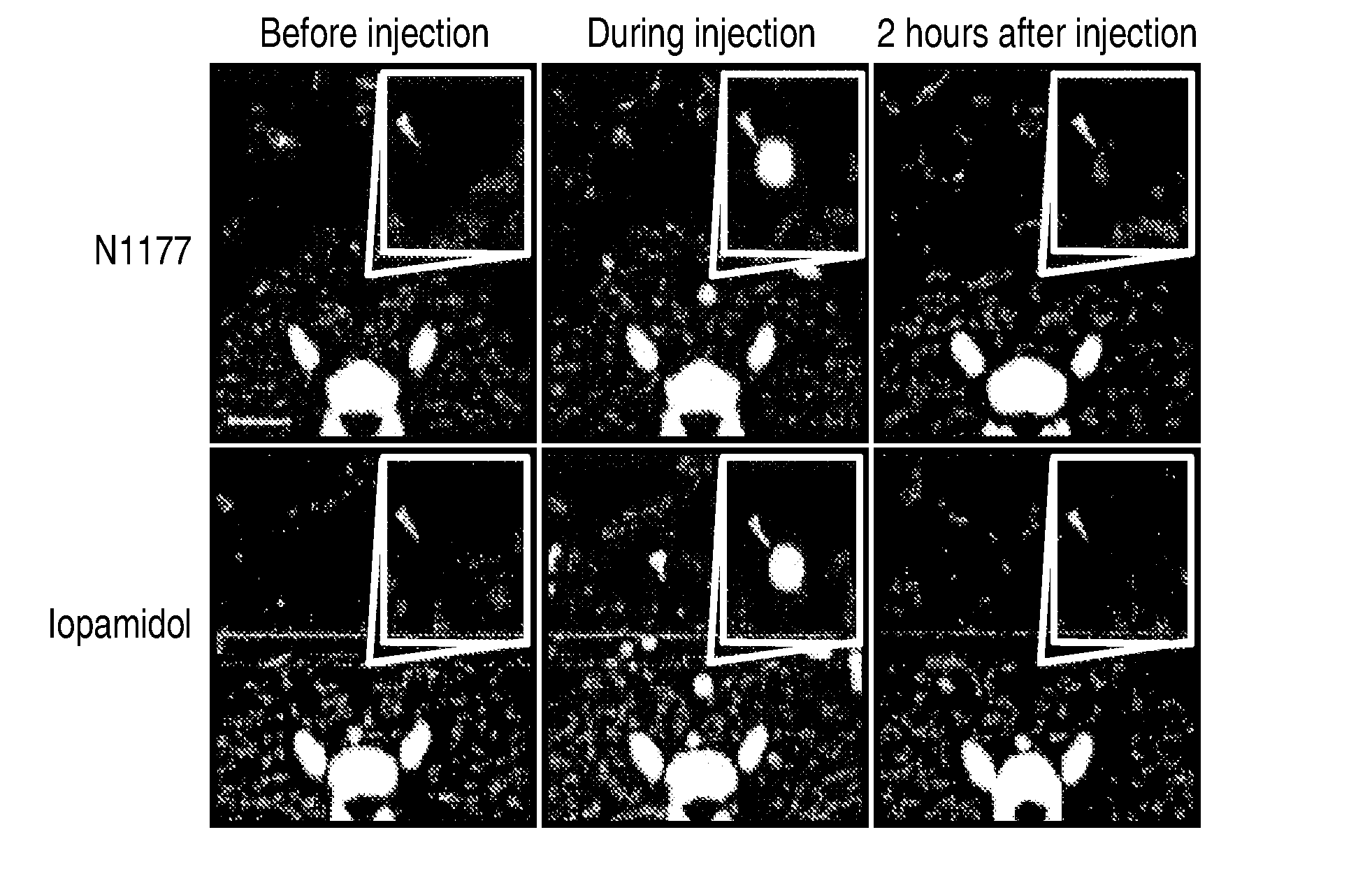
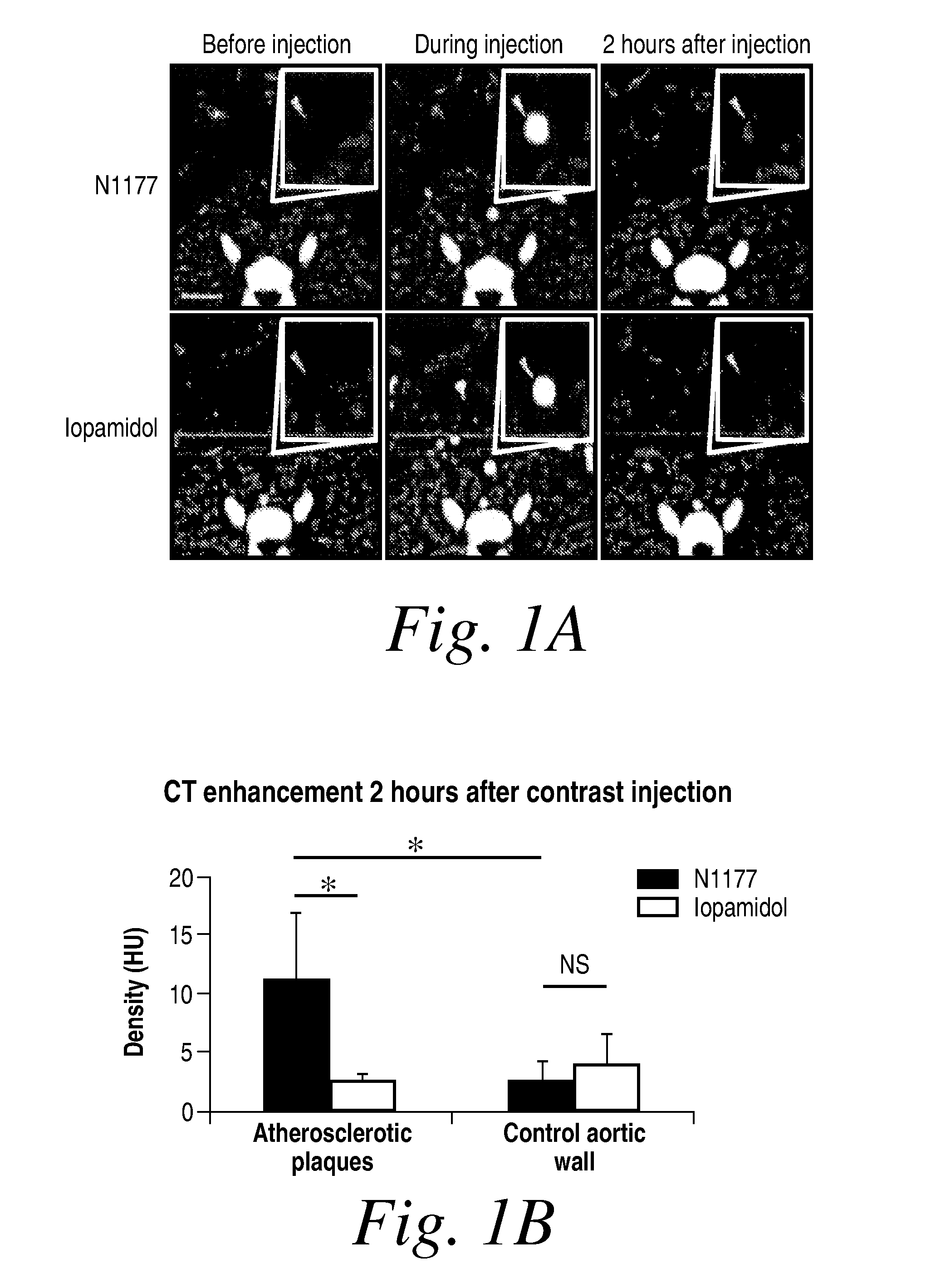
Detection of Macrophages in Atherosclerotic Plaques of Rabbits with N1177-iv-Enhanced Computed Tomography

[0206]Aortic atherosclerosis is induced in the male New Zealand White rabbits at a mean of 4 months and a mean weight of 3.3 kg. This is accomplished by 1) feeding the rabbits a high cholesterol diet for four months and by performing a double balloon denudation injury to the aorta. The same five rabbits are studied at three different doses of N1177: 125 mg / kg (dose 1), 250 mg / kg (dose 2, 1 week later), and 500 mg / kg (dose 3, 1 week after second dose).

[0207]Before imaging the animals are put under anesthesia by placing an intravenious access in the marginal vein of the ear with a 21-gauge line. Animals are kept in the same posture during all CT scans by placing them in a body-fitting thermosetting plastic holder. An initial localizer confirms the adequate position of the animal.

Image Acquisition and Analysis

[0208]All animals are imaged by computed tomography angiography many times...

example 4

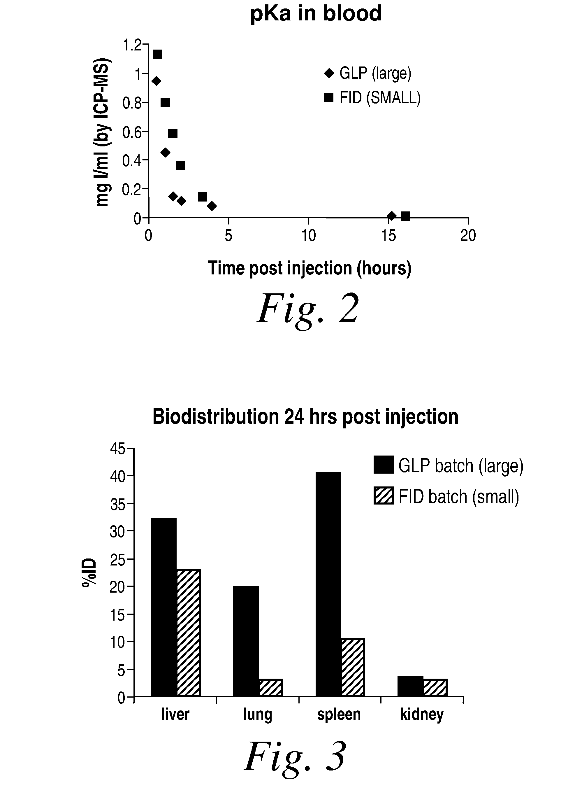
Biodistribution and pKa of Large (GLP) and Small (FID) Particle Formulations in NZW Rabbits

[0212]The experiments of this example evaluate the pharmacokinetics and biodistribution of the large and small N1177 formulations following bolus injection of 6-ml of either the large (150 mg I / ml) or small (160 mg I / ml) formulations in NWZ rabbits.

Materials and Methods: The large (GLP-batch N1177-2002001-A, 150 mg I / ml; referred to as Compound A) and small (batch FID 2470, 160 mg I / ml; referred to as Compound B) were obtained from Nanoscan. The hydrated particle size of each batch was determined using dynamic laser light scattering methods.

[0213]Two NZW rabbits (3.3-3.8 Kg, number 07-467 (Compound A) and 07-458(Compound B)) were used in the current study. The rabbits were imaged using CT imaging. For both rabbits, pre CT scans were obtained followed by CT imaging during bolus phase of injection, 2 minutes post injection, and 2 hours post injection. Six-ml of each formulation was administered ...


PUM

PropertyMeasurementUnit
Fractionaaaaaaaaaa
Diameteraaaaaaaaaa
Diameteraaaaaaaaaa

Abstract

The present invention also provides methods and compositions for imaging and evaluating, e.g., blood flow or inflammation in a subject. Such evaluations are important in a number of clinical diagnoses, including assessing organ damage associated with angina pectoris, heart attack, stroke, cancer, atherosclerosis, and the like, as well as assessing vessel leakages associated with aneurisms, diffuse bleedings after trauma, and the like.

Description

RELATED APPLICATION[0001]This application claims the benefit of U.S. Provisional Application Ser. No. 60 / 962,117, filed Jul. 26, 2007, the contents of which are hereby incorporated by reference.BACKGROUND OF THE INVENTION[0002]Contrast agents are injected into the vasculature of a subject in order to image blood flow in veins and arteries. Many of the agents currently used for imaging are water soluble. These agents provide contrast in the lumen of the vessel so that a positive image of the lumen can be made and the path of blood flow in the vessel can be detected. Images are made shortly after administration, before the agent is cleared from the lumen. Images made using such methods provide information regarding, for example, vessel occlusion, the presence of aneurisms, neovasculariztion, and blood flow.[0003]Different images can be made using nanoparticulate contrast agents. Certain of these agents can be taken up by phagocytic cells and, therefore, are useful in imaging sites of ...

Claims


Application Information

Patent Timeline
IPC IPC(8): A61K49/04B32B5/16B82Y5/00
CPCA61K49/0485Y10T428/2982B82Y5/00
Inventor SKERRETT, H. DONLONPATTERSON, JOSEPHLEE, ROBERT W.FAYAD, ZAHI A.
Owner NANOSCAN IMAGING
Why Eureka
Try Eureka