System And Method For Cell Analysis

Inactive Publication Date: 2012-04-12
MBIO DIAGNOSTICS
View PDF3 Cites 27 Cited by
  • Summary
  • Abstract
  • Description
  • Claims
  • Application Information

AI Technical Summary

Benefits of technology

[0065]One advantage of the present invention is that devices (cartridges) are processed independently of the reader instrument, enabling batch mode processing of cartridges. This provides a significant throughput advantage over competing technologies in which the instrument is occupied during cartridge processing. In one embodiment, the assay time on the reader instrument is less than 4 minutes, enabling up to 15 samples to be processed per hour on the reader.
[0066]In one embodiment, a cartridge rack may be used to simplify cartridge assays. For example, a cartridge rack with defined and labeled cartridge locations can improve organization of multiple cartridges being processed in parallel, reducing errors. In another embodiment, the cartridge rack can have active features further simplifying cartridge processing. For example, insertion of a cartridge into a rack position may actuate a timer and visual cues to the user such as lights and timer status that tell user that an assay step is in process or complete. Audible cues can also be used to alert the user to cartridge status, such as assay completion. In still another embodiment, the cartridge rack may have active feature that physically actuate processes on the cartridge. For example, a wash buffer may be incorporated in a blister pack on the cartridge during manufacturing. In one embodiment, insertion of a cartridge into a rack position may actuate a timer, and after a defined time the rack may further initiate a mechanical actuator (e.g., a ram or lever) that deploys the wash buffer blister pack into the cartridge, completing a timed assay step without any user interaction. An advantage of the present disclosure is that these simple features may be implemented in a highly parallel manner on an “active rack” that independently manages multiple cartridges. Simple logic circuitry and actuator motors may be incorporated at low cost in the rack, leaving the reader instrument available for processing multiple cartridges in series.
[0067]In one embodiment, different wavelengths may be used to illuminate the waveguide and the attached analytes or objects. One or more images may be taken of different fields of view on the attachment surface. The reader instrument may contain a translational mechanism to move the device relative to the lens and the image-capturing device so that images of different fields of view may be obtained.
[0068]In another embodiment, a brightfield microscopy image is captured for each field of view along with each fluorescence channel. The brightfield image may be used to resolve target objects via morphology analysis.
[0069]One of the advantageous features of the disclosed methods and systems is that a relatively small amount of the sample is required for each assay. In the context of blood based assays, it is desirable for the system to be compatible with both venous whole blood and capillary (finger stick) whole blood. In an embodiment, the sample has a specific volume in the range of 1 to 50 microliters, or preferably 1 to 20 microliters, or more preferable 1 to 10 microliters. In one embodiment, 10 microliters of blood sample is required to obtain an accurate count of T helper cells in a sample.
[0070]In one embodiment, a sample may be incubated with one or more labeling molecules, such as fluorescence tagged anti-CD4 and anti-CD14 antibodies, and the cells may be enumerated after being loaded onto the cartridge and subjecting to illumination in the reader instrument.

Problems solved by technology

After it gains access into an individual host, HIV may impair or eventually destroy the normal immune function of the infected individual.
As the immune system becomes weaker, the infected individual becomes more susceptible to other infections.
Over the last three decades or so, AIDS has spread globally and has become one of the biggest health challenges in many parts of the world, especially resource-limited settings.
Unfortunately, flow cytometry is a central lab-based technique; sample cold chain requirements as well as transport, equipment, and operational costs render the technique cost-prohibitive in limited resource settings where HIV prevalence is highest.

Method used


Image

  • System And Method For Cell Analysis
  • System And Method For Cell Analysis
  • System And Method For Cell Analysis

Examples


example 1

CD4 T Helper Cell Count from 10 Microliters of Whole Blood Sample

[0135]Biological Reagents. For the purposes of this disclosure, commercially available and commonly used antibodies were used. However, it is to be recognized that other commercial or customized antibodies may also be used. It is also to be understood that for any given antibody, a single batch or a mixture of antibodies directed against the same antigen may be used. Such antibodies may be obtained from different sources but are all reactive against the same antigen.

[0136]Assay Reagents. Some of the assay reagents include bovine serum albumin (“BSA”, Sigma Life Science, St. Louis, Mo.), phosphate buffered saline (“PBS”, Fisher Scientific, Rockford, Ill.).

[0137]Blood Samples. Because one of the important uses of the instant system and method will be in point-of-care settings, it is important to evaluate the performance of the system on whole blood samples. Whole blood was sourced under an IRB-approved protocol from HIV-...

example 2

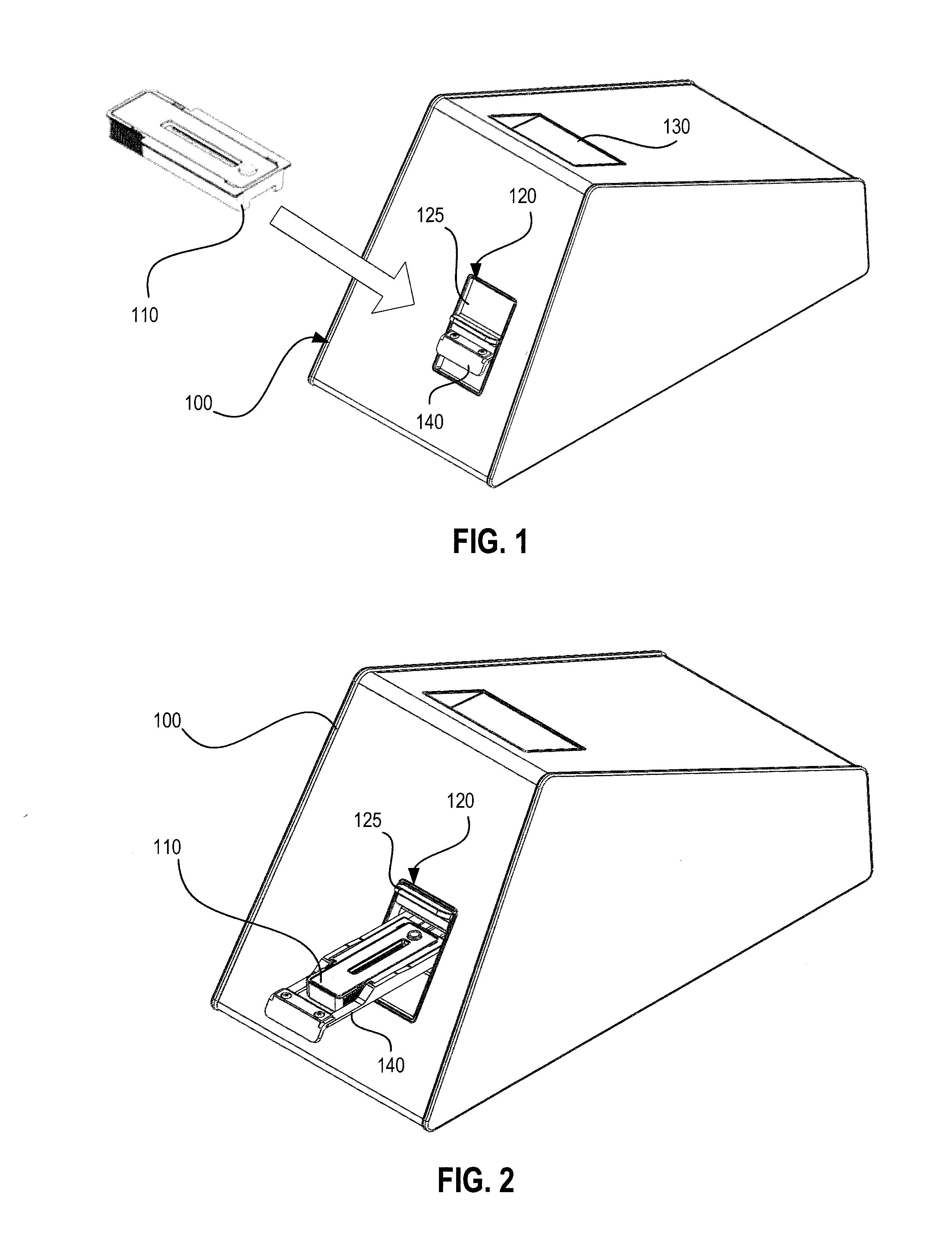
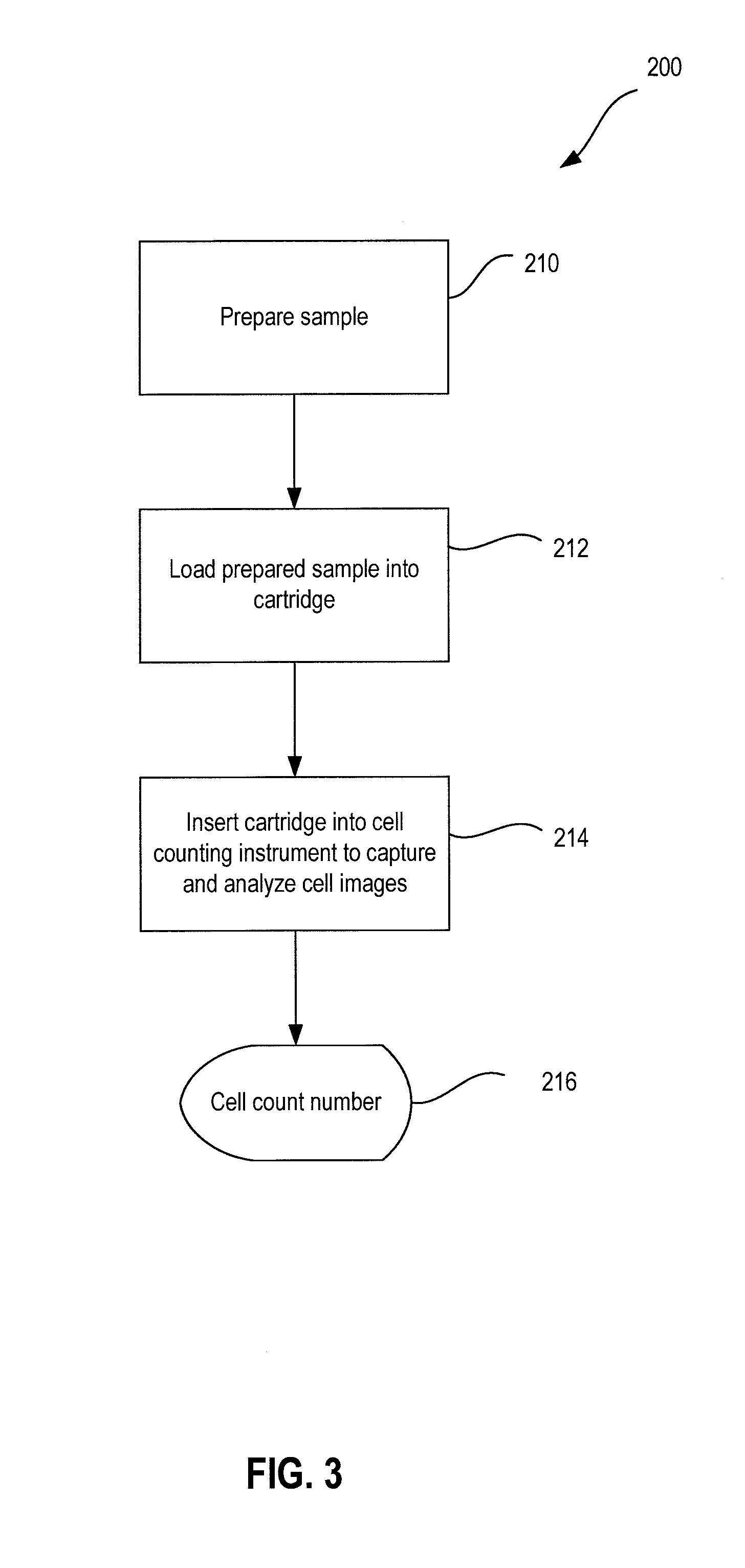
CD4 T Helper Cell Count from 10 Microliters of Whole Blood Sample Using a Combined Stain and Lysis Protocol

[0151]200 microliters of 10×RBC Lysis Buffer (Biolegend Catalogue #420301) were mixed with 1.8 ml deionized water to prepare 1× RBC Lysis Buffer. The 1×RBC Lysis Buffer was allowed to warm to room temperature. Cartridges were removed from packages and placed on a level surface. An antibody cocktail was prepared by mixing the following three antibodies: (a) Mouse anti-human CD4 Phycoerythrin, clone OKT4, (Biolegend Catalogue #317410); (b) Mouse anti-human CD4 Phycoerythrin, clone 13B8.2 (Coulter, Catalogue #IM0449U); and (c) Mouse anti-human CD14 AlexaFluor 647, clone HCD14 (Biolegend, Catalogue #32561). For example, the antibodies may be mixed by volume as 1 microliter of OKT4, 2 microliters of 13B8.2, and 1 microliter of HCD14; the exact mixture may be optimized according to assay performance. 4 microliters of the antibody cocktail were removed after vigorously mixing the cock...

example 3

Preparation and Use of Dried Reagents

[0156]For a point-of-care device, it would be desirable to offer kit components that do not require special controlled temperature storage. Dried kit reagent development is described briefly here. The most labile reagents in the presently described protocol may be the stain solutions, especially when stored at room temperature in facilities that may have inconsistent temperature control. Antibody stains in sample collection tubes have been successfully dried and assays with performance equal to direct addition of the liquid stains and flow cytometry measurements have been repeatedly demonstrated. Further, EDTA was incorporated into the dried antibody spot, resulting in direct anti-coagulant addition and staining of the 10 microliters blood sample. Experiments indicate high performance with only 5 to 10 minutes of incubation. These drying experiments have been based on selection of an appropriate sugar-based solution and overnight vacuum drying. H...


PUM


Abstract

A system for enumeration of objects such as cells in a sample is disclosed. The system uses a low-cost cartridge and a reader instrument, based on planar waveguide imaging technology. Cells of a blood sample may be stained with fluorescence-tagged antibodies and are loaded onto the cartridge where the differentially labeled cells may be distinguished and quantified.

Description

RELATED APPLICATIONS[0001]This application claims priority to U.S. Provisional Patent Application No. 61 / 391,909, filed Oct. 11, 2010 and entitled “System for Fluorescence Microscopy with Autofocus.” This application also claims priority to U.S. Provisional Patent Application No. 61 / 475,189, filed Apr. 13, 2011 and entitled “System and Method for Enumerating Cells in a Sample.” This application also claims priority to U.S. Provisional Patent Application No. 61 / 479,268 filed Apr. 26, 2011 and entitled “System and Method for Cell Analysis.” All of the aforementioned applications are incorporated by reference into the present application in their entireties and for all purposes.GOVERNMENT INTEREST[0002]This invention was made with Government support under the U.S. Department of Commerce National Institute of Standards (“NIST”) Advanced Technology Program (“ATP”), award number 70NANB7H7053, and National Institutes of Health (“NIH”) award number 2R44AI070052. The Government has certain r...

Claims


Application Information

Patent Timeline
IPC IPC(8): C12Q1/70C12Q1/02C12M1/34G01N21/75
CPCG01N21/0303G01N21/648G01N2015/1486G01N33/54373G01N2015/008G01N21/7703G01N2015/016
Inventor GIVENS, MONIQUEIVES, JEFFREYLOCHHEAD, MICHAEL J.DELANEY, MARIE J.MOLL, KEVIN D.ROWLEY, KEAGAN B.VOGEL, KURT R.MYATT, CHRISTOPHER J.
Owner MBIO DIAGNOSTICS
Why Eureka
Try Eureka