
Protein Structural Biomarkers to Guide Targeted Chemotherapies

Active Publication Date: 2012-05-17
KIM SUNYOUNG
View PDF0 Cites 2 Cited by
  • Summary
  • Abstract
  • Description
  • Claims
  • Application Information

AI Technical Summary

Benefits of technology

[0014]I have developed an infrared spectroscopic method to assess the efficacy of targeted chemotherapeutics against the structure of the polypeptide target based on the effect of natural polymorphic sequence variation on the conformation of the protein. This method has an advantage over the current genomics-based screening, as the new method provides a direct readout of the structural, and hence functional, outcome of polymorphisms to the protein region targeted by drugs. Second, it allows rapid measurement of tumor protein susceptibility to therapeutic targeted agents, prior to using the drug as treatment in the patient. Third, this method can be used to identify biomarkers for a response for a protein to a drug which could be readily tested, interpreted, and used in a clinical setting.

Problems solved by technology

However, the reliability of microarray biomarker assays for disease is unknown with few effective biomarkers or tools for assessment established.
No consensus of L5 residues key for transmitting allosteric information has been established, primarily due to interdependence of the allosteric and active-site interactions and the lack of rapid methods to observe structures of mutant motor proteins.
Yet, neither these reports nor X-ray structural analyses of allosterically-inhibited Eg5 proteins arrive at a common set of contact residues for binding many different chemical partners.
(See the website: http: / / www.resb.org / pdb) Although the atomic level detail of the structures is fairly complete, whether these static ‘snapshots’ accurately reflect functional states that these amino acid assemblies can achieve is more difficult to determine.
Thus, obtaining all needed conformational possibilities in kinesin mechanotransduction and its allosteric inhibition is a significant challenge.

Method used


Image

  • Protein Structural Biomarkers to Guide Targeted Chemotherapies
  • Protein Structural Biomarkers to Guide Targeted Chemotherapies
  • Protein Structural Biomarkers to Guide Targeted Chemotherapies

Examples


example 1

[0050]Materials and Methods

[0051]Generation of single- and double-mutants of Eg5. Starting cDNA was the truncated form of wildtype Eg5 as previously reported [residues 1-370; (28)]. Appropriate primers were synthesized and used for site-directed mutagenesis with the QuikChange II XL kit (Stratagene, La Jolla, Califf). Construction of the Eg5 motor domain with substitutions of Asp for E116 and E118 was performed using two rounds of mutagenesis. Expected mutations were confirmed by a single-read sequencing reaction. A subset of five mutations was completely sequenced on both strands to ensure that inadvertent coding errors were not introduced. The mutations produced are shown below in Table 1.

[0052]Motor protein expression and purification. The wildtype Eg5 kinesin motor domain, as well as single- and double-site polymorphisms, were expressed in BL21-Codon Plus (DE3)-RIL cell lines (Stratagene) and purified by cation exchange chromatography as previously described (28). Purity of wild...

example 2

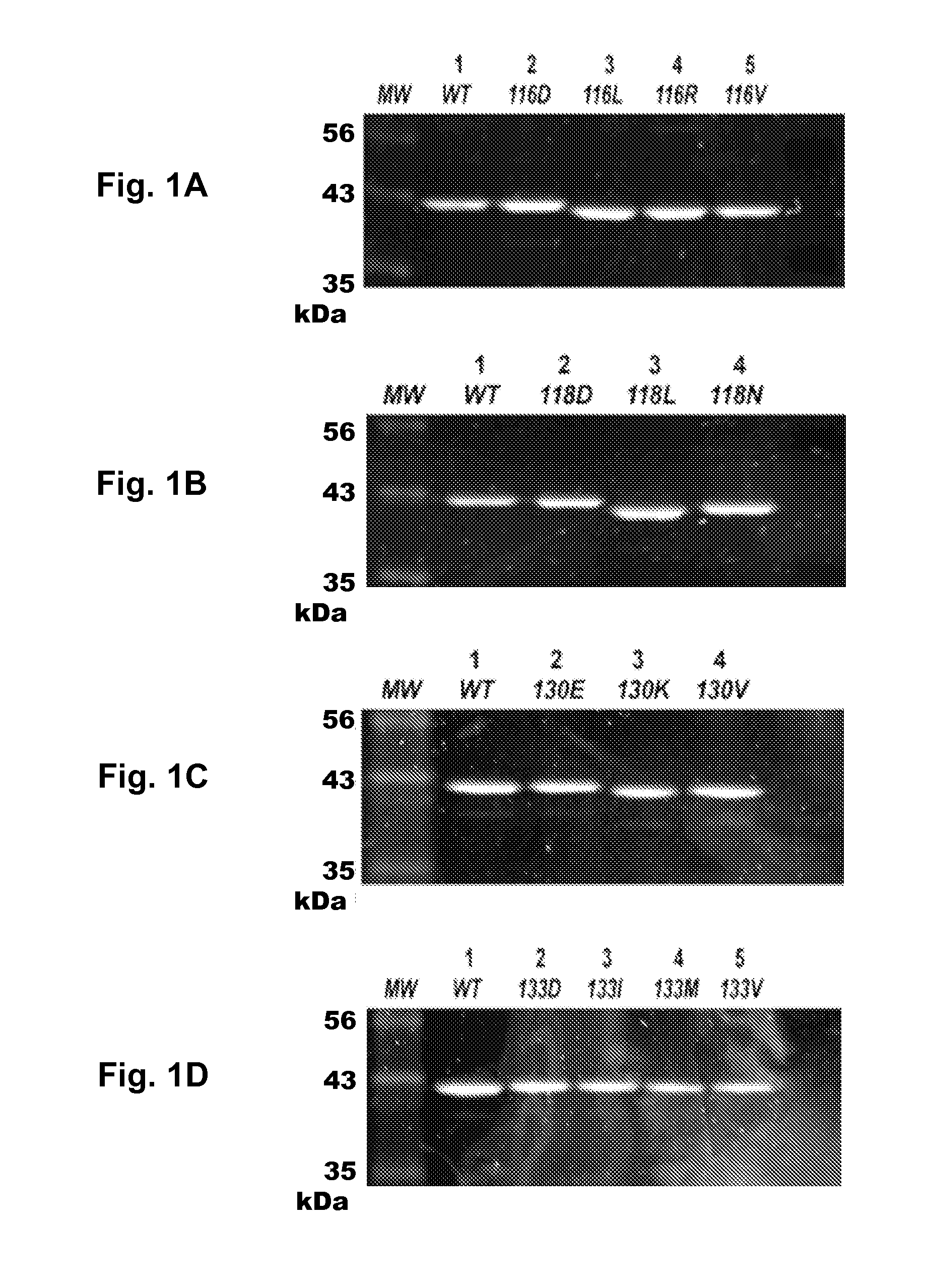
[0059]Substitutions of L5 Residues Result in Altered SDS-PAGE Mobility

[0060]The initial step of this study was to assess how perturbations of the L5 loop affect Eg5 steady-state kinetics and solution structure. The first type of perturbation involves alteration of sidechain chemistry. Fifteen (15) mutations were sampled at four sites in the L5 loop: two at the N-tenninus and two at the C-terminus of the insertion loop. Prior studies scrutinizing the allosteric pocket typically assumed that point mutations have little effect beyond the localized change of a particular functional group. However, this stance can underestimate the ability of the point mutation to remodel the binding surface or transmit long-range effects that are dependent on the nature of the substitution. Therefore, the mutations used herein ranged from conservative to pleiotropic alterations; this allowed a wider range of chemical and structural outcomes at specific positions to be seen.

[0061]The N-terminal residues ...

example 3

[0064]Preservation of N-Terminal Local Structure and C-Terminal Chemistry in the L5 Loop are Required for Allostery and Catalysis.

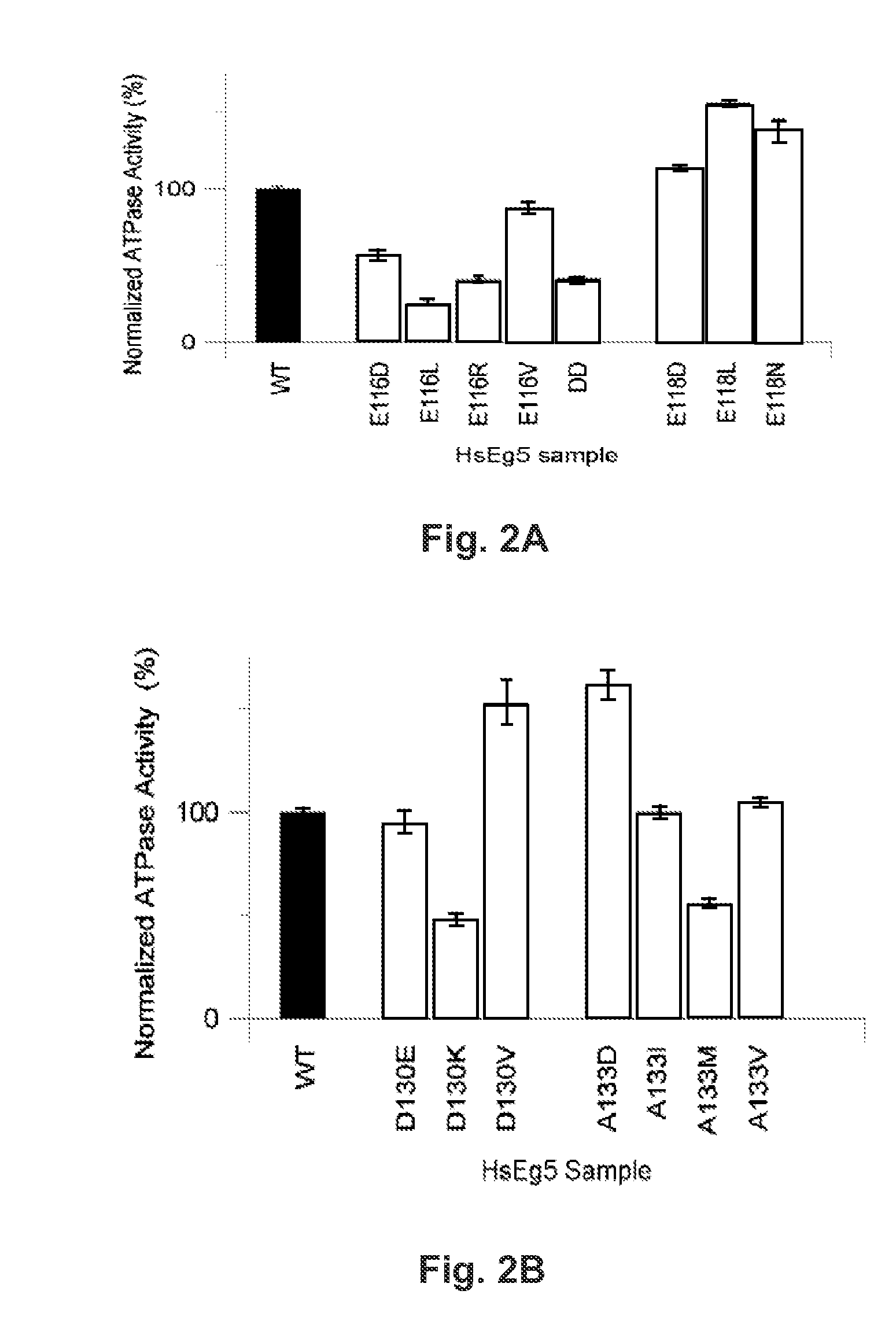
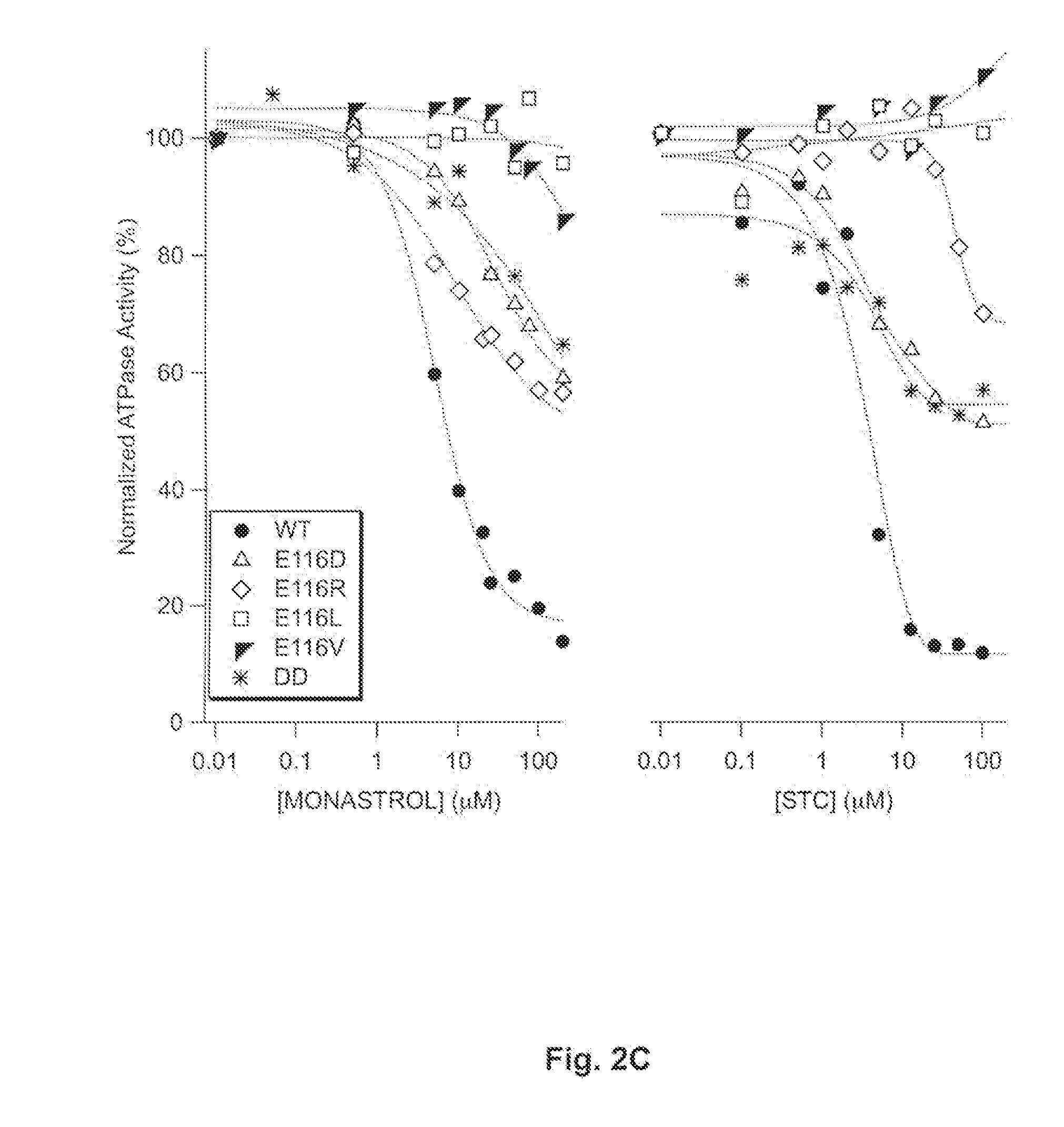
[0065]There are marked kinetic differences between the N-terminal and C-terminal substitutions of L5 loop residues. Compared with wildtype samples, single-site substitutions of E116 decreased the steady-state, basal ATP hydrolysis rate of Eg5 kinesin monomers (FIG. 2A, open boxes). However, mutants of E118 displayed increased activity (FIG. 2A, filled boxes). These results were consistent, whether or not the amino acid substitutions were synonymous with carboxylates. These data demonstrated that the native interactions of E116 and / or E118 in the motor domain influence ATP hydrolysis rates achievable by Eg5. Moreover, for mutants of these two positions in the L5 loop, the faithful difference in mutant kinetic behavior, despite conservative or pleiotropic changes, implicated the native sidechain of these N-terminal L5 residues not primarily in electrostatic...


PUM

PropertyMeasurementUnit
Massaaaaaaaaaa
Massaaaaaaaaaa
Fractionaaaaaaaaaa

Abstract

A rapid, infrared spectroscopic method has been developed to assess the efficacy of targeted chemotherapeutics against the structure of the polypeptide target, based on the effect of natural polymorphic sequence variation on the conformation of the protein. This method has an advantage over the current genomics-based screening, as the new method provides a direct readout of the structural, and hence functional, outcome of polymorphisms to the protein region targeted by drugs. It allows rapid measurement of a protein's susceptibility to therapeutic targeted agents, prior to using the drug as treatment in the patient. This method can be used to identify biomarkers for a response for a protein to a drug which can be readily tested, interpreted, and used in a clinical setting.

Description

[0001]The benefit of the filing dates of provisional U.S. application Ser. No. 61 / 154,121, filed 20 Feb. 2009, and of provisional U.S. application Ser. No. 61 / 261,939, filed 17 Nov. 2009, is claimed under 35 U.S.C. §119(e) in the United States, and is claimed under applicable treaties and conventions in all countries.[0002]The development of this invention was partially funded by the Government under grant number GM066328 from the National Institutes of Health. The United States Government has certain rights in this invention.TECHNICAL FIELD[0003]This invention pertains to a spectroscopic method to assess the efficacy of chemotherapeutics targeted to a specific protein in a patient, based on the effect of polymorphic sequence variation on the conformation pattern of the protein. This method views the structure of polypeptides and develops protein biomarkers that can be used to predict the protein's (and thus the patient's) response to a drug treatment. Using this method, protein bio...

Claims


Application Information

Patent Timeline
IPC IPC(8): C12Q1/34
CPCC12Q2600/156G01N33/574C12Q2600/106A61B5/0059C12Q1/6886G01N2800/52A61K31/198A61K31/505A61K31/506C12Q1/34G01N2333/914G01N2500/04
Inventor KIM, SUNYOUNG
Owner KIM SUNYOUNG
Learn More