Assay tools and methods of use
a technology of assay tools and methods, applied in the field of assay tools, can solve the problems of reducing the dynamic range of assays, unable to distinguish which specific proteases, and los assays may also have background problems
- Summary
- Abstract
- Description
- Claims
- Application Information
AI Technical Summary
Benefits of technology
Problems solved by technology
Method used
Image
Examples
example 1
Peptide Synthesis
[0190]Peptides were synthesized by manual Fmoc-based peptide synthesis in fritted syringes (Fields et al., Biochemistry. 1990 Jul. 17; 29(28):6670-7; Chan D et al., J Cell Physiol. 2000 December; 185(3):339-47) on Fmoc-glycine Wang resin solid support (Advanced Chemtech). Fmoc-Lpropargylglycine (Advanced Chemtech) was used at the first step of the peptide synthesis to introduce the alkyne group at the C-terminus of peptides. Fmoc-Lys(biotin)-OH (EMD Chemicals) was used as the last amino acid to introduce the N-terminal biotin residue. Peptide synthesis was followed by Fmoc deprotection and coupling of the fluorescent dye (6-carboxy-tetramethylrhodamine, TAMRA, EMD Chemicals). Molecular weight of peptides were confirmed using a ProteinPlex SELDI laser desorption / ionization TOF mass spectrometry based analytical system (Bio-Rad). We used peptide substrates GAENLYFQGA and GALVPRGSAG targeting TEV and thrombin proteases as model peptides. The protease recognition sites ...
example 2
Preparation of Substrate Slides
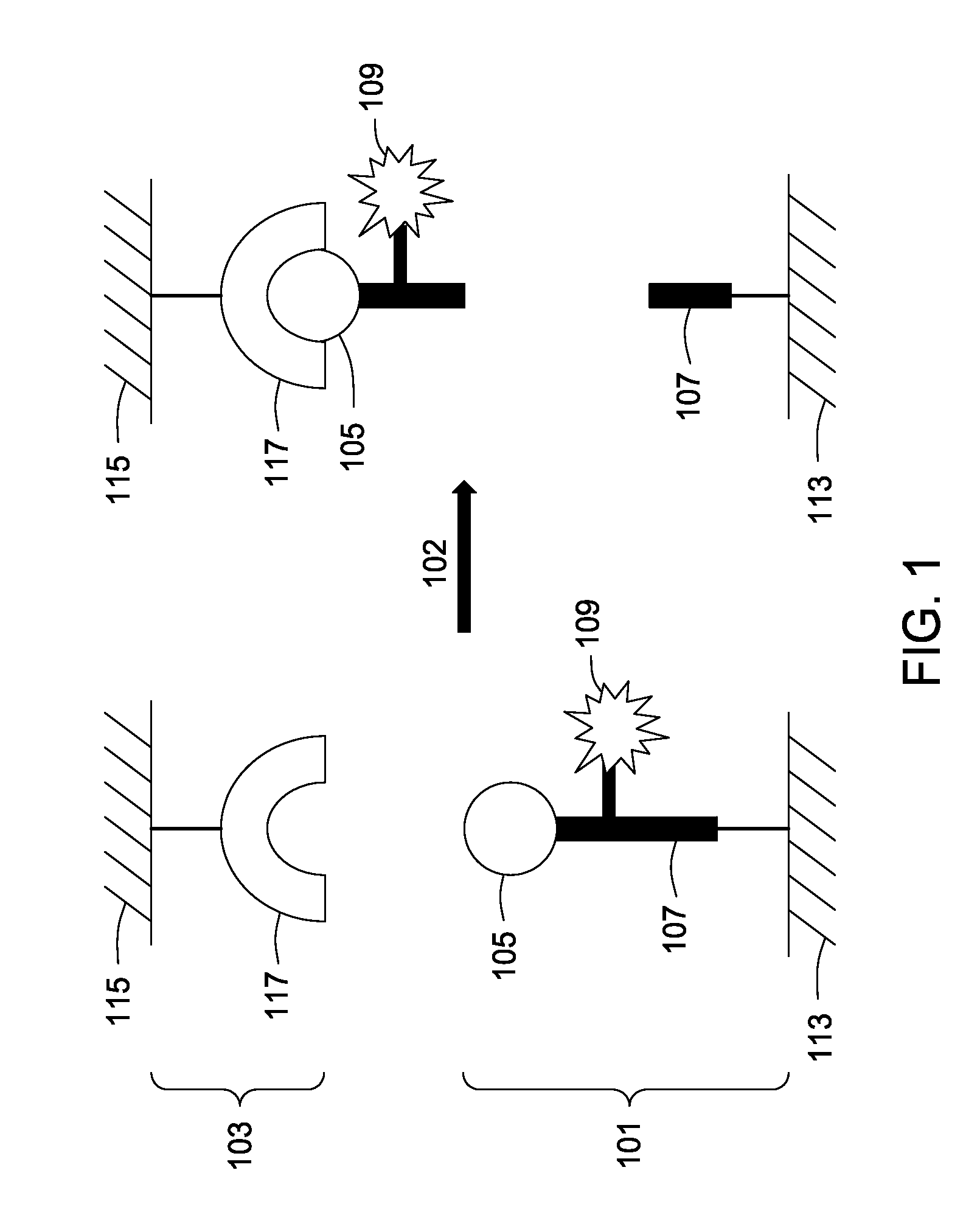
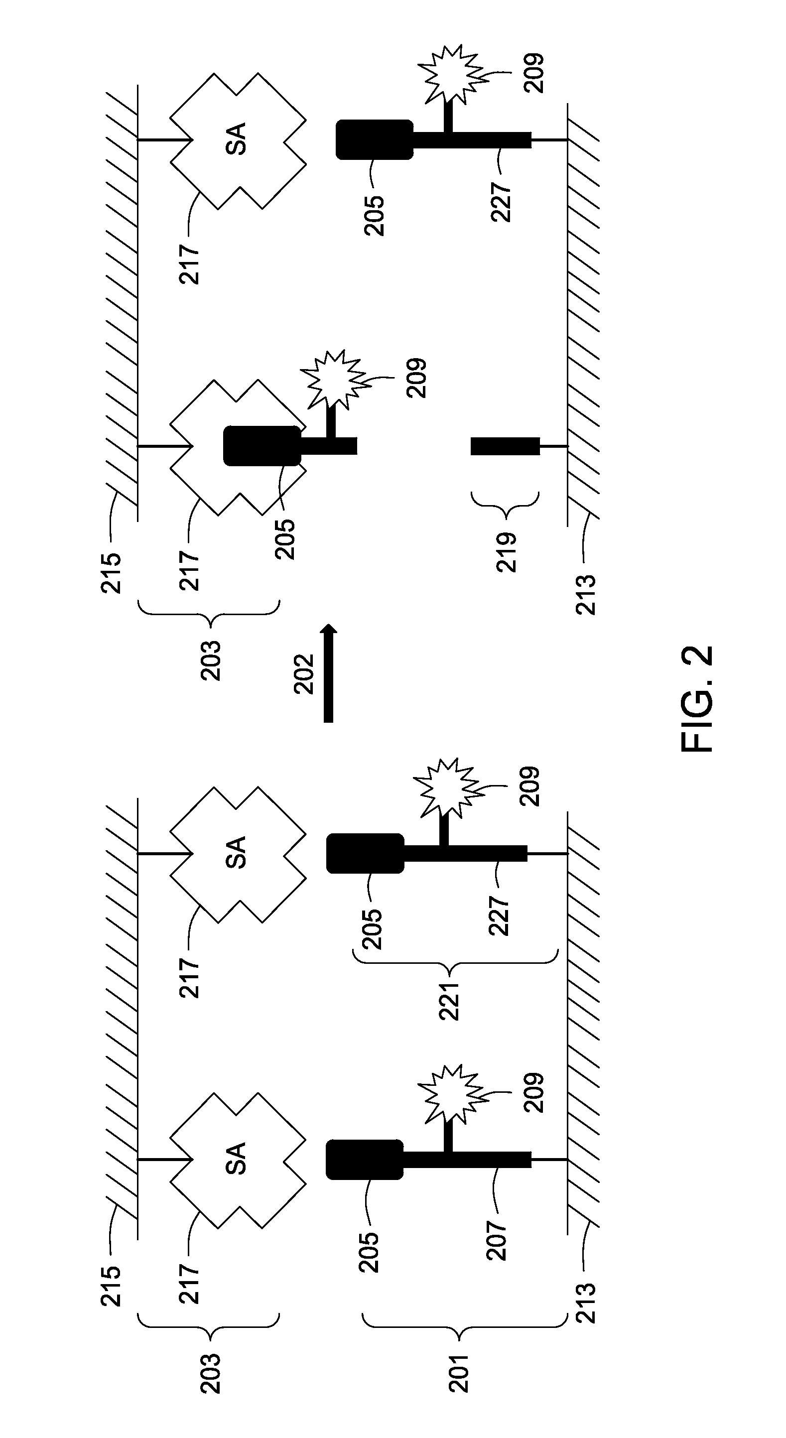
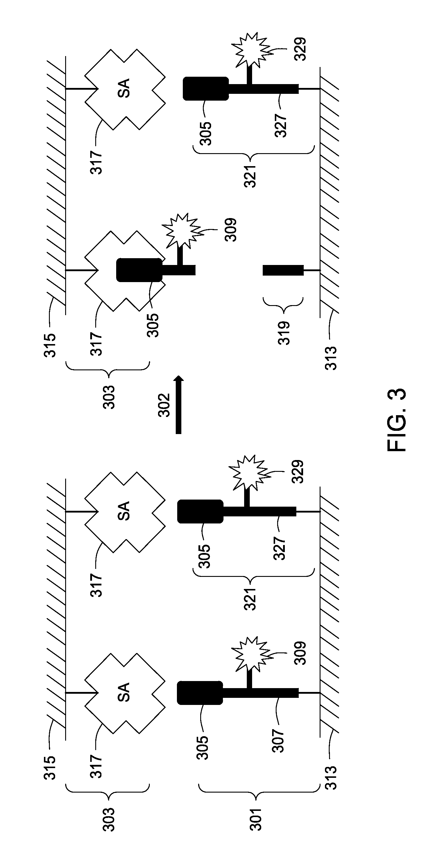
[0192]Standard 75×25 mm microscope slides were used to implement the two-surface assay tool. One slide comprises the peptide substrates (the “substrate” slide), and the other slide comprises the capture agents (the “reporter” slide). Together these comprise a two surface assay tool.
[0193]Microscope slides with a non-protein polyethyleneglycol (PEG) linker were is obtained in the following way. ES amino slides (Erie Scientific) were treated with 0.1M N3-(PEG)7-COOH(O-(2-Azidoethyl)-O′-(N-diglycolyl-2-aminoethyl) heptaethyleneglycol, EMD Chemicals) solution in DMF containing 0.1M PyBop (EMD Chemicals) and 0.2M N,N-diisopropylethylamine overnight at room temperature. Slides were subsequently washed with DMF (3 times), and water (3 times). This 33-atoms PEG-linker resulted in direct introduction of azide groups on the slide surface. Substrate slides were blocked with a non-protein blocking solution containing Ficoll 400, PVP 40 (polyvinylpyrrolidones), 40 ...
example 3
Immobilization of Peptides
[0197]The peptides were dissolved at 2 μm concentration in 0.1M Tris-HCl buffer pH 7.5 containing 20% DMSO, 10 mM CuSO4, and 50 mM Na-ascorbate. After spotting, the immobilization reaction was allowed to proceed for 12 hours at room temperature in a humidified chamber. The slides were washed with DMF, DMSO, and 50 mM Tris-HCl, pH 7.5 containing 0.01% Tween 20 for 30 min per washing solution, rinsed with water, ethanol, and dried. The peptides were spotted on the slides (0.1 μl per spot) using manual spotting with a standard 0.1-2 μl laboratory pipettor (Eppendorf). This yielded spots with a diameter of approximately 800 um.
PUM
 Abstract
Description
Claims
Application Information
 - R&D
- Intellectual Property
- Life Sciences
- Materials
- Tech Scout
- Unparalleled Data Quality
- Higher Quality Content
- 60% Fewer Hallucinations
Browse by: Latest US Patents, China's latest patents, Technical Efficacy Thesaurus, Application Domain, Technology Topic, Popular Technical Reports.
© 2025 PatSnap. All rights reserved.Legal|Privacy policy|Modern Slavery Act Transparency Statement|Sitemap|About US| Contact US: help@patsnap.com


