

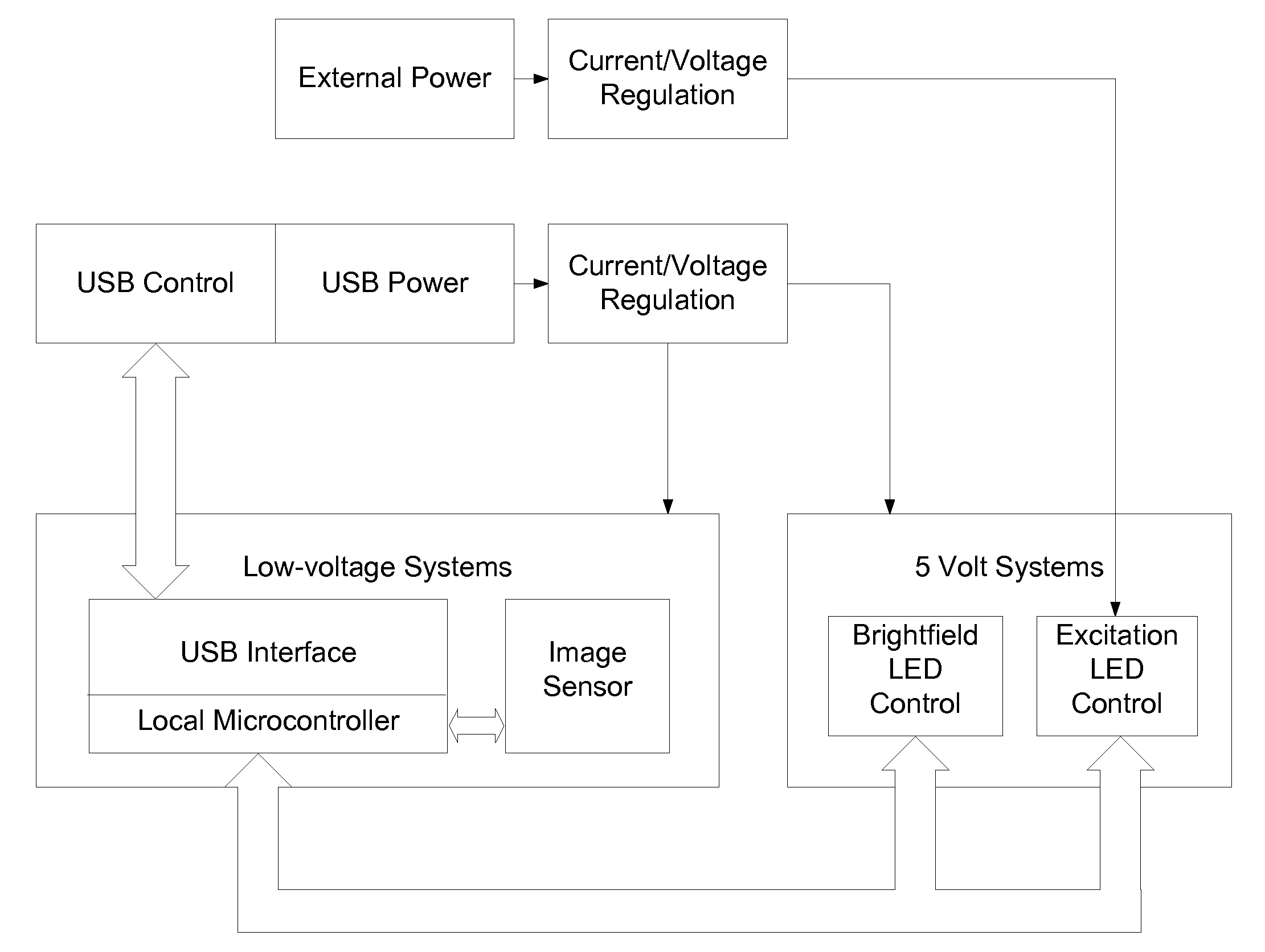
Compact, high-resolution fluorescence and brightfield microscope and methods of use
a fluorescence microscope and brightfield technology, applied in closed circuit television systems, instruments, television systems, etc., can solve the problems of large camera containers, weak light intensity of fluorescent samples, and low light throughput in optical systems, and achieve high light throughput.
- Summary
- Abstract
- Description
- Claims
- Application Information
AI Technical Summary
Benefits of technology
Problems solved by technology
Method used
Image

Examples
example 1
[0104]This example establishes the ease of use of a microscope of the present invention and computer interface and output with a microscope of the present invention.
[0105]Ease of use of the microscope is shown in FIG. 5A and FIG. 5B, which is a captured screen shot of the host computer running the control program for the preferred embodiment. An image of human neural stem cells derived from 13 week-old fetal telencephalon growing in a T25 polystyrene tissue culture flask with the growth surface coated with mouse laminins and observed with brightfield illumination through the top of the flask and with a Meiji 20× magnification 0.4 N.A. 7 mm working distance objective is shown in the image display area of the graphical user interface. The image displayed is the last one acquired during time lapse acquisition and saving of images once every 10 min. The acquisition sequence had been running continuously for 5 days when this screen capture was obtained. The four basic tools for operation...
example 2
[0106]This example establishes the application of a microscope of the present invention for educational purposes.
[0107]Brightfield images obtained with the educational set of microscope slides “The 5 Kingdoms” (cat. no. E2-70-4016, Neo / Sci, 80 Northwest Blvd., Nashua, N.H.) with the use of the Meiji 40× magnification 0.65 N.A. 0.5 mm working distance objective in the preferred embodiment is shown in FIG. 7, FIG. 8 and FIG. 9. FIG. 7 is an image of fixed Paramecium tetraurelia. Key definitive morphological features of the protozoans, including cilia and micronuclei, can be resolved. FIG. 8 is an image of a horizontal section through a leaf of Vicia fava (broad bean). The stomata that regulate water exchange between the leaf and the atmosphere and cell nuclei are clearly observed. FIG. 9 is an image of Spirogyra crassa, a filamentous freshwater green alga. To the left of the image, the helical arrangement of photosynthetic chloroplasts around prominent cell nuclei is visible in the ve...
example 3
[0108]This example establishes the application of a microscope of the present invention for research and educational purposes.
[0109]Usefulness of the preferred embodiment is demonstrated for research and education is demonstrated in the fluorescence images of bovine pulmonary arterial endothelial cells (BPAEC) shown in FIG. 10 and FIG. 11. BPAEC were cultured on a gelatin-coated no. 0 coverslip for 3 days, fixed with buffered 4% formaldehyde for 15 min, washed three times in buffer, incubated for 3 hrs in BODIPY FL-labeled phallacidin (Life Technologies, Cat. No. B607), washed 3 times in buffer and mounted on a microscope slide by techniques well-known to those skilled in the art. The slide was affixed to the x-y stage caliper with tape and mounted on the stage of the preferred embodiment with the coverslip facing the objective lens. Phallacidin binds to filamentous (F) actin, one of the predominant structural proteins of eukaryotic cells that maintain their shape and integrity, and...
PUM
 Abstract
Description
Claims
Application Information