Device and Methods for Epigenetic Analysis

a technology of epigenetic analysis and devices, applied in the field of devices and methods for epigenetic analysis, can solve the problems of imposing significant limitations on analysis throughput and sample quantity, suffering from two major limitations, and being incapable of assessing epigenetic changes in vanishing quantities, etc., to achieve high-throughput implementation, rapid data collection, and high detection system bandwidth

Active Publication Date: 2012-09-27
CORNELL UNIVERSITY
View PDF0 Cites 17 Cited by
  • Summary
  • Abstract
  • Description
  • Claims
  • Application Information

AI Technical Summary

Benefits of technology

[0012]In some embodiments, the present invention provides for assessing epigenetic variations at a single chromatin level. Single-molecule analysis provides several advantages over conventional approaches. First, the analysis provides information on individual molecules whose properties are hidden in the statistically averaged information that is recorded by ordinary ensemble measurement techniques. In addition, because the analysis can be multiplexed, it is conducive to high-throughput implementation, requires smaller amounts of reagent(s), and takes advantage of the high bandwidth of detection systems such as modern avalanche photodiodes for extremely rapid data collection. Moreover, because single-molecule counting automatically

Problems solved by technology

Existing approaches for analyzing epigenetic modifications of chromatin, such as chromatin immunoprecipitation (ChIP), can evaluate only one epigenetic mark at a time and are labor-intensive, serial processes that impose significant limitations on analysis throughput and sample quantity.
However, it suffers from two major limitations.
First, the analysis requires 104 to 107 cells and is incapable of assessing epigenetic changes in vanishing quantities, making studies of developing embryos, sorted cells or microdissected cells impossible.
Second, only one epigenetic mark can be isolated at a time, making detection of co-existent marks very difficult.
Sequential ChIPs against two different antibodies may circumvent this limitation, however this solution is impractical for whole

Method used


Image

  • Device and Methods for Epigenetic Analysis
  • Device and Methods for Epigenetic Analysis
  • Device and Methods for Epigenetic Analysis

Examples


example 1

A Nanofluidic Platform for Single Molecule Analysis

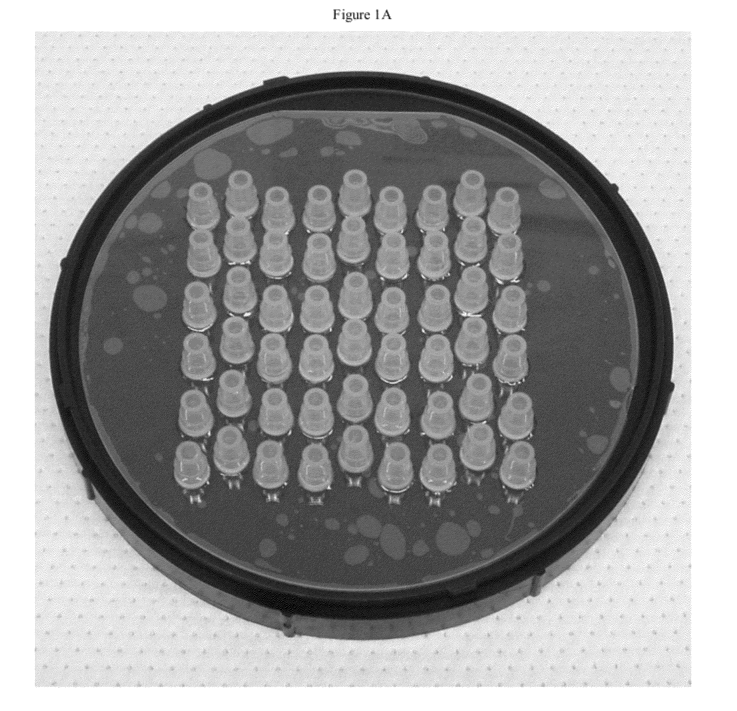
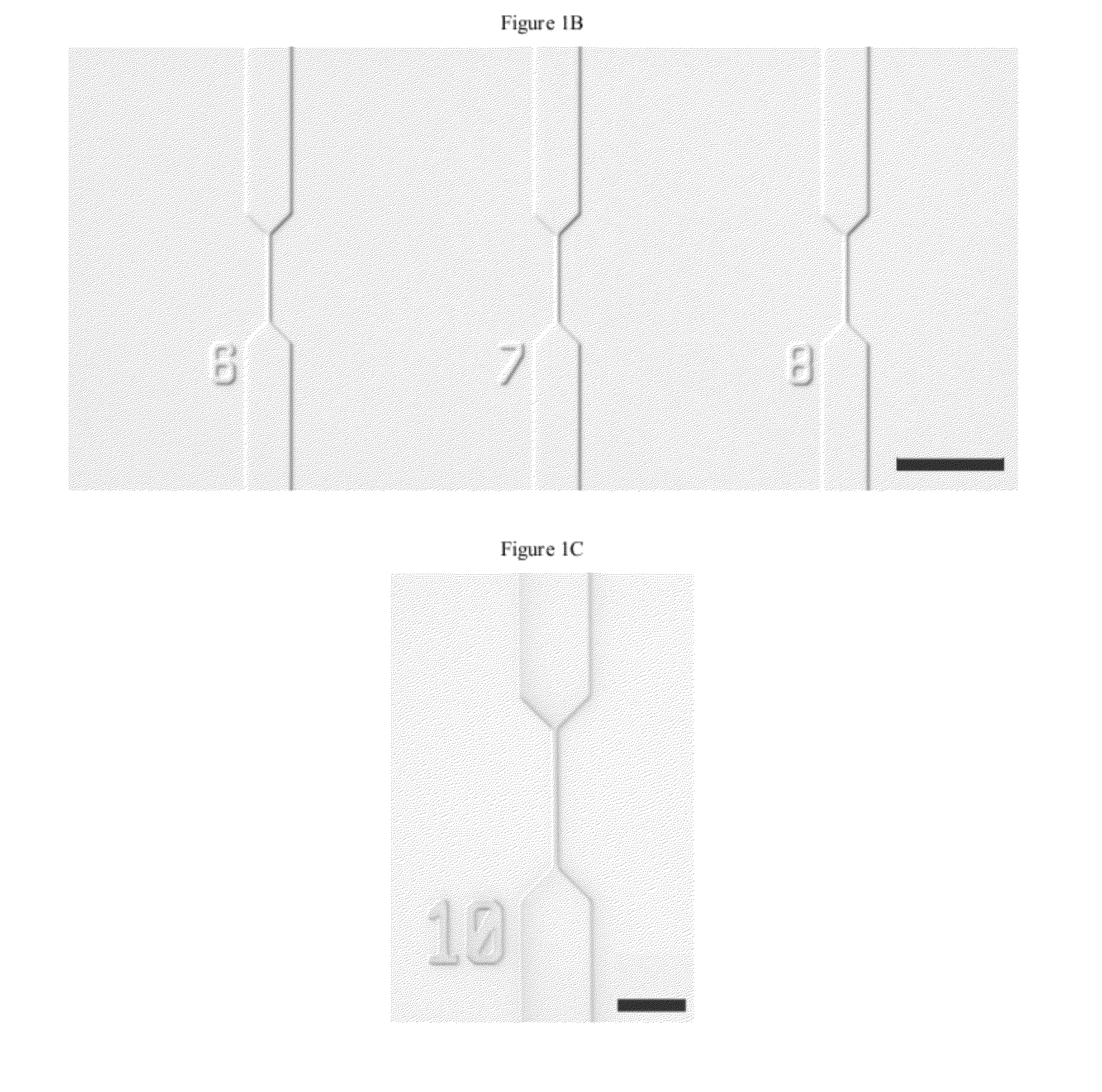
[0190]One strategy for high throughput analysis of single molecules of methylated DNA and chromatin can entail time-resolved detection and spectral identification of fluorescent labels bound to individual molecules containing epigenetic marks of interest. We flowed and detected DNA and chromatin in a solution confined within a nanofluidic channel. These channels reduce the optical excitation volume for fluorescent analysis, thus enabling us to interrogate individual molecules in solutions of relatively high concentration. The nanofluidic channels were fabricated in a fused silica substrate using photolithography and reactive ion etching. FIG. 1C a shows a single nanofluidic channel made by this process with cross-sectional dimensions of approximately 250 nm wide by 500 nm deep. We formed 27 separate fluidic channel arrays, each with 16 parallel channels, on a 100 mm diameter wafer. Each array also had access ports with a reservoir a...

example 2

Nucleosome Detection on Native Chromatin

[0192]This experiment demonstrates that chromatin can be directed through a nanofluidic channel and remain intact, having nucleosomes remain attached on the DNA. For this test we used chromatin extracted from HeLa cells bearing a transgene expressing an H2B-GFP (green fluorescent protein) fusion protein. H2B-GFP incorporated into nucleosomes allowed the chromatin to fluoresce. We prepared native chromatin from the cells using standard methods, treating isolated nuclei with micrococcal nuclease (MNase) and then extracting the soluble native chromatin using a high salt buffer. We next labeled the DNA within our chromatin preparations with TOTO-3, a red nucleic acid stain that is spectrally distinct from GFP. We wanted to analyze this dual labeled chromatin for two reasons. First, it permitted us to determine whether chromatin remained intact during nanofluidic electrokinetic flow, which is essential for any successful application of this method ...

example 3

Detection of DNA Methylation

[0198]Probes that have been used in epigenomic analysis were used to detect an epigenetic mark on our nanofluidic platform. We analyzed DNA methylation and used MBD1 as our probe, which has been shown to bind methylated DNA specifically. Our test material was HindIII digested lambda DNA from a methylation deficient host, which we left unmethylated, or methylated in vitro using SssI DNA methyltransferase. SssI can methylate all 3,113 CpGs in the 48.5 kbp genome. We verified the effectiveness of the methylation reactions by digesting the DNA with the methylation sensitive restriction enzyme HpaI (FIG. 25). Both DNA samples were stained with TOTO-3 and incubated with MBD1, which we labeled with Alexa Fluor 488. Alexa Fluor 488 is spectrally similar to GFP. The Alexa Fluor 488 labeled MBD1 retained its specificity for methylated DNA (FIG. 26). To facilitate optimum binding to methylated DNA, we added a molar excess of MBD1 to the stained DNA. We found that di...


PUM

PropertyMeasurementUnit
Timeaaaaaaaaaa
Volumeaaaaaaaaaa
Volumeaaaaaaaaaa

Abstract

Provided herein are methods and devices for single object detection. The methods and devices can be used to identify a plurality epigenetic markers on a genetic material, or a chromatin, encompassing fragments thereof. The invention provides for the characterization of the genetic material flowing through a channel in a continuous body of fluid based on detection of one or more properties of the genetic material. The methods and systems provided herein allow genome-wide, high-throughput epigenetic analysis and overcome a variety of limitations common to bulk analysis techniques.

Description

CROSS-REFERENCE[0001]This application is a continuation-in-part of PCT Patent Application No. PCT / US2010 / 044806, filed Aug. 6, 2010, which claims benefit of priority to U.S. Provisional Application No. 61 / 231,979, filed Aug. 6, 2009, U.S. Provisional Application No. 61 / 307,827, filed Feb. 24, 2010, U.S. Provisional Application No. 61 / 231,963, filed Aug. 6, 2009, U.S. Provisional Application No. 61 / 359,266, filed Jun. 28, 2010, and PCT Patent Application No. PCT / US2010 / 044810, filed Aug. 6, 2010, each of which is incorporated herein by reference in its entirety for all purposes.STATEMENT AS TO FEDERALLY SPONSORED RESEARCH[0002]This invention was made with the support of the United States government under Contract number R01DA025722 by the National Institutes of Health, Contract number ECS-9876771 by the National Science Foundation (NBTC), and Contract number 199-3166 by the Center for Vertebrate Genomics (Cornell University).BACKGROUND OF THE INVENTION[0003]Chromatin, located in the ...

Claims


Application Information

Patent Timeline
IPC IPC(8): G01N21/64C12M1/34
CPCB01L3/502761C12Q1/68B01L2300/0654C12Q1/6827B01L2200/0663C12Q2565/631C12Q2565/629C12Q2537/164C12Q2561/113
Inventor CRAIGHEAD, HAROLD G.CIPRIANY, BENJAMIN R.LEVY, STEPHENSOLOWAY, PAUL
Owner CORNELL UNIVERSITY
Why Eureka
Try Eureka