Multimodal Imaging System, Apparatus, and Methods

a multi-modal imaging and apparatus technology, applied in the field of medical treatment and diagnostics, can solve the problems of reducing the utility of existing integrated ivus and ffr diagnostic systems, increasing the clutter in the catheterization lab, and requiring time-consuming set-up procedures, so as to reduce the error and setup time, improve the flexibility of measurement equipment, and facilitate the effect of connection and disconnection quickly and easily

Active Publication Date: 2012-12-06
LIGHTLAB IMAGING
View PDF21 Cites 182 Cited by
  • Summary
  • Abstract
  • Description
  • Claims
  • Application Information

AI Technical Summary

Benefits of technology

[0042]In part, the invention relates to systems, methods and devices, such as input devices, controllers and interfaces that improve the process of initiating a measuring procedure, such as an OCT, IVUS, or pressure-wire based procedure and to provide procedures for configuring and installing a monitoring device that reduce error and setup time.
[0043]In part, the invention relates to systems, methods

Problems solved by technology

If more than one modality was to be used during an interventional procedure, this approach had the significant disadvantage of increasing clutter in the catheterization lab and requiring time-consuming set-up procedures.
Unfortunately, existing integrated IVUS and FFR diagnostic systems suffer from various limitations that reduce their utility.
One limitation is that existing integrated IVUS and FFR systems do not incorporate OCT imaging.
A second limitation is that existing integ

Method used


Image

  • Multimodal Imaging System, Apparatus, and Methods
  • Multimodal Imaging System, Apparatus, and Methods
  • Multimodal Imaging System, Apparatus, and Methods

Examples


Embodiment Construction

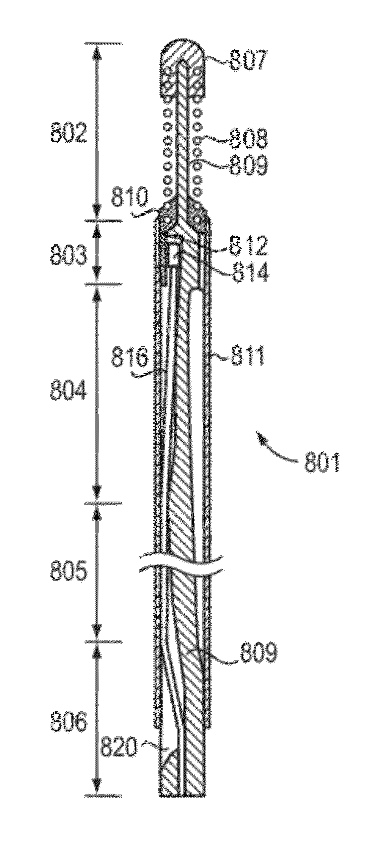
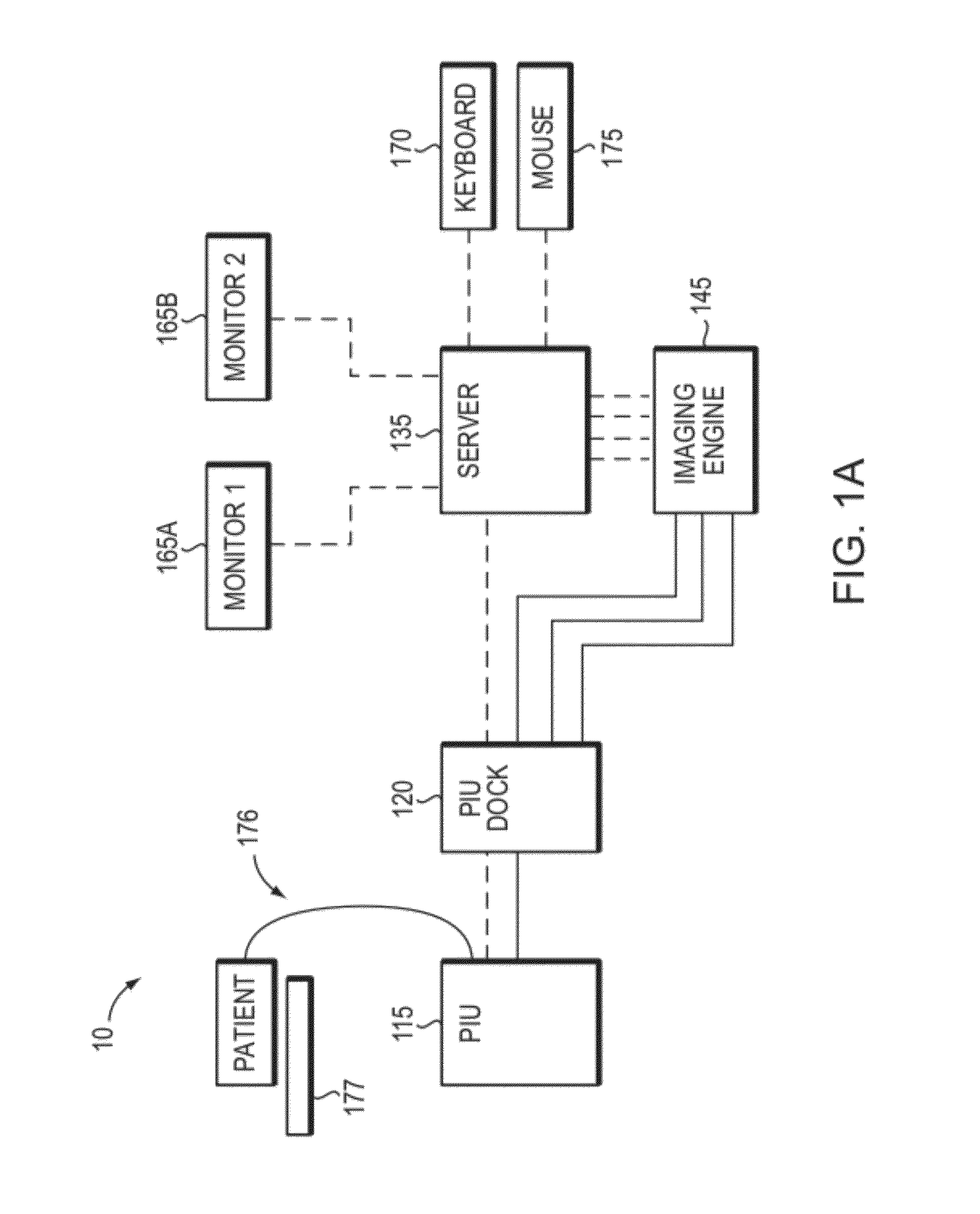
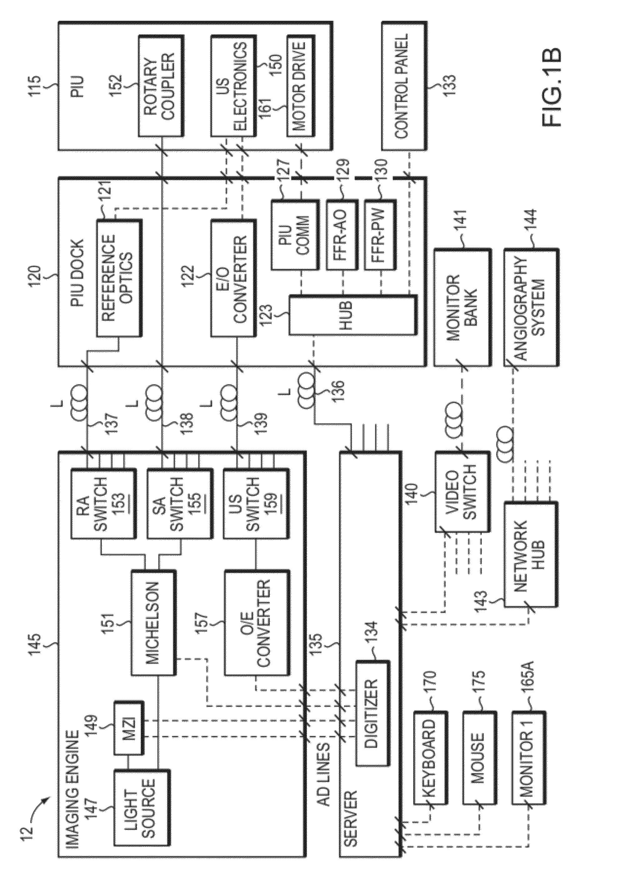
[0077]As described above, there are limitations to currently known intravascular diagnostic systems. In part, the invention relates to various systems and components thereof for use in a catheter lab or other facility to collect data from a patient and help improve upon one or more of these limitations. The data collected is typically related to the patient's cardiovascular or peripheral vascular system and can include image data, pressure and other types of data as described herein. In addition, in one embodiment image data is collected using optical coherence tomography (OCT) probes and other related OCT components. OCT is an imaging modality that uses interferometry to determine distances and other related measurements. As such, one or more embodiments of the invention relate to interferometer designs that are configured for longer sample and / or reference arms while maintaining image data levels within desirable quality levels or otherwise compensating for certain unwanted noise ...


PUM


Abstract

In part, the invention relates to an image data collection system. The system can include an interferometer having a reference arm that includes a first optical fiber of length of L1 and a sample arm that includes a second optical fiber of length of L2 and a first rotary coupler configured to interface with an optical tomography imaging probe, wherein the rotary coupler is in optical communication with the sample arm. In one embodiment, L2 is greater than about 5 meters. The first optical fiber and the second optical fiber can both be disposed in a common protective sheath. In one embodiment, the system further includes an optical element configured to adjust the optical path length of the reference arm, wherein the optical element is in optical communication with the reference arm and wherein the optical element is transmissive or reflective.

Description

CROSS REFERENCE TO RELATED APPLICATIONS[0001]This application claims priority to and the benefit of U.S. Provisional Patent Application No. 61 / 491,701, filed on May 31, 2011, and U.S. Provisional Patent Application No. 61 / 555,663, filed on Nov. 4, 2011, the disclosures of which are herein incorporated by reference in their entirety.FIELD OF THE INVENTION[0002]The invention relates generally to devices and methods suitable for use in the fields of medical treatment and diagnostics and more specifically to devices and methods that support one or more modes of data collection relevant to cardiology.BACKGROUND[0003]Interventional cardiologists incorporate a variety of diagnostic tools during catheterization procedures in order to plan, guide, and assess therapies. These tools typically include optical coherence tomography (OCT), intravascular ultrasound, (IVUS), fractional flow reserve (FFR), and angiography. Intravascular OCT, IVUS, and FFR are invasive catheter-based systems that coll...

Claims


Application Information

Patent Timeline
IPC IPC(8): A61B5/0215A61B8/12A61B6/02
CPCA61B5/0035A61B8/5261A61B5/0046A61B5/0066A61B5/0084A61B5/0215A61B5/6852A61B6/5247A61B8/0891A61B8/12A61B8/4416A61B8/4433A61B2560/0437A61B2560/045A61B2560/0456A61B6/504A61B8/54A61B5/0044
Inventor ADLER, DESMONDSCHMITT, JOSEPH M.DAHLBERG, MATTIASGUSTAFSSON, PARHUBINETTE, ULRIKSAMUELSSON, MAGNUSSVANERUDH, JOHAN
Owner LIGHTLAB IMAGING
Why Eureka
Try Eureka