Trans-catheter ventricular reconstruction structures, methods, and systems for treatment of congestive heart failure and other conditions
- Summary
- Abstract
- Description
- Claims
- Application Information
AI Technical Summary
Benefits of technology
Problems solved by technology
Method used
Image
Examples
experiment 1
Implantation in a Pig Cadaver Heart #1
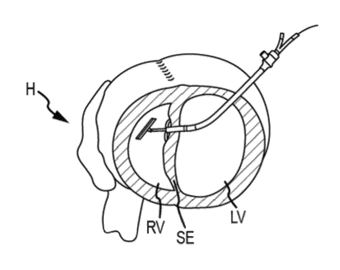
[0071]A frozen heart was thawed and placed into expanding foam in a position representative of that in the body seen via a median sternotomy. A variety of baskets were provided for grasping a guide wire passed into the right ventricle across the septum from the left ventricle. The shape of the catheters and baskets varied with target locations along the right ventricular septum that the guide wire would be was to enter.
[0072]As shown in FIGS. 25a-c, basket configured to retrieve a wire from the apex of the right ventricular septum, from the mid-portion of the right ventricular septum, and from the infundibulum (pulmonary outflow tract) of the right ventricular septum were provided, respectively.
[0073]The apex basket was placed into the right ventricle through the opened right atrium and imaged via fluoroscopy. A curved needle was passed through the eipcardial surface of the left ventricle (LV) lateral to the LAD, through the LV cavity, across th...
experiment 2
Implantation in a Human Cadaver Heart
[0086]67-year-old male. Cause of death: Heart failure
[0087]Serology: NEG
[0088]Height: 71 inches; Weight: 237 lbs
[0089]A frozen heart was thawed, placed into expanding foam in a position representative of that in the body seen via a median sternotomy. A variety of baskets were provided for grasping a guide wire passed into the right ventricle across the septum from the left ventricle, as described above.
[0090]The surgical approach was from the right atrium (RA). The basket was placed into the right ventricle through the opened right atrium and imaged via fluoroscopy. A curved needle was then passed through the epicardial surface of the left ventricle (LV) lateral to the LAD, through the LV cavity, across the ventricular septum and into the right ventricle (RV) in the vicinity of the basket. Following positioning of the needle, a guide wire is passed through the needle and into the basket via fluoroscopic control. The wire position is confirmed in ...
experiment 3
Implantation in a Live Sheep Heart
Weight: 84.7 kg
[0101]Age: adult
Sex: male
[0102]With the animal under general anesthesia, arterial and venous lines were placed. The chest was opened through a median sternotomy and the pericardium was opened in the midline. A pericardial cradle was created and the right atrial (RA) appendage was exposed. A purse-string suture (4-0 Prolene) was placed on the RA and a 14 Fr. sheath was passed into the right ventricle (RV) through the RA under fluoroscopic guidance.
[0103]In FIG. 33a, the heart is exposed through a mid-sternotomy and pericardiotomy. The LAD and apex are labeled, and (see FIG. 33b) a tourniquet is place on the RA. A sheath (blue) is passed and under fluoroscopy and (see FIG. 33c) positioned in the RV (in circle).
[0104]An apical basket was passed through the sheath in the RA into the RV, and a needle was passed through the LV epicardium near the apex and aimed toward the basket under fluoroscopic control. A guide wire was passed through th...
PUM
 Abstract
Description
Claims
Application Information
 - R&D
- Intellectual Property
- Life Sciences
- Materials
- Tech Scout
- Unparalleled Data Quality
- Higher Quality Content
- 60% Fewer Hallucinations
Browse by: Latest US Patents, China's latest patents, Technical Efficacy Thesaurus, Application Domain, Technology Topic, Popular Technical Reports.
© 2025 PatSnap. All rights reserved.Legal|Privacy policy|Modern Slavery Act Transparency Statement|Sitemap|About US| Contact US: help@patsnap.com


