Plants with altered cell wall biosynthesis and methods of use
Inactive Publication Date: 2013-04-25
UNIV OF GEORGIA RES FOUND INC
3 Cites 0 Cited by
AI-Extracted Technical Summary
Problems solved by technology
However, cost effectiveness is one of the major limitations for this industry and therefore many researchers are working to tackle this problem.
The major ba...
View moreBenefits of technology
[0006]The goal of using bioenergy crops for bio-ethanol production in the United States is well established. However, cost effectiveness is one of the major limitations for this industry and therefore many researchers are working to tackle this problem. The major barrier is the cost of the bacterial and fungal enzymes needed to degrade...
View moreAbstract
Provided herein are plants having altered expression of a GAUT polypeptide. Such plants have phenotypes that may include decreased recalcitrance, increased growth, decreased lignin content, or a combination thereof. Also provided herein are methods of making and using such plants.
Application Domain
Technology Topic
Cell wall biosynthesisPhenotype +1
Image
Examples
- Experimental program(2)
Example
Example 1
Methods
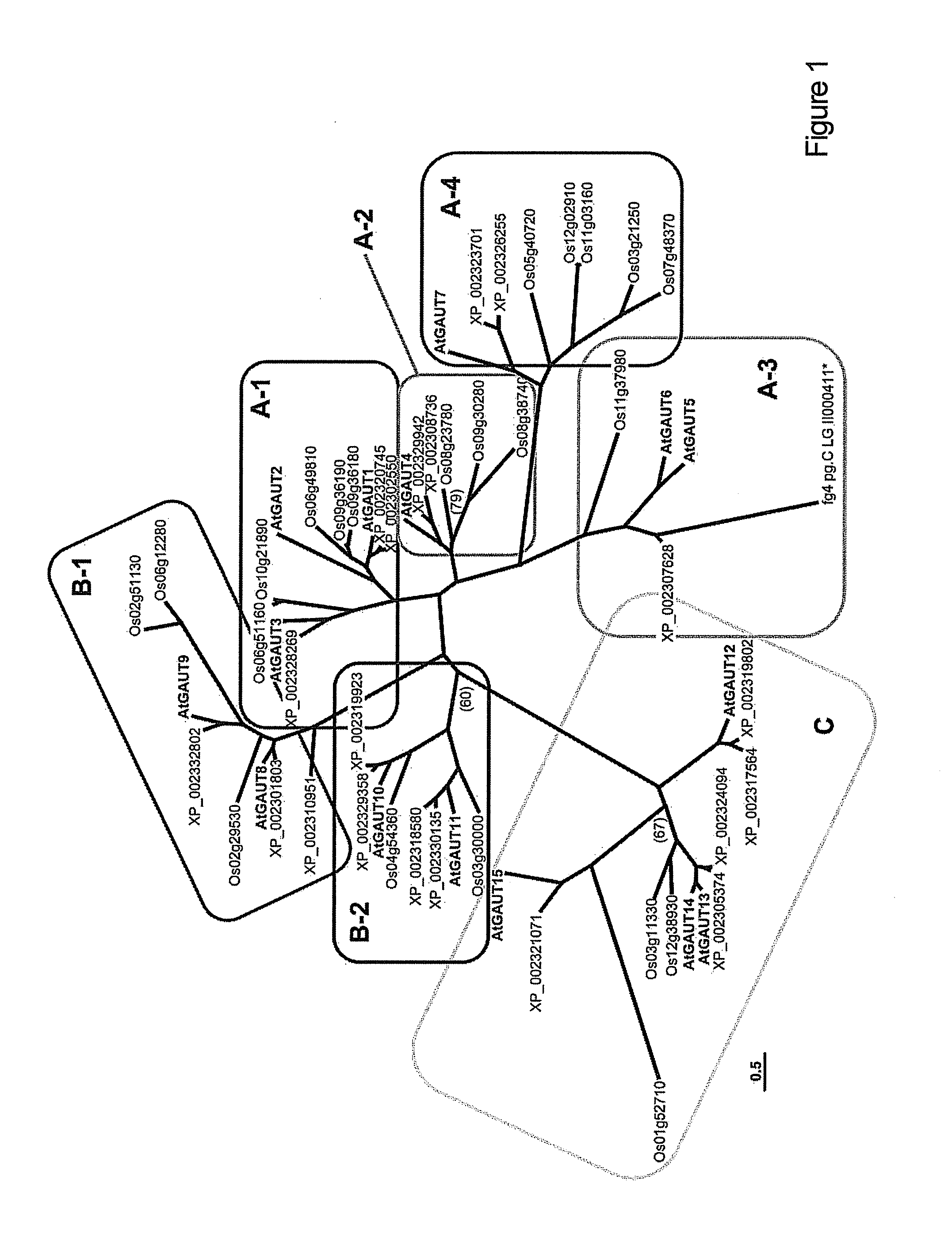
[0137]Sequence Alignment of GAUT Family Proteins and Phylogenetic Analysis
[0138]Protein sequences were identified by BLASTsearch of Arabidopsis thaliana (www.Arabidopsis.org/index.jsp), Oryza sativa (www.tigr.org/tdb/e2k1/osa1/), and Populus trichocarpa (http://genome.jgi-psf.org/Poptr1—1/Poptr1—1.home.html) genomes, using AtGAUT1 as the search probe. The GAUT protein sequences were aligned using ClustalX (Thompson et al., 1997, Nucleic Acids Res. 24, 4876-4882) and suggested protein alignment parameters (Hall, B. G. 2004, Phylogenetic Trees Made Easy: A How-To Manual, 2nd ed, (Sunderland, M A: Sinauer Associates, Inc.), pp 29-30). Phylogenetic Bayesian analysis was carried out employing MrBayes (Huelsenbeck and Ronquist, 2001, Bioinformatics. 17, 754-755; Ronquist and Huelsenbeck, 2003, Bioinformatics, 19, 1574). Full-length protein sequences were used in the analysis for all proteins except Os09g36180, whose C-terminal 404 amino acid extension was excluded.
[0139]Plant Materials and Growth Conditions
[0140]Arabidopsis thaliana var. Columbia S6000 T-DNA insertion mutant seeds were obtained from the Arabidopsis Biological Resource Center (www.biosci.ohio-state.edu/pcmb/Facilities/abrc/abrchome.htm). Arabidopsis WT and gaut mutant seeds were sown on pre-moistened soil and grown to maturity under 60% constant relative humidity with a 14/10 light/dark cycle (14 h (19° C.; 150 microEi m−2 s−1)/10 h (15° C.)). The plants were fertilized (Peters 20/20/20 with micronutrients) once a week or as needed. WT and T-DNA insert mutant seeds were sown in ‘growth sets’ of 20 plants. Walls were harvested from multiple 8-week-oldWT and PCR-genotyped mutant plants and pooled, respectively, together for wall glycosyl residue composition analysis. The following tissues were harvested for the wall analyses: the apical inflorescence excluding the young siliques; the young fully expanded leaves approximately 3 cm long; green siliques; and the top 8 cm of actively growing stem minus the inflorescence and siliques.
[0141]DNA Extraction and Mutant Genotyping
[0142]Fresh, flash-frozen leaf tissue (100-200 mg) was ground with a mortar and pestle and suspended in 0.5 ml extraction buffer (100 mM Tris-HCl pH 8.0, 100 mM EDTA pH 8.0, 250 mM NaCl, 100 lg ml−4 proteinase K and 1% (w/v) n-lauroylsarcosine) and extracted with an equal volume of phenol:chloroform:isoamyl alcohol (49:50:1, v/v). RNA was degraded by addition of 2 microliter of DNase-free RNase A (10 mg ml−1) for 20 min at 37° C. The DNA was precipitated twice with 70% (v/v) ethanol and suspended in a final volume of 50 microliter. Primers used for mutant genotyping were designed by ISECT tools (http://signal.salk.edu/isects.html). The genotype of mutant plants was determined based on the ability of the LB primers to anneal and produce T-DNA-specific PCR products when combined with the appropriate GAUT gene-specific primer. Gene-specific primer pairs were similarly used to determine the presence of intact GAUT genes (see Table 1).
TABLE 1 Primer sequences used in the GAUT analyses. Primer 5′ to 3′ primer sequence Locus GAUT name (SEQ ID NO: 69-159) Notes At3g61130 1 gs ATG GCG CTA AAG CGA GGG CTA TCT For RT- At1g61130 GGA (69) PCR F At3g61130 1 gs TCG TTC TTG TTT TTC AAT TTT GCA ATC For RT- At1g61130 (70) PCR R At2g46480 2 gs ATG ACT GAT GCT TGT TGT TTG AAG For RT- At2g46480 GGA PCR F At2g46480 2 gs ATC AGA GAA GAG AGC GTA GTG GTA For RT- At2g46480 AAG PCR R At4g38270 3 gs ATG TCG GTG GAG CCA TTT TAG AGT For RT- At4g38270 CAC PCR F At4g38270 3 gs TTG AAG GAA GGT CAG CAT CAG AGG For RT- At4g38270 TTG PCR R At5g47780 4 gs ATG ATG GTG AAG CTT CGC AAT CTT For RT- At5g47780 GTT PCR F At5g47780 4 gs GGA GCA TAG CAC GTA GCT TCT TGA For RT- At5g47780 CCA PCR R At2g30575 5 gs ATG AAT CAA GTT CGT CGT TGG CAG For RT- At2g30575 AGG PCR F At2g30575 5 gs TGT GAA AGG CAC GGC TGA CCT TGT For RT- At2g30575 ATA PCR R At1g06780 6 gs ATG AAA CAA ATT CGT CGA TGG CAG For RT- At1g06780 AGG PCR F At1g06780 6 gs CTT CTG TGT TAT AAT TCA TGG CAC For RT- At1g06780 GGA PCR R At2g38650 7 gs ATG AAA GGC GGA GGC GGT GGT GGA For RT- At2g38650 GGA PCR F At2g38650 7 gs CTT CAC AAG TTC TCC AAG TTT CAT For RT- At2g38650 CAC CA PCR R At3g25140 8 gs ATG GCT AAT CAC CAC CGA CTT TTA For RT- At3g25140 CGC PCR F At3g25140 8 gs GTA AAG ATT CGG ATC CTC GAG CTC CC For RT- At3g25140 G PCR R At3g02350 9 gs ATG GGC AAC GCA TAT ATG CAG AGG For RT- At3g02350 ACG PCR F At3g02350 9 gs CAC CTT CAT GGC TGC GAG ATT CAT For RT- At3g02350 CCG PCR R At2g20810 10 gs ATG AGA AGG AGA GGA GGG GAT AGT For RT- At2g20810 TTC PCR F At2g20810 10 gs CCA CAA CAG AAG TAG CAA TAA TGT For RT- At2g20810 TAT PCR R At1g18580 11 gs ATG AGG CGG TGG CCG GTG GAT CAC For RT- At1g18580 CGG PCR F At1g18580 11 gs CTC ATC TGC CAG TTC ATG GCG AGA For RT- At1g18580 TGG PCR R At5g54690 12 gs ATG CAG TTA CAT ATA TCT CCG AGC For RT- At5g54690 TTG PCR F At5g54690 12 gs TAG CCA CAA CCG AAG CTG CAA GAA For RT- At5g54690 TAT PCR R At3g01040 13 gs ATG CAG CTT CAC ATA TCG CCT AGC For RT- At3g01040 ATG PCR F At3g01040 13 gs TTC TTG TCT GTG ATA ACA TGG AAG For RT- At3g01040 ACA PCR R At5g15470 14 gs ATG CAG CTT CAC ATA TCG CCT AGC For RT- At5g15470 ATG PCR F At5g15470 14 gs CAG CAG ATG AGA CCA CAA CCG ATG For RT- At5g15470 CAG PCR R At3g58790 15 gs ATG AAG TTT TAC ATA TCA GCG ACG For RT- At3g58790 GGG AT PCR F At3g58790 15 gs CGA GCC ATT GCA TTT ACA GAG TAC For RT- At3g58790 TCT TC PCR R L23alpha F CCA TGT CTC CGG CTA AAG TTG ATA C For RT- PCR L23alpha R CAG CAC GAA TGT CAA CAA TGA AAA For RT- CA PCR At2g46480 2 122209 F tcagaagaagtttgaactgagttagccac iSECT tools T- DNA insertion site At2g46480 2 122209 R atgtttaacaagcccaataaggcataatc iSECT tools T- DNA insertion site At4g38270 3 001920 F TTTGAAAACTCAGTCATAGGGAAATA iSECT tools T- DNA insertion site At4g38270 3 001920 R GAAGGATGATTTGCTTTGAAATAGTA iSECT tools T- DNA insertion site At4g38270 3 113167 F Accaggttaaagccattgtagagtgaaat iSECT tools T- DNA insertion site At4g38270 3 113167 R atgtagcactactacctgcaaatcgtc iSECT tools T- DNA insertion site At2g30575 5 050186 F GATCATTATAACTTTGTTGCAAAAGCTGC iSECT tools T- DNA insertion site At2g30575 5 050186 R AATGCGGAGGTACGTAGTTTAATCCAGTT iSECT tools T- DNA insertion site At2g30575 5 058223 F taatgttgagatacagatatagtgcggcg iSECT tools T- DNA insertion site At2g30575 5 058223 R aaaattcaaagctagctgaagtaaaagtg iSECT tools T- DNA insertion site At1g06780 6 007987 F ttatctaagggtgaaaagaacacaagggt iSECT tools T- DNA insertion site At1g06780 6 007987 R acattgagattgctgggtaattaagtgaa iSECT tools T- DNA insertion site At1g06780 6 056646 F cagggaagaacaagtgattgtttca iSECT tools T- DNA insertion site At1g06780 6 056646 R gaaatgcatgatacctttgatgaaga iSECT tools T- DNA insertion site At1g06780 6 073484 F catagtcaacgttaacacccatttgactt iSECT tools T- DNA insertion site At1g06780 6 073484 R ctcttaagccgattcgatacgaaaataag iSECT tools T- DNA insertion site At2g38650 7 015189 F atatcaaggtcccaaaggggagataagt iSECT tools T- DNA insertion site At2g38650 7 015189 R ctcaagagaagctttgatgtgtagaatcc iSECT tools T- DNA insertion site At2g38650 7 046348 F ttcggatacatctctctgcaaaacc iSECT tools T- DNA insertion site At2g38650 7 046348 R cttgcaccagattgaacctaaatgg iSECT tools T- DNA insertion site At3g25140 8 030075 F gatcaaagagaagtttaatcccaaagcat iSECT tools T- DNA insertion site At3g25140 8 030075 R taattggagtcaaaacttgagagcaagag iSECT tools T- DNA insertion site At3g25140 8 102380 F tctcttctaatgatctaatcccacaataa iSECT tools T- DNA insertion site At3g25140 8 102380 R ggtttgttaatcagatccgtgtaattcct iSECT tools T- DNA insertion site At3g25140 8 041919 F tctcttctaatgatctaatcccacaataa iSECT tools T- DNA insertion site At3g25140 8 041919 R ggtttgttaatcagatccgtgtaattcct iSECT tools T- DNA insertion site At3g02350 9 135312 F acagcctgttgtaacaaagcccata iSECT tools T- DNA insertion site At3g02350 9 135312 R ctcgctgtcttcaccttatccttca iSECT tools T- DNA insertion site At3g02350 9 115588 F tctctgataatgtcattgctgtgtctgtt iSECT tools T- DNA insertion site At3g02350 9 115588 R tcatgtttccattgtaatgaatcactcct iSECT tools T- DNA insertion site At3g02350 9 040287 F acacagcttaaaatccagaagttgaaaga iSECT tools T- DNA insertion site At3g02350 9 040287 R agttaaacaatggacttaccaggttctgc iSECT tools T- DNA insertion site At2g20810 10 029319 F ctcttctttctcattctctccaaagctg iSECT tools T- DNA insertion site At2g20810 10 029319 R atgagaaatcctcgaacttctgaacct iSECT tools T- DNA insertion site At2g20810 10 082273 F atgggtttttaaccaatacccgaattact iSECT tools T- DNA insertion site At2g20810 10 082273 R agcaagagcaatctgatcattaacttgac iSECT tools T- DNA insertion site At1g18580 11 104761 F ccaaatcaaacgaaatgaaagtagacaaa iSECT tools T- DNA insertion site At1g18580 11 104761 R cgaacattagcagttataaacactcaccc iSECT tools T- DNA insertion site At1g18580 11 148781 F tatttcgtttgatgaggctaaaccg iSECT tools T- DNA insertion site At1g18580 11 148781 R tttcgatcagacggttatcgatgtt iSECT tools T- DNA insertion site At5g54690 12 044387 F ggtttgcttcttgcttccgct iSECT tools T- DNA insertion site At5g54690 12 044387 R tttgggacattgacatgaatgga iSECT tools T- DNA insertion site At5g54690 12 014026 F ttttagtgagaatcgaatgttttgtc iSECT tools T- DNA insertion site At5g54690 12 014026 R cttcaacataaagccaaatcctaaa iSECT tools T- DNA insertion site At3g01040 13 122602 F aaaaggcttgatttttcttcttctcctct iSECT tools T- DNA insertion site At3g01040 13 122602 R ccttaacttgatagttgaacaaaatgcca iSECT tools T- DNA insertion site At5g15470 14 000091 F TTAAGTCTCCCTGGACAACTATATCAT iSECT tools T- DNA insertion site At5g15470 14 000091 R CAATTGTCAAGTTGGTTTCTTTTCT iSECT tools T- DNA insertion site At5g15470 14 029525 F ttgggtccgctactgatctga iSECT tools T- DNA insertion site At5g15470 14 029525 R gcagtgatccactacaatgggc iSECT tools T- DNA insertion site At3g58790 15 113194 F agcactatgtgcaagtgttgagattttt iSECT tools T- DNA insertion site At3g58790 15 113194 R tgtttttgatgaactgatagtggagatca iSECT tools T- DNA insertion site At3g58790 15 117272 F ttttctaaagaagccaagcggacat iSECT tools T- DNA insertion site At3g58790 15 117272 R tgttatccacagctgacaatgtttttg iSECT tools T- DNA insertion site At3g58790 15 070957 F tggcatctatagtaatccatacgacgatt iSECT tools T- DNA insertion site At3g58790 15 070957 R ttgaatgctatgtgcttgtcatctttaat iSECT tools T- DNA insertion site Left Border TGGTTCACGTAGTGGGCCATCG pROK T- a F DNA insertion seq Left Border GCGTGGACCGCTTGCTGCAACT pROK T- b F DNA insertion seq Left Border GGTGATGGTTCACGTAGTGGGCCATCGC pROK T- c F DNA insertion seq
[0143]Isolation of Cell Walls
[0144]Cell wall samples were harvested from selected tissues of multiple 8-week-old plants from WT and mutant lines (n=4). The plant tissues for cell wall extraction were weighed (100-200 mg), flash frozen in liquid N2 and ground to a fine powder. The tissues were consecutively extracted with 2 ml of 80% (v/v) ethanol, 100% ethanol, chloroform:methanol (1:1, v/v), and 100% acetone. Centrifugation in a table-top centrifuge at 6000 g for 10 min was used to pellet the sample between all extractions. The remaining pellet was immediately treated with a-amylase (Sigma, porcine Type-I) in 100 mM ammonium formate pH 6.0. The resulting pellet was washed three times with sterile water, twice with acetone, and dried in a rotary speed-vac overnight at 40° C. and weighed.
[0145]Mucilage Extraction
[0146]Mucilage was extracted from 200 Arabidopsis seeds incubated with sterile water at 60° C. over the course of 6 h as follows. Each hour during the 6-h period, the seeds were centrifuged and the supernatant was transferred to a sterile tube. The combined supernatants were lyophilized and re-suspended in 600 microliter of sterile water. Phenol-sulfuric (Dubois et al., 1956, Anal. Chem. 28, 350-356) and m-hydroxybiphenyl (Blumenkrantz and Asboe-Hansen, 1973, Anal. Biochem. 54, 484-489) assays, to quantify total sugars and uronic acids, respectively, were carried out using 100 microliter of the mucilage extracts. Duplicate 200 microliter aliquots of the mucilage extract were used for glycosyl residue composition analyses. To analyze the seed coat material remaining after extraction, the water-extracted seeds were aliquoted in water to glass tubes and 20 microgram of inositol was added. The seeds were lyophilized to dryness and used for glycosyl residue composition analyses.
[0147]TMS GC-MS Glycosyl Residue Composition
[0148]The cell walls were aliquoted (1-3 mg) as acetone suspensions to individual tubes and allowed to air dry. Inositol (20 microgram) was added to each tube and the samples were lyophilized and analyzed for glycosyl residue composition by combined gas chromatography-mass spectrometry (GC-MS) of the per-O-trimethylsilyl (TMS) derivatives of the monosaccharide methyl glycosides produced from the sample by acid methanolysis basically as described by York et al. (1985, Methods Enzymol. 118, 3-40). The dry samples were hydrolyzed for 18 h at 80° C. in 1 M methanolic-HCl. The samples were cooled and evaporated under a stream of dry air and further dried two additional times with anhydrous methanol. The walls were derivatized with 200 mircrol of TriSil Reagent (Pierce-Endogen, Rockford, Ill., USA) and heated to 80° C. for 20 min. The cooled samples were evaporated under a stream of dry air, re-suspended in 3 ml of hexane, and filtered through packed glass wool. The dried samples were re-suspended in 150 microliter of hexane and 1 microliter of sample was injected onto an HP 5890 gas chromatograph interfaced to a 5970 MSD using a Supelco DB1 fused silica capillary column.
[0149]Statistical Analyses
[0150]The variance ratio test (α=0.05) was used to compare the variances of standards and samples. ANOVA analyses, standard deviation, variance, t, and the mean of sample were calculated using SAS 9.1.3 software (SAS Institute Inc., Cary, N.C., USA). Significant differences between WT and mutant compositions were determined with ta(2)=0.1 (90% confidence), but was set to 0.05 (95% confidence) for all other analyses. The appropriate sample size was predicted using equation 7.7, p. 105 of Biostatistical Analysis, 4th edn (Zar, 1999, Biostatistical Analysis, 4th edn (Englewood Cliffs, N.J.: Prentice Hall) (Table 2).
TABLE 2 Determination of the number of replicate TMS GC-MS samples required or 90% or greater statistical confidence. sam- GalA mean d = n at mean d = n at plea mol % of 3 15%b tα(2) = 0.1c of 4 15%b tα(2) = 0.1c 1 22.69 20.98 3.15 4.82 20.25 3.04 2.80 2 21.98 3 18.28 4 18.05 16.32 2.45 3.24 5 15.61 18.47 2.77 0.90 6 15.29 7 22.35 22.10 3.31 1.45 8 20.62 9 23.32 19.56 2.93 3.17 10 15.40 18.30 2.74 7.65 11 20.42 12 19.08 13 16.92 16.54 2.48 0.20 16.44 2.47 1.47 14 16.16 15 16.56 16 16.14 18.29 2.74 32.68 17 24.40 18 14.33 mean 18.76 aThe arbitrarily assigned sample number for each independent replicate is listed with the corresponding GalA mole % composition used for the determination of the minimum number of replicates necessary for a statistical confidence of 90%. The data shown are from pooled walls of 10 week old inflorescence samples, although comparable variation was also obtained from leaf, silique, stem and inflorescence tissue samples from 8 week old plants. b‘d’ refers to a margin of difference from the mean of 15%. Analysis of WT walls showed that natural variation was within 15% of the mean. Variation greater than 15% was indicative of mutation-associated changes in wall composition. The equation used to calculate ‘d’ is: Sample size = n = 2(S21 * t2)/d2 where n = sample size, d = [Xave − (t − se)] = difference from mean, S2 = (Xave − Xi)2 = sum of squares and α = 0.05 for a 2 tailed analysis. X is the value of the sample in whatever units used and se = standard error. c‘n’ = the number of replicates necessary to obtain a 90% confidence level in a two tailed analysis (tα(2) = t0.1(2)). For example, if n > the actual number of replicates used in the analysis, then it is false that a 15% difference (d) can be detected with 90% confidence. In this analysis, when 3 replicates were used, n is greater than 3 in four out of six cases, which means that a 15% difference (d) was detected with 90% confidence in only 2 out of 6 experiments. Conversely, when 4 replicates were used, n was less than 4 in all experiments and thus a 15% difference was detected with 90% confidence in all experiments.
[0151]RNA Extraction and RT-PCR
[0152]Total RNA was extracted from 0.5 g of stem, inflorescence, silique, and leaf tissue from 8-week-old plants. The tissues were homogenized in 10 ml of Homogenization Buffer (2% (w/v) SDS in 50 mM Tris-HCl pH 7.8 and 40% water-saturated phenol) and shaken for 15 min at 25° C. Tissue samples were centrifuged for 10 min at 8000 g and 4° C., and the supernatant removed to a clean tube. The samples were extracted two times with phenol:chloroform:isoamyl alcohol (25:24:01, v/v) and the aqueous phases were pooled. RNA was precipitated overnight with 0.1 vol. of 3 M sodium acetate and 2.5 vol. of cold ethanol. The samples were DNase-treated with RQ1 RNase-Free DNase (Promega, Madison, Wis., USA) according to the manufacturer's instructions.
[0153]RT-PCR products were generated using primer sequences unique to each of the 15 GAUT genes (Table 2). Each GAUT gene primer set was designed to span at least one intron such that unique PCR products were produced from RNA for each GAUT gene. Control RT reactions were carried out alongside GAUT-specific reactions, utilizing primers designed to the small ribosomal protein L23 alpha, wherein the primers do not produce a product in genomic DNA (Volkov et al., 2003, J. Exp. Bot., 54, 2343-2349). Qualitative RT-PCR was carried out using 5 lg of total RNA in a 20-microliter RT first-strand synthesis reaction that contained oligo(dT) primers. The RT first-strand reaction (2 microliter) was added to a PCR reaction mix containing the respective GAUT gene-specific primers and amplified for 30 cycles. Semi-quantitative RT-PCR was done using 2 microgram of total RNA in a 20-microliter RT first-strand synthesis reaction containing oligo(dT) primers. An aliquot (1.5 microliter) of the RT first-strand reaction was amplified through 26 cycles of PCR using GAUT genespecific primers. The PCR parameters were: Step 1: 95° C. for 5 min; Step 2: 95° C. for 0.5 min; Step 3: 55° C. for 0.5 min; Step 4: 72° C. for 1.5 min; Step 5: Return to step 2 (29 or 25) times; Step 6: 72° C. for 2 min; and Step 7: 4° C. forever.
TABLE 3 The Arabidopsis GAUT Family and T-DNA Insertion Seed Lines. Locus Gene Cladea I/Sb SALK Mutant Name Lc KO/KD/Wd At3g61130 GAUT1 A-1 100/100 Not available At2g46480 GAUT2 A-1 65/78 122209 gaut2-1 P Not detected At4g38270 GAUT3 A-1 68/84 001920 gaut3-1 I KO 113167 gaut3-2 5′ KD At5g47780 GAUT4 A-2 66/83 034472 gaut4-1 5′ Not recovered 001026 gaut4-2 5′ Not recovered At2g30575 GAUT5 A-3 45/67 050186 gaut5-1 E KO 058223 gaut5-2 P KD At1g06780 GAUT6 A-3 46/64 007987 gaut6-1 E KO 056646 gaut6-2 E KO 073484 gaut6-3 5′ KD At2g38650 GAUT7 A-4 36/59 015189 gaut7-1 E KD 046348 gaut7-2 P KD At3g25140 GAUT8 B-1 58/77 030075 gaut8-1 3′ KD 039214 gaut8-2 E HM lethal 041919 gaut8-3 I HM lethal 102380 gaut8-4 I HM lethal At3g02350 GAUT9 B-1 57/76 135312 gaut9-1 E W 115588 gaut9-2 E W 040287 gaut9-3 E KD At2g20810 GAUT10 B-2 50/72 029319 gaut10-1 E KO 082273 gaut10-2 E KD At1g18580 GAUT11 B-2 51/71 104761 gaut11-1 5′ KD 148781 gaut11-2 3′ KD At5g54690 GAUT12 C 40/61 044387 gaut12-1 I KO 014026 gaut12-2 E KO 038620 gaut12-5 P HM lethal At3g01040 GAUT13 C 43/62 122602 gaut13-1 E W At5g15470 GAUT14 C 43/62 000091 gaut14-1 E KO 029525 gaut14-2 3′ KO At3g58790 GAUT15 C 37/56 113194 gaut15-1 I W 117272 gaut15-2 P W 070957 gaut15-3 I KO aGAUT clades based on phylogenetic analysis (Sterling et al., 2006, PNAS USA, 103, 5236-5241). bThe amino acid sequence identity and similarity (I/S) of each GAUT gene to GAUT1 (Sterling et al., 2006, PNAS USA, 103, 5236-5241). cThe tentative location of the T-DNA insertion site is in one of the following gene structures; exon (E), 5′ untranslated region (5′), intron (I), promoter (P), or 3′ untranslated region (3′). dTranscript levels of GAUT T-DNA insertion mutant lines: Knockout, KO; Knockdown, KD; WT-like, W. Transcript for GAUT2 was not detectable in WT; therefore, the status of the mutant transcript was not able to be determined.
[0154]Mutant transcript levels were assessed as follows: knockouts (KO) were defined as mutants with RT-PCR reactions that yielded no detectable PCR product using gene-specific primers. Knockdown (KD) mutants were those that yielded a PCR product with significantly decreased intensity compared to the WT.
Results
[0155]The GAUT Family of Arabidopsis, Poplar, and Rice The Arabidopsis GAUT1-related gene family encodes 15 GAUT and 10 GATL proteins with 56-84 and 42-53% amino acid sequence similarity, respectively, to GAUT1 (Sterling et al., 2006, PNAS USA, 103, 5236-5241). Previous phylogenetic analyses of the Arabidopsis GAUT1-related gene family resulted in the designation of three GAUT clades, clades A through C, and one GATL clade (Sterling et al., 2006, PNAS USA, 103, 5236-5241). The GATL clade, which consists of genes that cluster tightly and somewhat independently of the GAUT genes, was not included in the study reported here. It was previously determined that some Arabidopsis GAUT genes had conserved orthologs among species of both vascular and non-vascular plants (Sterling et al., 2006, PNAS USA, 103, 5236-5241). The genomes of rice (Oryza sativa) and poplar (Populus trichocarpa) have now been sequenced and a BLAST search of Arabidopsis GAUT motifs against the poplar and rice genomes revealed GAUT1-related gene families of 21 members in poplar and 22 members in rice (FIG. 1). Due to a recent genome duplication event in Populus (Tuskan et al. 2006, Science. 313, 1596-1604), there are one to two apparent poplar orthologs for each Arabidopsis GAUT. A similar distribution of GAUTs in poplar and Arabidopsis is observed, except for the absence of a GAUT2 ortholog in poplar. In contrast, rice has major distinctions from Arabidopsis and poplar in the distribution of GAUT gene orthologs. Rice does not have apparent orthologs of GAUT2 or GAUT12. In addition, there are multiple apparent isoforms of GAUTs 1, 4, 7, and 9, suggesting an expansion of the role of these GAUT genes in rice.
[0156]The rice and poplar genes included in this comparative phylogenetic analysis resolved the GAUT genes into seven clades. In order to preserve previous clade identity between the original three Arabidopsis clades (Sterling et al., 2006, PNAS USA, 103, 5236-5241) and the more finely resolved seven clades presented here, the following clade identities are assigned. Arabidopsis GAUT clade A is subdivided into clades A-1, A-2, A-3, and A-4; GAUT clade B is subdivided into clades B-1 and B-2; and GAUT clade C remains undivided. The corresponding GAUTs in each clade are: A-1 (1 to 3); A-2 (4), A-3 (5 and 6) and A-4 (7); B-1 (8 and 9), B-2 (10 and 11) and C (12 to 15).
[0157]GAUT Gene Transcript Expression in Arabidopsis Tissues
[0158]Available transcript expression of AtGAUTs compiled from the Whole Genome Array, Massively Parallel Signature Sequence, and Genevestigator bioinformatic databases (Table 4) was used to select tissues used for the cell wall analyses reported here. In addition, total RNA from 8-week-old Arabidopsis WT inflorescence, silique, stem, and leaf tissues was used for qualitative and semi-quantitative RT-PCR using GAUT genespecific primers. PCR products corresponding to the transcripts of 14 GAUT genes, excluding GAUT2, were detected in the WT inflorescence, leaf, stem, silique, and root tissues tested. GAUT2 may be expressed at a very low level or at different stages of development that have not yet been tested (FIG. 2). Qualitative RT-PCR results partially agree with the published transcript expression data (see Table 4). In several instances, we detected GAUT transcript in tissues where it had not been previously reported. The data available from the Whole Genome Analysis (Yamada et al., 2003, Science. 302, 842-847) did not detect GAUT5, while the Massively Parallel Signature Sequence data did not indicate detection of GAUTs 7, 10, 11, and 12 in leaf, GAUTs 1, 3, and 7 in stem, and GAUTs 1, 3, 4, 8, 9, 10, 13, and 15 in silique (Meyers et al., 2004, Plant Physiol., 135, 801-813). Overall, the data supplied by Whole Genome Analysis and Massively Parallel Signature Sequences under-reported GAUT gene transcript expression. The relative transcript expression of the GAUT genes, however, more closely agrees with that reported by Genevestigator (Zimmermann et al., 2004, Plant Physiol. 136, 2621-2632). Genevestigator does not list a probe for GAUT5, and therefore has no expression data for this gene, while the MPSS database reports low to moderate expression of GAUT5, in agreement with the result reported here.
TABLE 4 Bioinformatic Arabidopsis GAUT Gene Transcript Expression Data. Locus potentiald Genea WGAb INFc LEF LES ROF SIF SIS CAF CAS Expression At3g61130 GAUT1 + 114 48 46 42 22 25 18 0 14 093 At2g46480 GAUT2 − 0 0 0 0 0 0 0 0 1493 At4g38270 GAUT3 + 0 11 12 2 13 58 31 50 6851 At5g47780 GAUT4 + 87 161 0 142 154 0 152 0 18 061 At2g30575 GAUT5 − 11 19 1 14 7 18 5 20 — At1g06780 GAUT6 + 0 4 0 0 0 0 0 0 11 224 At2g38650 GAUT7 + 68 69 111 62 40 218 53 236 7126 At3g25140 GAUT8 + 405 125 72 230 285 664 117 329 27 875 At3g02350 GAUT9 + 74 78 28 450 249 106 93 69 15 384 At2g20810 GAUT10 + 39 29 50 42 13 0 42 0 7087 At1g18580 GAUT11 + 19 1 5 22 29 38 17 26 12 6915 At5g54690 GAUT12 + 44 5 2 19 37 3 0 0 12 028 At3g01040 GAUT13 + 24 11 8 58 4 1 22 10 9670 At5g15470 GAUT14 + 5 14 15 25 4 46 3 9 5386 At3g58790 GAUT15 + 0 0 0 0 16 0 4 12 6717 aGAUT gene designation (Sterling et al., 2006, PNAS USA, 103, 5236-5241) bExpression of GAUT gene transcript was detected (+) or not (−) according to the Whole Genome Analysis (WGA) of Arabidopsis (Yamada et al., 2003, Science. 302, 842-847). cRelative expression of the designated GAUT gene transcript in different tissues, available through the Massively Parallel Signature Sequences (MPSS) website (http://mpss.udel.edu/at/) (Meyers et al., 2004, Plant Physiol., 135, 801-813): INF (Inflorescence-mixed stage, immature buds, classic MPSS), LEF (Leaves-21 d, untreated, classic MPSS), LES (Leaves-21 d, untreated), ROF (Root-21 d, untreated, classic MPSS), SIF Silique-24-48 h post-fertilization, classic MPSS), SIS (Silique-24-48 h post-fertilization, signature MPSS), CAF (Callus-actively growing, classic MPSS), CAS (Callus-actively growing, signature MPSS). dGENEVESTIGATOR Expression Potential is the average of the top 1% signal value of a probe for the designated GAUT gene across all tissue expression arrays (Zimmermann et al., 2004, Plant Physiol. 136, 2621-2632).
[0159]In general, RT-PCR indicated that relative transcript expression in Arabidopsis was highest for GAUTs 1, 4, 8, 9, and 12, moderate for GAUTs 3, 5, 6, 10, 14, and 15, and low for GAUTs 2, 7, 11, and 13. It should be noted that RT-PCR of GAUT7 repeatedly produced two bands, one of the expected size and a minor band of a smaller size. Whether the smaller band represents a splice variant has not been investigated. The RT-PCR data indicated that the GAUT genes were expressed at some level in all tissues tested; therefore, inflorescence, silique, leaf, and stems were used for the chemical and biochemical studies of the GAUT mutants.
[0160]Isolation of Homozygous Mutants of 13 of the 15 GAUT Genes
[0161]Twenty-six Arabidopsis homozygous T-DNA insertion seed lines in 13 distinct GAUT genes were isolated from mutagenized seed obtained from the SALK Institute (http://signal.salk.edu/cgi-bin/tdnaexpress) through the Arabidopsis Biological Resource Center (Alonso et al., 2003, Science. 301, 653-657). Mutant seed lines were preferentially selected with the T-DNA insertion site in an exon, 5′ UTR, or intron of the GAUT gene, if such lines were available. SALK insertion seed lines of GAUT1 were not available and neither homozygous nor heterozygous mutants were recovered from the SALK insertion seed lines for GAUT4. RT-PCR of total RNA isolated from homozygous gaut mutant lines identified 10 knockout mutants and 10 knockdown mutants (Table 3).
[0162]Growth Phenotypes of gaut Mutants
[0163]The gaut mutants plants were initially inspected visually for obvious growth phenotypes, such as dwarfing and/or organ malformation, compared to WT plants. Major abnormalities were not observed in plant growth or morphology for most gaut mutants isolated in this study, with the exception of gaut8 and gaut12. The presence of subtle growth phenotypes may require more sensitive methods than those applied here. Indeed multiple stem elongation phenotypes are observed with multiple gaut mutants. Functional redundancy among the GAUT proteins may contribute to the lack of severe phenotypes observed among gaut mutants. Estimates put forth by Østergaard and Yanofsky (2004, Plant J. 39, 682-696) predict that mutations in only approximately 10% of genes may result in detectable mutant phenotypes due to gene redundancy among large gene families in higher organisms. Thus far, two out of 13 GAUT genes (;15%) have yielded mutants with severe growth phenotypes, which is in line with the predicted outcome (Østergaard and Yanofsky, 2004, Plant J. 39, 682-696).
[0164]Previously analyzed qua1-1 insertion mutants (insertion in the 5#UTR) had severe dwarfmg, sterility, and bumpy epidermal surfaces as a result of reduced cell adhesion (Bouton et al., 2002, Plant Cell, 14, 2577-2590). Mutants allelic to qua1-1 (gaut8-2, gaut8-3, and gaut8-4) produced only heterozygous and WT progeny, suggesting an embryo-lethal phenotype. A single homozygous mutant was isolated, gaut8-1, with a predicted insertion in the 3#UTR that did not show the expected qua1-1 phenotype and was experimentally determined to have detectable GAUT8 transcript by RT-PCR, which may account for the WT like phenotype of these plants.
[0165]The irx8-1/gaut12-1 and irx8-5/gaut12-2 mutant plants were severely dwarfed and sterile, which necessitated recovery of homozygous plants from the progeny of heterozygous parental plants, as previously reported (Persson et al., 2007, Plant Cell. 19, 237-255). The phenotype of irx8-1/gaut12-1 and irx8-5/gaut12-2 was recognized in plants at least 4 weeks old. Such plants were small and with darkened leaves compared to WT. Surprisingly, the gaut12-5 promoter mutant (SALK—038620) did not produce homozygous progeny. In addition, gaut12-5 heterozygous mutants were dwarfed compared to WT, and more severely dwarfed compared to the irx8-1/gaut12-1 or irx8-5/gaut12-2 heterozygotes. RT-PCR of RNA from homozygous irx8-1/gaut12-1 and irx8-5/gaut12-2 plants did not yield PCR products using 5#- and 3#-end coding region-specific primers, showing that the full-length GAUT12 transcript was not produced. Because of the lethal phenotype, only heterozygous gaut12-5 was obtained and therefore was not included in our analyses of gaut homozygous mutants.
[0166]Strategy to Identify Glycosyl Residue Composition Differences between gaut Mutant and WT Walls
[0167]Gas chromatography-mass spectrometry (GC-MS) has been used to detect the changes in glycosyl residue composition in cell walls arising from mutations in cell wall-related genes (Reiter et al., 1997, Plant J. 12, 335-345). Analysis of wall glycosyl residue composition by GC-MS of trimethylsilyl (TMS) derivatives allows detection of acidic and neutral sugars in a single analysis (Doco et al., 2001), in contrast to composition analysis by formation of alditol acetate derivatives that detects neutral but not acidic sugars (Reiter et al., 1997, Plant J. 12, 335-345). Since uronic acids make up the largest proportion of glycosyl residues in the non-cellulosic wall polysaccharides of WT Arabidopsis tissues (FIG. 3), the TMS method was chosen to analyze gaut mutant walls. A statistical assessment of the TMS method showed that at least four independent TMS analyses per wall sample are necessary to detect a 15% difference between the glycosyl residue composition of different wall samples with 90% or greater statistical confidence (Table 2). The mutant glycosyl residue composition results were normalized to the composition of WT plants grown in the same experiment, in order to minimize the variability observed in the glycosyl residue compositions of plants grown in different experiments. Thus, for example, rhamnosyl compositions would be normalized according to the following foiinula: [0168] Normalized Rha=[(mutant mol % Rha/WT mol % Rha)×100].
[0169]Normalization of mutant glycosyl residue composition to WT controls allowed mutant wall composition phenotypes to be compared between experiments. The tissues chosen for the cell wall analyses of each specific gaut mutant were based on transcript expression of the corresponding GAUTs in WT tissues according to the Whole Genome Array (Yamada et al., 2003, Science. 302, 842-847) and Massively Parallel Signature Sequences (Meyers et al., 2004, Plant Physiol., 135, 801-813) databases (see Table 4). To identify gaut mutant wall glycosyl residue compositions that were statistically different from those of WT walls, the normalized compositions were evaluated by ANOVA procedures (ta(2)=0.1). As an extra measure of stringency, a 15% point or greater departure from the normalized WT mean, in addition to a statistically different outcome by ANOVA, was required for declaration of a real difference from WT.
[0170]Wall Glycosyl Residue Composition is Altered in Multiple gaut Gene Mutants
[0171]TMS glycosyl residue composition analyses of walls from two or more tissues of WTand mutant lines, representing 13 GAUT genes, revealed that specific gaut mutants have unique wall composition changes, which include increases and decreases in GalA, as well as significant changes in other glycosyl residues (Table 5). The wall glycosyl residue compositions that were statistically different in the gaut mutants compared to WT are shown in bold italics in Table 5. Reproducible mutant phenotypes were identified by comparing the natural log transformed data for all mutants that had statistically different mol % GalA, Xyl, Rha, Gal, and Ara levels compared to WT in at least two mutant alleles of the same gene or in at least two tissues of the same mutant allele (FIG. 4).
TABLE 5 Percent Cell Wall Glycosyl Residue Composition of Arabidopsis gaut Mutants Compared to Wild-Type.a (mutant mol %/WT mol %*100) Mutant Tissueb Ara Rha Fuc Xyl GalUA Man Gal Glc gaut2-1 S 116 108 114 103 103 L 152 98 91 89 112 123 gaut3-1 I 74 111 104 108 S 90 62 72 110 104 126 gaut3-2 I 99 102 181 99 89 97 S 132 112 109 118 87 98 86 gaut5-1 I 117 112 110 105 85 102 73 S 109 132 117 130 94 112 gaut5-2 I 98 99 97 97 106 103 93 97 S 102 118 43 95 105 78 gaut6-1 I 193 154 80 162 128 S 123 141 153 L 168 75 167 89 107 gaut6-2 I 126 95 122 114 133 99 S 87 137 108 126 112 150 73 L 103 115 129 87 131 79 153 gaut6-3 I 113 111 102 100 88 111 98 92 S 161 114 104 103 86 L 139 106 109 92 102 102 112 gaut7-1 I 91 113 104 110 102 89 93 126 L 114 130 117 90 96 107 93 114 gaut7-2 I 100 96 87 98 114 89 96 105 L 112 102 100 110 113 102 108 51 gaut8-1 I 81 111 106 119 S 59 137 102 102 95 111 gaut9-1 I 130 99 159 S 89 113 118 122 92 99 136 ST 101 131 146 127 gaut9-2 I 100 106 100 99 S 82 72 103 104 282 99 85 ST 100 90 96 105 81 58 106 gaut9-3 I 139 130 151 108 102 91 114 S 147 137 100 98 112 ST 100 100 100 100 100 100 100 100 gaut10-1 I 103 98 107 89 120 112 86 S 103 103 110 116 113 92 108 gaut10-2 I 152 128 115 87 94 92 S 104 85 103 85 110 gaut11-1 I 110 96 99 137 85 100 105 146 S 135 125 109 86 117 L 222 128 133 86 90 131 124 gaut11-2 I 86 108 99 125 S 110 73 76 108 91 112 114 95 L 75 83 88 115 95 83 gaut12-1 I 148 120 97 101 89 102 142 S 147 115 112 114 100 121 ST 179 124 130 103 66 132 gaut12-2 I 163 137 105 130 82 80 91 115 S 65 67 102 169 ST 198 154 126 117 60 148 109 gaut13-1 I 62 111 120 123 S 117 89 99 110 gaut14-1 I 88 135 113 S 70 98 117 110 97 L 81 78 gaut14-2 I 84 105 90 S 136 86 204 104 86 121 111 64 L 102 102 61 88 98 98 gaut15-1 I 87 89 104 105 S 107 117 118 85 86 96 gaut15-2 I 111 67 134 99 213 72 S 98 147 90 156 60 112 gaut15-3 I 109 111 109 98 S 130 95 112 93 103 109 84 aData represent four independent TMS GC-MS reactions from four independent wall extractions. Residues are abbreviated according to FIG. 3. SALK T-DNA seed lines were unavailable for gaut1 and were unable to be isolated from SALK seed received for gaut4. bThe walls used for glycosyl residue analysis were harvested from inflorescence (I), silique (S), leaf (L), and stem (ST). cBold highlighted italicized values indicate mutant glycosyl residue compositions that were statistically and ±15% different from the WT mean.
[0172]Eight gaut mutants had statistically different mol% levels of GalA, Xyl, Rha, Gal, or Ara in at least two mutant alleles of the same gene or in at least two tissues of the same mutant allele compared to WT, resulting in distinguishable patterns of glycosyl residue composition changes in the walls of gaut mutants (summarized in Table 6). The silique tissues of gaut6-1 and gaut6-3 were consistently reduced in GalA, increased in Xyl, Rha, and Fuc, and similar to WT in Gal and Ara wall composition. Viable gaut8 homozygous knockout mutants were not isolatable, and, therefore, the wall composition of qua1-1 is used to establish a phenotype grouping for gaut8 mutants. The leaves of qua1-1 that were previously analyzed (Bouton et al., 2002, Plant Cell, 14, 2577-2590) were decreased in GalA and Xyl, but were not changed in Rha or other sugars. The gaut9-1 stems were reduced in wall GalA and increased in Xyl and Fuc. The gaut10-1, gaut10-2, and gaut11-1 were consistently reduced in silique GalA only. The irx8-1/gaut12-1 and irx8-5/gaut12-2 mutant stems were severely reduced in Xyl, coincident with elevated Ara, Rha, and Gal content. The gaut12-1 and gaut12-2 are analogous to irx8-1 and irx8-5, and, consequently, show similar stem glycosyl residue composition as previously reported (Brown et al., 2005, Plant Cell. 17, 2281-2295; Pena et al., 2007, Plant Cell., 19, 549-563; Persson et al., 2007, Plant Cell. 19, 237-255). Gaut13-1, gaut14-1, and gaut14-2 had increased GalA and Gal and reduced Xyl, Rha, Ara, and Fuc, with greater mol% changes in gaut14-1 (T-DNA insertion in an exon) than gaut14-2 (T-DNA insertion in the 3′ region). There were also some changes in Fuc, Man, and Glc in walls of several gaut mutants. For example, increased Fuc was observed in gaut6-1, gaut6-2, gaut6-3, gaut9-1, gaut9-2, and gaut9-3; decreased Fuc in gaut8-1, gaut11-2, gaut14-1, and gaut14-2; increased Man in gaut5-1 and gaut5-2; increased Glc in gaut3-1, gaut3-2, and gaut6-2; and decreased Glc in mutants of gaut5-1, gaut5-2, and gaut10-2. Few significant changes were found in the walls of gauts 2, 3, 5, 7, and 15, and those that did occur were not consistent between two or more mutants or in more than one tissue of a single mutant.
TABLE 6 Phenotypic Grouping of gaut Mutants.a gaut GalA Xyl Rha Gal Ara 6 Down Up Up Down No change 8b Down Down No change No change No change 9 Down Up Variable Variable No change 10 Down No change No change No change No change 11 Down No change Variable Variable Variable 12 Upc Down No change Up No change 13 Up Down Down Up Down 14 Up Down Down Up Down aChanges in the relative amount of the designated glycosyl residues compared to WT. bDue to the lethality of gaut8 homozygous mutants, the qual-1 leaf compositions were used for the phenotypic grouping of gaut8 (Bouton et al., 2002, Plant Cell, 14, 2577-2590). cThe GalA composition of gaut12 stems and siliques was increased, but was reduced in inflorescences.
[0173]Survey of Seed Mucilage Reveals GAUT11 Involved in Mucilage Extrusion
[0174]The seeds of myxospermous species, such as Arabidopsis, extrude mucilage from the seed coat epidermal cells when hydrated to protect against desiccation and to aid in seed dispersal. The mucilage of WT and gaut mutant seeds was investigated by ruthenium red staining as a facile method to determine whether specific GAUT genes are involved in mucilage polysaccharide extrusion or synthesis. The mucilage extruded from Arabidopsis seeds is enriched in the pectic polysaccharide RG-I, which efficiently binds ruthenium red stain due to the negative charge on the GalA residues in mucilage. This method has been successfully employed to identify mucilage or testa polysaccharide biosynthesis mutants (Western et al., 2001). The seed mucilage was evaluated by observing the staining intensity of mucilage and measuring the mucilage thickness under a dissecting microscope after application of aqueous 0.05% ruthenium red to the seeds of WT and the 26 gaut mutant lines. A single mutant (gaut11-2) was identified that displayed a reproducible reduced mucilage thickness phenotype compared to WT seed mucilage thickness.
[0175]Ruthenium red staining of WT and gaut11-2 seeds (FIG. 5A-5C) revealed that; 68% of gaut11-2 seeds had little extruded mucilage, while the remaining gaut11-2 seeds (˜32%) had reduced thickness of the mucilage layer to approximately half that of WT. Samples of WT and gaut11-2 seed were tested three separate times independently, with similar results obtained in seed derived from different parental plants (Table 7). Analysis of the uronic acid content of the hot water-extracted mucilage (WEM) of gaut11-2 and WT seed indicated that WEM of WT had 59 microgram uronic acid per 200 extracted seeds, while gaut11-2 mucilage had 48 microgram uronic acid per 200 extracted seeds (Table 6). The total carbohydrate extracted, as detected by a phenol sulfuric acid assay, was similar for WT and gaut11-2 WEM. This suggests that even though very little mucilage was observed by ruthenium red staining, a similar amount of carbohydrate was able to be extracted over several hours, but that the uronic acid content of that mucilage was reduced by 19%. The gaut11-2 WEM was subjected to glycosyl residue composition analysis (FIG. 5) and found to have statistically significant reductions in GalA and Xyl content and increases in Man and Gal content, as determined by ANOVA (tα2=0.05). The glycosyl residue composition of residual gaut11-2 seed material that represents the remaining mucilage, some testa wall, and possibly some storage polysaccharide was also reduced in GalA (69%) and Gal (68%) and increased in Ara (110%), Man (128%), and Glc (138%) compared to WT.
TABLE 7 WT and gaut11.2 Mucilage Expansion and Uronic Acid Content. Mucilage (% seeds)a UA (ug UA/200 seeds)b Experiment WT gaut11-2 WT gaut11-2 Experiment # 1 92 16 59 46 Experiment # 2 100 41 58 46 Experiment # 3 87 39 56 45 Experiment # 4 61 48 Experiment # 5 58 53 Average 93.0 ± 7 31.8 ± 14 58.8 ± 2 47.8 ± 3 P = 2.3−3 P = 2.2−4 aThe data are the average (%) seeds with expanded mucilage after staining with aqueous ruthenium red. bThe data are the uronic acid content of hot water-extracted mucilage per 200 seeds of WT and gaut11-2 as assayed by the m-hydroxylbiphenyl reagent assay.
[0176]Newly Resolved GAUT Gene Clades in Arabidopsis, Poplar, and Rice
[0177]The relatedness of GAUT genes has been re-evaluated based on the analysis of phylogenetic relationships of Arabidopsis, poplar, and rice GAUT genes. This comparative phylogenetic analysis distinguished seven GAUT clades (FIG. 1), instead of three, as previously proposed by Sterling et al. (2006, PNAS USA, 103, 5236-5241). The previous Arabidopsis GAUT clade A that included AtGAUT1-GAUT7 has been subdivided into four clades; GAUT clade A-1 (AtGAUT1 through 3), GAUT clade A-2 (AtGAUT4), clade A-3 (AtGAUT5 and AtGAUT6), and GAUT clade (AtGAUT7). The former Arabidopsis clade B has been subdivided into GAUTclade B-1 (AtGAUT8 and AtGAUT9) and GAUT clade B-2 (AtGAUT10 and AtGAUT11). The former Arabidopsis GAUTclade C has not been subdivided and contains AtGAUT12 through AtGAUT15.
[0178]GAUT2 does not appear to have a direct ortholog in either rice or poplar. It is possible that GAUT2 may not be a complete copy of a GAUT1 duplication event, based on a shorter N-terminus compared to GAUTs 1-7; however, its length is comparable to the other GAUTs. GAUT2 also does not have detectable transcript in the tissues tested and GAUT2 T-DNA insertion mutants did not have reproducible phenotypes. These data, combined with the phylogenetic analysis of GAUT2, support the hypothesis that GAUT2 may be a nonfunctional truncated homolog. It cannot be ruled out, however, that GAUT2 may have a very low abundance transcript and a unique function in Arabidopsis alone, although this seems unlikely based on the current data.
[0179]The Arabidopsis and poplar genomes have one (At2g38650) and two (XP—002323701, XP—002326255) copies of GAUT7, respectively, while the rice genome contains five GAUT7-like sequences. There is considerable evidence that the AtGAUT7 protein resides in a complex with AtGAUT1, a complex that has homogalacturonan a1,4-GalAT activity. GalAT activity was detected in immunoprecipitates from HEK cells transiently transfected with GAUT1, but not in HEK cells transiently transfected with GAUT7 (Sterling et al., 2006, PNAS USA, 103, 5236-5241). Based on these data, GAUT7 may be expressed in an inactive state with limited activity itself or may function as an ancillary protein necessary for GAUT1-associated GalAT activity. Whatever the role of GAUT7, its function appears to be dramatically expanded in rice. Because the role of GAUT7 in wall polysaccharide biosynthesis is currently unknown, the underlying biological reason for five copies of GAUT7 in rice remains to be determined.
[0180]Poplar and rice each have putative orthologs of GAUT9: XP—002332802 (poplar), Os06g12280 (rice), and Os02g51130 (rice). Poplar also has at least one putative ortholog of GAUT8 (XP—002301803). There is not an obvious ortholog of GAUT8 in rice, although there is one rice gene (Os02g29530) positioned between GAUT8 and GAUT9. Phylogenic analyses using additional sequenced plant genomes may clarify the relatedness of the latter gene to GAUT8 and GAUT9.
[0181]GAUT12 has two poplar orthologs but no orthologs in rice (FIG. 1). GAUT12 has been linked xylan synthesis. The putative functions that have been hypothesized for GAUT12 include an a1,4-GalAT that adds GalA into a primer or cap for xylan synthesis or as a novel linkage in xylan or pectic polysaccharides (Brown et al., 2005, Plant Cell. 17, 2281-2295; Pena et al., 2007, Plant Cell., 19, 549-563; Persson et al., 2007, Plant Cell. 19, 237-255). GAUT12 has been shown to be essential for normal growth and more specifically for the synthesis of secondary wall glucuronoxylan and/or wall HG synthesis. Rice does not have an apparent homolog of GAUT12, and appears to produce secondary wall xylan and glucuronoarabinoxylan, but not 4-O-methylglucuronoxylan (Ebringerova and Heinze, 1999, Macromol. Rapid Commun. 21, 542-556). Thus, GAUT12 may have a specialized function in glucuronoxylan synthesis of dicot plants. GAUT12transcript has been shown to be localized closely with glucuronoxylan-rich vascular tissues, suggesting that GAUT12 has a specialized role in the synthesis of secondary wall glucuronoxylan of dicot walls (Persson et al., 2007, Plant Cell. 19, 237-255). GAUT12 has an expression profile distinct from that of other GAUT genes according to semi-quantitative RT-PCR; it is much more highly expressed in stem than in other tissues compared to other GAUT transcripts. The unique transcript expressionprofile, role in secondarywall 4-O-methylglucuronoxylan synthesis, and exclusivity among the dicot species suggest that GAUT12 has undergone a differentiation that has rendered it essential in dicots and nonessential in monocots.
[0182]GAUT Gene Transcripts are Expressed Ubiquitously in Arabidopsis Tissues
[0183]The transcript expression of GAUT8 and GAUT12 has been associated with vascular tissues in Arabidopsis stem (Orfila et al., 2005, Planta. 222, 613-622; Persson et al., 2007, Plant Cell. 19, 237-255). The GAUT12 results described here agree with previous analyses of GAUT12/IRX8 gene expression by RT-PCR analysis (Persson et al., 2007, Plant Cell. 19, 237-255) and GAUT8 RT-PCR data agree with reports of QUA1 expression (by Northern blot) in ‘Flowers II’ and ‘Rosette Leaves II’ RNA, but do not agree with the low transcript expression reported in ‘Stems II’ by Bouton and colleagues (2002, Plant Cell, 14, 2577-2590). We report high relative expression of GAUT8 in stems. In situ PCR of QUA1/GAUT8 in WT stems (Orfila et al., 2005, Planta. 222, 613-622), however, did reveal prominent expression in that tissue, which is more closely aligned with our results. The detectable expression of all of the GAUT genes in all of the tissues tested correlates with a function in wall biosynthesis, as this is a process required by all plant cells. GUS reporter gene studies have shown that QUA2, a putative pectinmethyltransferase involved in pectin biosynthesis, also has ubiquitous expression (Mouille et al., 2007, Plant J. 50, 605-614).
[0184]The Wall Compositions of Multiple gaut Mutants are Altered Compared to WT
[0185]Analysis of the walls of gaut mutants using the TMS method (Doco et al., 2001, Carbohydr. Polym., 46, 249-259) allowed the GalA content of the walls to be quantified. An accurate quantification of wall GalA content is important when attempting to identify mutants of putative pectin biosynthesis genes, because GalA is a major component of the pectic polysaccharides (Ridley et al., 2001, Phytochemistry, 57, 929-967). Mutants of GAUTs 6, 9, 10, and 11 had statistically significant reductions in GalA content in more than one mutant sampling. Two other gaut mutants, gaut13 and gaut14, had statistically significant increased wall GalA content. The wall compositional phenotypes of the gaut mutants are discussed below.
[0186]The wall glycosyl residue composition phenotype of gaut6 provides compelling evidence that GAUT6 is a putative pectin biosynthetic GalAT. GAUT6 has 64% amino acid similarity to GAUT1 and gaut6 has reduced wall GalA that coincides with higher levels of Xyl and Rha wall compositions. It is possible that the increased Xyl and Rha content signifies the compensatory reinforcement of the wall by xylans and an apparent enrichment of RG-I in proportion to reduced HG polymers. Further work is necessary to test this hypothesis; however, preliminary results are in agreement with this hypothesis (Caffall, K. H., Ph.D. thesis, University of Georgia, 2008).
[0187]GAUTs 8, 9, 10 and 11 have been placed in two separate subclades (B-1 and B-2). However, all mutants in the two B clades show marked reductions in wall GalA content. Qual-1 mutant plants have walls with both reduced GalA and Xyl, and microsomal membrane protein preparations from qual-1 stems had reduced GalAT and xylan synthase activity compared to WT (Orfila et al., 2005, Planta. 222, 613-622; Brown et al., 2007, Plant J., 52, 1154-1168). The QUAl cumulative experimental evidence argues in favor of a putative pectin biosynthetic GalAT, based on the significant reduction in homogalacturonan and the strong defect in cell adhesion (Bouton et al., 2002, Plant Cell, 14, 2577-2590; Leboeuf et al., 2005, J. Exp. Bot., 56, 3171-3182). Deficiencies in cell adhesion have been associated with changes in pectin synthesis (Iwai et al., 2002, PNAS USA. 99, 16319-16324) and pectin localization (Shevell et al., 2000, Plant Cell. 12, 2047-2059). In addition, the transcript expression of a pair of Golgi-localized putative pectinmethyltranserfases is strongly correlated with QUA1/GAUT8 expression, as well as with the expression of GAUT9 and GAUT1 (Mouille et al., 2007, Plant J. 50, 605-614). The gaut9, gaut10, and gaut11 mutant plants did not have any obvious physical growth or cell adhesion defects, but the wall compositional phenotypes of these gaut plants, and the high amino acid similarity with QUA1/GAUT8, suggest that these GAUTs are putative pectin biosynthetic GalATs. The mutant alleles of GAUT9, GAUT10, and GAUT11 have reduced wall GalA content but were not decreased in Xyl, which has been observed in some mutants thought to be involved in xylan synthesis (Brown et al., 2007, Plant J., 52, 1154-1168; Lee et al., 2007; Pena et al., 2007, Plant Cell., 19, 549-563; Persson et al., 2007, Plant Cell. 19, 237-255). Based on the evidence, a role for the genes in GAUT clades A as well as a role for the genes in clade B and C in pectin biosynthesis is proposed.
[0188]In contrast to QUA1/GAUT8, IRX8/GAUT12 is believed to function in glucuronoxylan synthesis essential for secondary wall function. The irx8-1/gaut12-1 and irx8-5/gaut12-2 mutant plants have reduced Xyl content with increases in the GalA content in stem and silique walls, consistent with previous reports and consistent with the proposed function of IRX8/ GAUT12 in the synthesis of an oligosaccharide essential for xylan synthesis. Mutants of IRX8/GAUT12 and other putative xylan biosynthetic genes, IRX7, IRX8, IRX9, IRX14, and PARVUS, have similar wall compositional phenotypes (Pena et al., 2007, Plant Cell., 19, 549-563; Persson et al., 2007, Plant Cell. 19, 237-255). IRX8/GAUT12 may play a specialized role, among the GAUTs, in secondary wall synthesis and vascularization in dicot species (Brown et al., 2007, Plant J., 52, 1154-1168). Xylans are abundant in stem and silique tissues, where the Xyl compositional phenotype is observed; however, reductions in Xyl are not observed in inflorescence where IRX8/GAUT12 is also expressed. In inflorescences, irx8/gautl2 mutants show a reduction in GalA to 82% that of WT. Thus, the changes brought about by the lesion in GAUT12 additionally impact the pectin component of the wall. The underlying causes for the reduced GalA content in the inflorescence may be of significance to understand how pectin and xylan synthesis are regulated and connected.
[0189]The walls of gaut13 and gaut14 have increased GalA and Gal content and reduced Xyl and Rha content compared to WT. It seems unlikely that a mutant showing an increased wall GalA phenotype is involved in the synthesis of HG. However, reduced Rha, primarily a component of RG-I, may lead to walls enriched in HG, driving up GalA content. A Gal containing wall component is increased in the walls of gaut13 and gaut14 (and also gaut12). Pectic galactans have been associated with wall strengthening (McCartney et al., 2000) and are also increased in irx8/gaut12 walls (Persson et al., 2007, Plant Cell. 19, 237-255). A galactan in gaut13 and gaut14 may be up-regulated in response to wall weakening in a similar manner. GAUT13 and GAUT14 are very closely related to GAUT12, which would also suggest that the Xyl containing polysaccharide that is reduced in mutants of these genes is also a xylan and that GAUT13 and GAUT14 share overlapping function with GAUT12. Based on the strong transcript expression of GAUT12, most notably in the stem tissues of 8-week-old Arabidopsis plants, it is conceivable that gaut13 or gaut14, which have WT-like growth phenotypes, may be partially rescued by existing GAUT12 expression, if function is shared between GAUT12, GAUT13, and GAUT14, thus resulting in mild or undetectable growth phenotypes.
[0190]GAUT11 Effects Mucilage Extrusion
[0191]The composition and linkage analysis of gaut11-2 mucilage suggests a minor reduction in RG-I-like extractable polysaccharides. The gaut11-2 mutant has reduced mucilage expansion and reduced GalA content of extracted mucilage and testa, suggesting a role in the synthesis of mucilage polysaccharides. The gaut11-2 mutant has reduced GalA in silique walls, while gaut11-1 has reduced GalA in inflorescence, silique, and leaf walls. The gaut11-1 seeds, however, did not appear to have inhibited mucilage expansion. The predicted insertion site location of the T-DNA insertion present in gaut11-2 is in the 3#UTR, a location that may alter the targeting or regulation of GAUT11 expression rather than knocking out function (Lai, 2002) and account for the difference in phenotype between gaut11-1 and gaut11-2. The visible phenotype of gaut11-1 is similar in character to the mucilage modified (mum) mutants (Western et al., 2001, 2004). Three types of mum mutants have been described: mutants of pectin modification (mum2 and mum1), mutants affecting cytoplasmic rearrangement (transparent testa glabra-1; ttg1, glabra-2; g12), and mutants of mucilage biosynthesis (mum3, mum5, and mum4) (Western et al., 2001). Preliminary data suggest a role for GAUT11 in wall modification or biosynthesis based on the reduction in GalA in the extractable mucilage and based on the observation that the majority of the polysaccharides may be extracted over time, but are inefficiently released from the seed epidermal cells. It is known that unbranched RG-I, or reductions in intact RG-I, may lead to increased Ca2+cross-linking of HG in the wall (Jones et al., 2003, PNAS USA, 100, 11783-11788), and thus inhibit expansion and release of mucilage by hydration. Additionally, accumulation of less RG-I in the epidermal cells of the seed coat may prevent extrusion of the mucilage by reducing the internal pressure that is required to break through the epidermal cell wall necessary to release mucilage (Western et al., 2000, Plant Physiol., 122, 345-355).
[0192]Lethality of gaut Mutants: Something Lost, Something Gained
[0193]GAUT1 is an HG-GalAT. GAUT1 was the most abundant glycosyltransferase isolated from Arabidopsis suspension culture microsomal membrane fractions (Sterling et al., 2006, PNAS USA, 103, 5236-5241). In addition, GAUT1 and GAUT4 are expressed highly in the tissues of 8-week-old plants according to semi-quantitative RT-PCR and to the GENEVESTIGATOR and MPSS databases (FIG. 2 and Table 1) (Meyers et al., 2004, Plant Physiol., 135, 801-813; Zimmermann et al., 2004, Plant Physiol. 136, 2621-2632). Proteins that share high amino acid similarity often have a similar function and it is likely that GAUT4 (83% amino acid similarity to GAUT1) also has a function in synthesizing HG in the walls of Arabidopsis similar to that of GAUT1. The lack of recoverable mutants for GAUT1 and GAUT4 may speak to the importance of these genes in plant growth and development. Indeed, a gautl SAIL mutant yielded only heterozygous and WT progeny; homozygotes were not obtained. More vigorous attempts to isolate and characterize GAUT1 and GAUT4 and their respective mutants will undoubtedly aid in the clarification of their roles in pectin and wall biosynthesis. A degree of lethality has also been demonstrated in gaut8 and gaut12 mutants, both in this report and elsewhere (Bouton et al., 2002, Plant Cell, 14, 2577-2590; Persson et al., 2007, Plant Cell. 19, 237-255). Qual-1, irx8-1, and irx8-5 mutants are severely dwarfed and semi-sterile (Brown et al., 2005, Plant Cell. 17, 2281-2295; Orfila et al., 2005, Planta. 222, 613-622).
[0194]The data presented establish the foundation for multiple hypotheses regarding GAUT gene function. The rigorous testing of these hypotheses is expected to lead to the identification of additional genes involved in specific pectin and wall biosynthetic pathways. The wall compositional phenotypes support the proposition that (1) GAUT proteins play a role in wall biosynthesis, (2) GAUTs 6, 9,10, and 11, which have the highest amino acid similarity to GAUT1, have putative functions in pectin biosynthesis, and (3) GAUTs 13 and 14 are likely to have putative functions in xylan biosynthesis like GAUT12, or in pectin RG-I biosynthesis. The mutant wall composition phenotypes presented here are not sufficient to prove GAUT function, but serve to support hypotheses regarding GAUT function. The data demonstrate that mutants corresponding to more than half of the gaut mutants have significantly altered wall polysaccharides and strongly support a role for the family in pectin and/or xylan synthesis and function. Potential gene redundancy could explain the lack of wall phenotypic changes in some of the gaut mutants, and the generation of double mutants might uncover phenotypes masked by such potential redundancy.
Example
Example 2
Materials and Methods
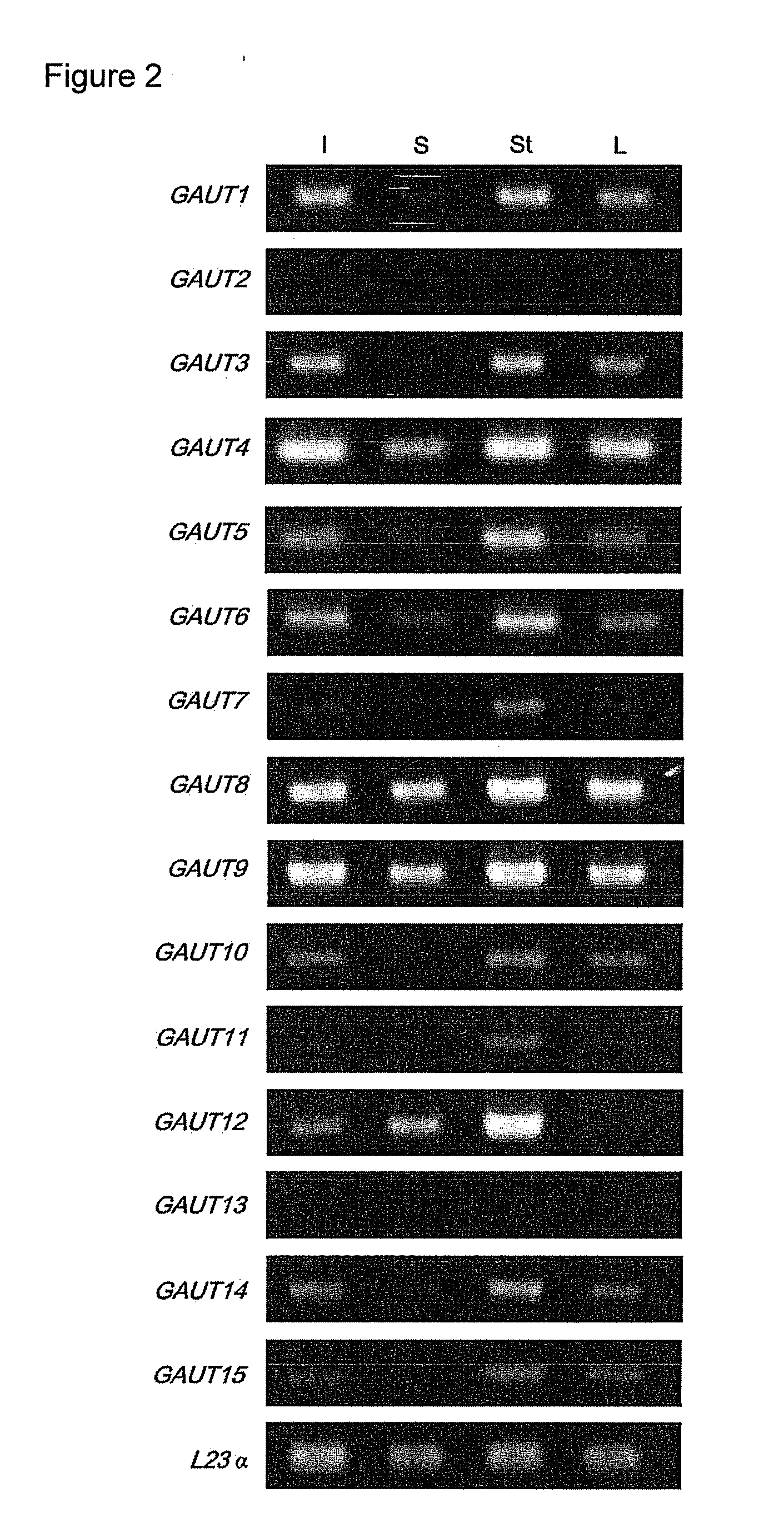
[0195]Plant Materials and Growth Conditions. Two independent T-DNA insertion lines (00091 and 02925) in GAUT14 were obtained from the Arabidopsis Biological Resource Center (www.biosci.ohio-state.edu/pcmb/Facilities/abrc/abrchome.htm). Arabidopsis WT (Arabidopsis thaliana var. Columbia S6000) and gaut14 mutant seeds were sown on pre-moistened soil in a growth chamber with 60% constant relative humidity with a photoperiod 14/10 light/dark cycle (14 h 19° C. and 10 h 19° C.) and fertilized as described (Example 1). The 7-weeks old WT and PCR-genotyped mutant plants were harvested used for glycome profiling and as a carbon source for bacterial growth analyses.
[0196]DNA Extraction, mutant genotyping and identification of two T-DNA insertion lines in GAUT14. Approximately 100 mg of leaf tissue was ground with a mortar and pestle to fine powder. The ground leaf tissue was suspended in 0.5 ml extraction buffer (100 mM EDTA pH 8.0, 100 mM Tris-HCl pH 8.0, 250 mM NaCl, 100 μg ml−1 proteinase K and 1% (w/v) n-lauroylsarcosine). The suspension was extracted with an equal volume of phenoLchloroformn:isoamyl alcohol (49:50:1, v/v). DNase-free RNase A (1 μl) was used to degrade RNA for 20 min at 37° C. and the DNA was precipitated twice with 70% (v/v) ethanol.
[0197]The genotype of gaut14 mutant plants was determined by the appropriate GAUT14 gene-specific primer with T-DNA-specific primers based on the ability of the LB primers to anneal.
[0198]The GAUT14 gene-specific primer pairs used for genotyping were AtGAUT14 (forward, 5′-ATGCAGCTTCACATATCGCCTAGCATG (SEQ ID NO:160)′; reverse, 5′-CAGCAGATGAGACCACAACCGATGCAG (SEQ ID NO:161)). Following T-DNA-specific primer pairs were used for genotyping like gaut14-1 (forward, 5′-TTAAGTCTCCCTGGACAACTATATCAT (SEQ ID NO:162); reverse, 5′-CAATTGTCAAGTTGGTTTCTTTTCT(SEQ ID NO:163)), gaut14-2 (forward, 5′-TTGGGTCCGCTACTGATCTGA (SEQ ID NO:164); reverse 5′-GCAGTGATCCACTACAATGGGC (SEQ ID NO:165)). Homozygous lines were identified by PCR for further characterization of the gaut14 mutants. The two mutant lines are designated gaut14-1 and gaut14-2.
[0199]Quantitative Real-Time PCR. For expression analysis wild type, Arabidopsis leaf, flower, upper stem, middle stem, lower stem, hypocotyls, silique and seeds were harvested and frozen immediately in liquid nitrogen and stored at −80° C. until use. All the tissues were ground to a fine powder using N2(l) in a chilled mortar and pestle. Total RNA was extracted using the RNeasy Plant Mini Kit (Qiagen) followed by DNAse (DNA-free kit, Ambion) treatment to remove genomic DNA contamination. First strand cDNA synthesis was performed using 1 μg of total RNA with a blend of oligo (dT) and random primers in the iScript™cDNA Synthesis Kit (Bio-Rad, Hercules, Calif., USA) according to the manufacturer's instructions. The primers used to amplify the GAUT14 transcripts of the above tissues were as follows: AtGAUT14 (forward, 5′-CAAGGCAGTCTGCAGATATTAC (SEQ ID NO:166); reverse, 5′-CTTATGCAACCTTCCCTTCG (SEQ ID NO:167)), with two primers (forward, 5′-AGTGTCTGGATCGGTGGTTC (SEQ ID NO:168); reverse, 5′-ATCATACTCGGCCTTGGAGA (SEQ ID NO:169)) to amplify the actin2 transcript were also designed as an internal standard for quantification. PCR reactions were performed in a 96-well plate with a Bio-Rad iCycler MyiQ Real-Time PCR Detection System. Detection of products was by binding of the fluorescent DNA dye SYBR Green (iQ SYBR Green Supermix) to the PCR products. All assays were carried out in triplicate, and one-set of no-template controls was included per gene amplification. A PCR reaction contained a total volume of 25 μl with appropriate cDNA, SYBR Green, and both forward and reverse primers. Thermal cycling conditions were as follows: initial activation step 3 min at 95° C., followed by 15 s at 95° C., 30 s at 55° C., 30s at 72° C. for 45 cycles, 1 min 95° C., 1 min 55° C., a melting curve program (80 cycles, 10 s each of 0.5° C. elevations starting at 55° C.) and a cooling step to 4° C. The presence of one product per gene was confirmed by analysis of the disassociation curves. The iCycler MyiQ software 1.0 (Bio-Rad, Hercules, Calif., USA) was used to calculate the first significant fluorescence signal above noise, the threshold cycle (Ct). The PCR efficiencies (E) of each amplicon were determined by using pooled cDNA originating from the assayed tissues in 4-fold serial dilutions and the calculation was performed in the iCycler MyiQ software 1.0 (Bio-Rad). The relative transcript levels (RTL) was calculated as follows: 100 000×ECT Control/ECT Target, thus normalizing target gene expression to the control gene expression.
[0200]Isolation of cell wall, cell wall (AIR) fractionation and ELISA assay. The walls from leaves and stem of WT and two gaut14 mutants were sequentially extracted from frozen ground tissue with 80% ethanol, 100 ml ethanol, chloroform:methanol (1:1) (Example 1) and the resulting AIR (alcohol insoluble residue) was washed with acetone. The cell walls (AIR) were then de-starched with alpha amylase (Sigma) in 50 mM ammonium formate, pH 6.5, for 24 hrs. In the next step the AIR walls were sequentially fractionated enzymatically and chemically. The enzyme treatments were carried out in ammonium formate, pH 6.0 for 24 hours at room temperature with Aspergillus niger EPG and Aspergillus niger PME. The walls were then sequentially extracted with 50 mM sodium carbonate (pH 10.0) and then with 1M KOH and 4M KOH. Each fraction was neutralized (if necessary), dialyzed and lyophilized for analysis. The extracted cell walls were dissolved in deionized water (0.2 mg/mL) and the total amount of sugar measured. Equal amounts of sugar (500 ng) were applied to the wells of ELISA plates (Costar 3598) and a series of 152 monoclonal antibodies directed against plant cell wall carbohydrate epitopes were used for this analysis. The data are presented as a heat map on a hierarchical clustering (Pattathil et al., 2010, Plant Physiol., 153:514-525).
[0201]Microorganisms and bacteria growth medium in WT and gaut14-1 and gaut14-2 mutants in Arabidopsis.
[0202]Microorganisms: Caldicellulosiruptor bescii DSM 6725 (former Anaerocellum thermophilum DSM 6725) was obtained from the DSMZ (http://www.dsmz.de/index.htm). Caldicellulosiruptor saccharolyticus DSM 8903 was a gift from Robert Kelly of North Carolina State University.
[0203]Growth medium. C. bescii DSM 6725 and C. saccharolyticus DSM 8903 were grown in the 516 medium (Svetlichnyi et al., 1990, Microbiology (Translation of Mikrobiologia) 59:598-604) except that vitamin and trace mineral solutions were modified as follows. The minerals solution contained per liter: NH4Cl0.33 g, KH2PO4 0.33 g, KCl 0.33 g, MgCl2×6 H2O 0.33 g, CaCl2×2 H2O 0.33 g, yeast extract 0.5 g, resazurin 0.5 mg, vitamin solution 5 ml, trace minerals solution 1 ml. The vitamin solution contained (mg/l): biotin 4, folic acid 4, pyridoxine-HCl 20, thiamine-HCl 10, riboflavin 10, nicotinic acid 10, calcium panthotenate 10, vitamin B12 0.2, p-aminobenzoic acid 10, lipoic acid 10. The trace minerals solution contained (g/l) FeCl3 2, ZnCl2 0.05, MnCl2×4H2O 0.05, H3BO3 0.05, CoC2×6H2O 0.05, CuCl2×2H2O 0.03, NiCl2×6H2O 0.05, Na4EDTA (tetrasodium salt) 0.5, (NH4)2MoO4 0.05, AlK(SO4)2.12H2O 0.05. The medium was prepared anaerobically under a N2/CO2 (80:20) atmosphere, NaHCO3 (1 g/l) was added and it was reduced using (per liter) 0.5 g cysteine and 0.5g N2S. Finally, 1 ml/L of 1M potassium phosphate buffer (pH 7.2) was added. The final pH was 7.2. The medium was filter-sterilized using a 0.22 micron pore size sterile filter (Millipore Filter. Corp., Bedford, Mass.). Arabidopsis (wild type and two gaut14 mutants) dried stems were used as a growth substrate at a final concentration of 0.5% (wt/vol). The dried intact biomass was added directly to each bottle. Growth was at 78° C. (A. thermophilum) or at 71° C. (C. saccharolyticus) as static cultures in 50 ml serum bottles with 20 ml medium with shaking (150 rpm) for 24 hours. The culture media containing the insoluble substrates without inoculation were used as controls. All growth experiments were run in triplicate. Cell density was monitored by cell count using phase-contrast microscope with 40× magnification and expressed as cells per ml. Samples of growing cultures were taken each three hours and cell count was done immediately.
Results
[0204]Endogenous expression of GAUT14 in Arabidopsis. The level of GAUT14 transcripts in various WT tissues was investigated using qRT PCR as described in the materials and methods. Acting used as a control. GAUT14 mRNA was detected in stem, leaf, flower, hypocotyl, silique and seeds in all major tissues, suggesting a role in plant growth and development (FIG. 8). However, transcript expression was more prominent in upper and lower stem in Arabidopsis.
[0205]Position of T-DNA insertion, phenotypes and growth measurement of T-DNA knock-out mutants in gaut14-1 and gaut14-2. The two T-DNA insertional mutants for GAUT14 (At5g15470) were obtained from the Salk collection as described in materials and methods. The T-DNA is inserted in the fourth exon in gaut14-1 (Salk—000091) and in the 3′ untranslated region (UTR) in gaut14-2 (Salk—029525) mutants (FIG. 9). Five week old homozygous gaut14 mutants exhibited a clear visible phenotype when grown on soil, with reduced stem length and leaf blade length (FIG. 10). There is a 10% and 36% decrease in stem length in gaut14-1 and gaut14-2 mutants, respectively in comparison to their wild type plants (FIG. 11). Similarly there is a 10% and 24% decrease in leaf blade length in gaut14-1 and gaut14-2 mutants, respectively (FIG. 11). Interestingly, the reduced growth phenotype in these two gaut14 mutants caught up to WT within 7-weeks.
[0206]Glycome profile of WT and gaut14 mutants in Arabidopsis. A method recently developed by Pattathil et al. (2010, Plant Physiol., 153:514-525) was used to determine how the release of sequentially extracted cell wall polymers from the stem and leaf cell walls of WT are different from those of the gaut14 mutants based on detection of released wall material using 150 cell wall carbohydrate-directed monoclonal antibodies. Both the gaut14 mutant leaf walls retain less polysaccharide in the insoluble pellet in comparison to the WT leaves (FIG. 12). The release of more cell wall polymers was detected in the 4M KOH fractions in gaut14-1 than WT, especially in the case of RG-I/AGP directed antibodies. However, more significant differences were exhibited by gaut14-2 mutants with more release of polysaccharides in the early stages of fractionation, for example in the 1M KOH fraction (FIG. 12). The same pattern of less polysaccharide material being retained in the insoluble pellet of the gaut14-1 and gaut14-2 mutant stem was obtained (FIG. 13). The EPG/PME and carbonate fractions in gaut14-1 showed different binding patterns from WT, especially in the case of HG/RG-I backbone, AGP and RG-I/AGP directed antibodies (FIG. 13). The glycome profiles suggest that the absence of GAUT14 products have profound effects on the cell wall extractability which makes the wall more easily extractable.
[0207]Growth of two pectin degrading bacteria in Arabidopsis WT and gaut14 mutants. Growth of Caldicellulosiruptor bescii DSM 6725 was quite efficient on Arabidopsis wild type and on the gaut14-1 and gaut14-2 mutants (FIG. 15). After 24 hours, the cultures were still growing, although they reached middle stationary phase. Cell densities upon growth on Arabidopsis WT, gaut14-1 and gaut14-2 mutants were over >4e+8 with slightly at 26 hours. C. bescii grew somewhat better on the Arabidopsis gaut14 mutants than on the Arabidopsis WT. Growth of C. saccharolyticus DSM 8903 on Arabidopsis WT, gaut14-1 and gaut14-2 mutants was much different than the growth of C. bescii on the same walls (FIG. 15). The bacterium grew less well on WT, and grew better on the two gaut14 mutants, approaching stationary phase growth after 24 hours. The growth was more efficient on the gaut14-1 and gaut14-2 mutants than on the WT, with final cell densities of 3.5e+8, 3.4e+8 and 1.6e+8 cells/ml, respectively.
Discussion
[0208]C. bescii and C. saccharolyticus are thermophilic anaerobic bacteria capable of growing on different polysaccharides including crystalline cellulose, xylans, starch and pectin (Rainey et al., 1994, FEMS Microbiol Lett 120: 263-266; Yang et al., 2009, Appl. Environ. Microbiol., 75:4762-4769). The genome of C. saccharolyticus has been available for about three years. The genome of C. bescii was sequenced and analyzed recently (Kataeva et al., 2009, J. Bacteriol., 191: 3760-3761). Both genomes are very similar and encode sets of enzymes acting on polysaccharides and metabolizing multiple sugars. Both bacteria are able to process cellulose and xylan simultaneously and grow on Arabidopsis plant biomass. However, comparison of the growth of C. bescii and C. saccharolyticus on Arabidopsis WT and on the gaut14 knockouts mutants, mutants that appear to modify the pectin biosynthesis pathway, revealed differences. In particular, C. bescii grew well on all Arabidopsis samples but showed somewhat better growth on the gaut14 mutants with a final cell density exceeding 4e+8 cells/ml, which is a high density for anaerobic thermophiles. C. saccharolyticus also grew on the three different Arabidopsis biomass sources, however, the cells reached stationary phase in shorter time and the cell densities were lower for C. saccharolyticus compared to C. bescii. Moreover, the growth of C. saccharolyticus on WT Arabidopsis biomass was much less efficient compared to growth on gaut14-1 and gaut14-2 mutant biomass, with lower final cell densities when grown on WT.
[0209]These differences could be attributed to the different pectin degrading systems produced by these bacteria (FIGS. 14A and 14B). C. bescii has a unique enzymatic system related to pectin degradation. It is composed of 3 polysaccharide lyases (PL) of different PL families (encoded by Cbes—1853 -1855 genes). In addition, the genome encodes two glycoside hydrolases of family 28 (GH28, see CAZy database) capable of hydrolysis of unsubstituted polygalacturonic acid as part of pectin backbone (FIG. 13A). Search within 25 genomes of anaerobic thermophilic bacteria (our data, not published) revealed that only two of them encode sets of 3 PLs of different families (C. bescii and Cl. thermocellum, although the latter does not encode GH28s). In contrast to C. bescii, all PLs are missing from the genome of C. saccharolyticus. The genome encodes only two GH28s with limited activity against pectin (FIG. 13B). This genome analysis suggests that better growth of C. bescii on Arabidopsis vs. C. saccharolyticus is related to a comprehensive set of pectin degrading enzymes while C. saccharolyticus has a truncated set composed of just two GH28s. The gaut14-1 and gaut14-2 mutants have either less content of pectin or modified pectin. As a result, C. saccharolyticus grows better on the mutants than on WT Arabidopsis without interruptions in pectin content/structure.
[0210]The present data suggest that the pectin, similar to lignin, is a “recalcitrance factor” of plant biomass decreasing accessibility of cellulose and hemicelluloses to the corresponding degrading enzymes. The data also are very promising for the development a novel approach to test recalcitrance of plant biomass. This “microbial recalcitrance test” would be based on a limited ability of a given microorganism to degrade a particular constituent(s) of plant biomass, so that genetically modified plants with the decreased amounts of, or simplified structures of, the relevant wall polymer will serve as better growth substrates in comparison to wild type plants.
[0211]The complete disclosure of all patents, patent applications, and publications, and electronically available material (including, for instance, nucleotide sequence submissions in, e.g., GenBank and RefSeq, and amino acid sequence submissions in, e.g., SwissProt, PIR, PRF, PDB, and translations from annotated coding regions in GenBank and RefSeq) cited herein are incorporated by reference in their entirety. Supplementary materials referenced in publications (such as supplementary tables, supplementary figures, supplementary materials and methods, and/or supplementary experimental data) are likewise incorporated by reference in their entirety. In the event that any inconsistency exists between the disclosure of the present application and the disclosure(s) of any document incorporated herein by reference, the disclosure of the present application shall govern. The foregoing detailed description and examples have been given for clarity of understanding only. No unnecessary limitations are to be understood therefrom. The invention is not limited to the exact details shown and described, for variations obvious to one skilled in the art will be included within the invention defined by the claims.
[0212]Unless otherwise indicated, all numbers expressing quantities of components, molecular weights, and so forth used in the specification and claims are to be understood as being modified in all instances by the term “about.” Accordingly, unless otherwise indicated to the contrary, the numerical parameters set forth in the specification and claims are approximations that may vary depending upon the desired properties sought to be obtained by the present invention. At the very least, and not as an attempt to limit the doctrine of equivalents to the scope of the claims, each numerical parameter should at least be construed in light of the number of reported significant digits and by applying ordinary rounding techniques.
[0213]Notwithstanding that the numerical ranges and parameters setting forth the broad scope of the invention are approximations, the numerical values set forth in the specific examples are reported as precisely as possible. All numerical values, however, inherently contain a range necessarily resulting from the standard deviation found in their respective testing measurements.
[0214]All headings are for the convenience of the reader and should not be used to limit the meaning of the text that follows the heading, unless so specified.
Login to view more PUM
Property | Measurement | Unit |
Fraction | 0.8 | fraction |
Volume | 8.42E-16 ~ 8.47E-16 | L |
Volume | 5.236E-15 ~ 5.241E-15 | L |
tensile | MPa | |
Particle size | Pa | |
strength | 10 |
Description & Claims & Application Information
We can also present the details of the Description, Claims and Application information to help users get a comprehensive understanding of the technical details of the patent, such as background art, summary of invention, brief description of drawings, description of embodiments, and other original content. On the other hand, users can also determine the specific scope of protection of the technology through the list of claims; as well as understand the changes in the life cycle of the technology with the presentation of the patent timeline. Login to view more.
Login to view more Similar technology patents
Disk Brake Having a Pad-Retaining Clip and a Securing Device, and Brake Pad Set
ActiveUS20180252280A1Easily releaseEnhance rangeAxially engaging brakesBraking elementsDisc brakeBackplane
Owner:KNORR-BREMSE SYST FUER NUTZFAHRZEUGE GMBH
Stripper plate mounting apparatus
ActiveUS20100162866A1Easily releasePerforating toolsStripping-off devicesEngineeringLocking mechanism
Owner:AMADA CO LTD
Lever-Fitting Type Connector
InactiveUS20080194129A1Improve release operabilityEasily releaseEngagement/disengagement of coupling partsEngineeringMechanical engineering
Owner:YAZAKI CORP +1
Circuit board fall-out prevention structure and numerical control device
ActiveUS20200305299A1Easily releaseCasings with display/control unitsPrinted circuit board receptaclesStructural engineeringElectrical and Electronics engineering
Owner:FANUC LTD
Cellular Phone Provided with Key Lock Function
InactiveUS20080119234A1Easily releaseLow electric power consumptionUnauthorised/fraudulent call preventionTransmissionMobile telephonyKey pressing
Owner:KYOCERA CORP
Classification and recommendation of technical efficacy words
- Easily release
- Cost effectiveness
USB connector
InactiveUS6902432B2Excellent fit propertyEasily releaseElectrically conductive connectionsTwo-part coupling devicesEngineeringUSB
Owner:YAZAKI CORP
Medical article anchoring system
ActiveUS20060276752A1Different configurationEasily releaseCatheterInfusion needlesBiomedical engineeringMechanical engineering
Owner:VENETEC INT INC
Foam bodied gasket and gasket tape and method of making and using the same
Owner:THE PATENT WELL LLC
Method For Releasing a Thin-Film Substrate
ActiveUS20100279494A1Easily releaseLow porositySemiconductor/solid-state device manufacturingSemiconductor devicesPorous semiconductorsThin film semiconductors
Owner:BEAMREACH SOLAR INC
Adhesive film
InactiveUS20130040093A1Easily releaseFacilitate positioningLaminationLamination apparatusEngineering
Owner:HITACHI CHEM CO LTD
User authentication
ActiveUS9122866B1Cost effectivenessDigital data processing detailsDigital data authenticationUser authenticationInternet privacy
Owner:EMC IP HLDG CO LLC
Fabrication of array pH sensitive EGFET and its readout circuit
InactiveUS20060148118A1Simple fabrication equipmentCost effectivenessMaterial analysis by electric/magnetic meansSemiconductor/solid-state device manufacturingPhysicsIon sensitive
Owner:CHUNG YUAN CHRISTIAN UNIVERSITY
Secondary air supply system for internal combustion engine
InactiveUS20070079604A1Lower emissionCost effectivenessInternal combustion piston enginesExhaust apparatusExhaust gasClutch
Owner:MACALUSO ANTHONY
Depressible key structure
InactiveUS20110114461A1Cost effectivenessSimplify configurationEmergency actuatorsContact mechanismsEngineeringKey pressing
Owner:PRIMAX ELECTRONICS LTD
Wave filter for rotary filter
InactiveUS20020074278A1Cost effectivenessEasily removableFrozen sweetsConfectioneryPolymer meltWave shape
Owner:ALTER INVEST FUND LP