Methods And Compositions For Detecting Fungi And Mycotoxins

a technology of mycotoxins and compositions, applied in the field of methods and compositions for detecting or identifying fungi or detecting or identifying mycotoxins, can solve the problems of affecting the health of patients, affecting the quality of life of patients, so as to achieve reliable, sensitive, rapid and reliable results

Inactive Publication Date: 2013-07-18
ADVATECT DIAGNOSTICS LLC
View PDF2 Cites 16 Cited by
  • Summary
  • Abstract
  • Description
  • Claims
  • Application Information

AI Technical Summary

Benefits of technology

The patent text discusses the need for a reliable and rapid method for detecting molds and their toxic metabolites (mycotoxins) in patient body fluids and tissues. This method can help identify patients at risk for developing diseases related to mold infections and quickly determine the cause of the disease so that effective treatment regimens can be developed. The invention also involves developing a sensitive and specific method for detecting fungal DNA in patient body fluids and tissues.

Problems solved by technology

Exposure to molds can cause a number of adverse effects including allergic reactions, asthma attacks, and infections, particularly in individuals with immune system deficiencies.
Mycotoxins have toxic effects ranging from severe irritations, such as allergic reactions and asthma, to immuno-suppression and cancer.
As a result, mycotoxins may be damaging to the skin, the lungs, the gut, and the like.
The mold infections in such patients are often fatal with a documented fatally rate of 92% (Paterson and Singh, 1999).
There are no current methods that have been developed for determining the presence of mycotoxins in patient tissues or body fluids.
The reasons for the late diagnosis of fungal infections include the lack of good clinical specimens, the difficultly in differentiating invasive mold infections from other types of infections, the lack of identification of molds with special stains in pathological specimens (i.e., these assays have a high error rate, a low sensitivity, and low specificity), the lack of an ability to obtain an antibody-based diagnosis in immuno-compromised patients, and the lack of assays to determine the presence of mycotoxins or fungal DNA in the tissue or fluids of those patients.

Method used


Image

  • Methods And Compositions For Detecting Fungi And Mycotoxins
  • Methods And Compositions For Detecting Fungi And Mycotoxins
  • Methods And Compositions For Detecting Fungi And Mycotoxins

Examples


example 1

Samples and Sample Preparation

[0061]Human urine was received in 5-10 ml quantities as first in the morning voided urines. Serums were received with the blood clot removed prior to receipt and a minimum of 1 ml of serum was frozen or used. Nasal secretions were obtained from hospital patients or out-patients. Fixed autopsy and surgical biopsy specimens were obtained from patients who had a history of exposure to mycotoxins or fungi. These samples were obtained from hospital pathology departments or coroners' offices. Tissue samples and body fluid samples were also obtained from patients who had no exposure to mycotoxins or fungi and were sampled as a negative control group. Tissue specimens were cut using procedures described in Examples 2 and 7.

[0062]All specimens were placed into two groups. Group 1 comprised samples from individuals with no reported symptoms or known fungi or mycotoxin exposure. These samples served as negative controls and n values differed in each group of speci...

example 2

Preparation of Tissues for Mycotoxin Extraction

[0065]Preparation of tissues for myctotoxin extraction from formalin fixed tissue and paraffin-embedded tissue from humans or animals was accomplished using the following procedure.

Specimens

[0066]Tissue was received as either tissue fixed in a 10% formalin solution or in a paraffin-embedded tissue block. Tissue can be stored indefinitely in either form. However, because of cross-linking of formalin and proteins which may give false negative readings for DNA, the tissue was not stored in formalin for greater than 6 months. A minimum of 25-35 mg of formalin-fixed tissue was required for mycotoxin extraction. A maximum of 3 grams of formalin-fixed tissue can be used.

Materials

[0067]Phosphate Buffered Saline (PBS; 0.9%), acid-washed silica beads (Cat # G1277; obtained from Sigma-Aldrich), collection tubes (2 ml) screw cap, methanol (reagent grade, Sigma), and microcentrifuge tubes (2 ml) were used.

Procedure

[0068]For silica beads, 0.3 g±0.01 ...

example 3

Preparation of Body Fluids for Mycotoxin Detection

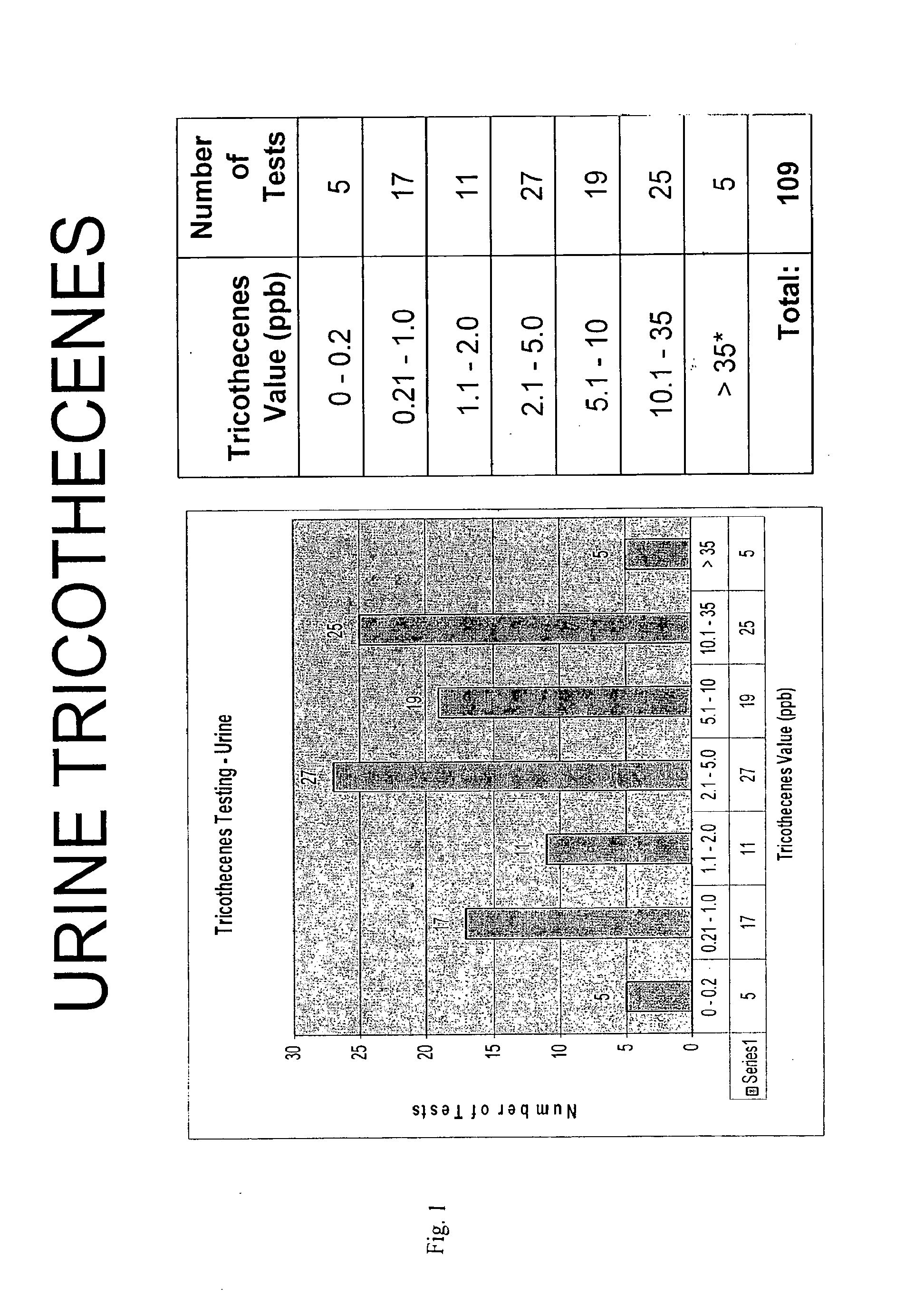
[0070]Urine was received from a morning fresh first-voided specimen and stored at 1-6 degrees centrigrade in a glass container. A urine analysis was conducted using a dipstick to measure pH, specific gravity, glucose, nitrates, ketones, and blood. The urine was examined for sediment and was centrifuged at 2500 rpm for 5 minutes if sediment was present. The supernatant was saved in a glass container for mycotoxin testing (storing in plastic was avoided to avoid a decrease in the detection level of tricothecenes). The urine was then used as indicated in Example 4 for aflatoxin determination, Example 5 for ochratoxin determination, and Example 6 for tricothecene determination.

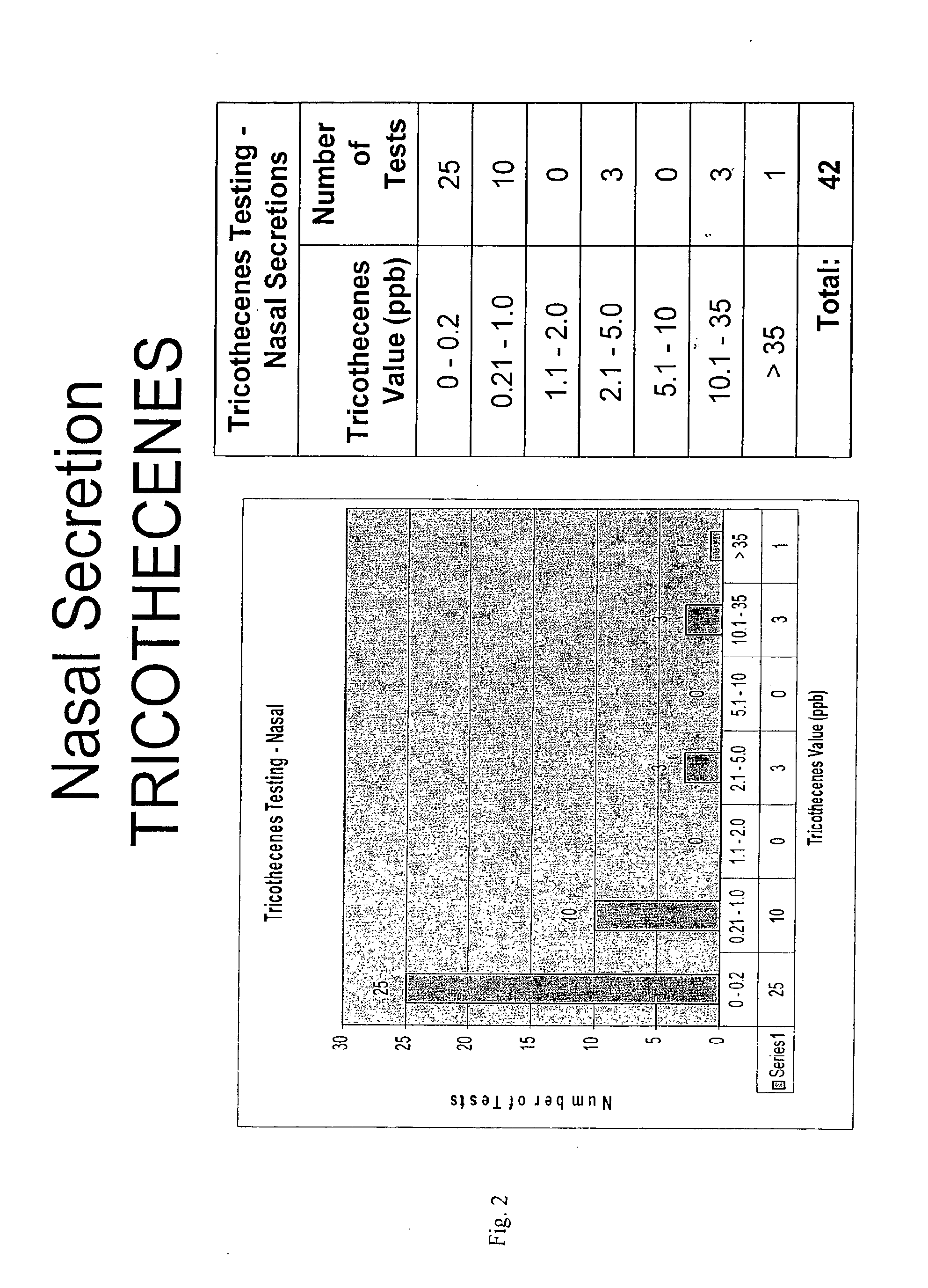
[0071]Nasal secretions and mucous samples as well as washes were observed for mucous presence. If mucous was present, a solution of MUCOSOL™ (Alpha Tec Systems, Inc. Vancouver, Wash.) was prepared and added in equal amounts of body fluid to MUCOSOL™ in the secretio...


PUM


Abstract

The invention relates to a method of identifying a specific fungal species in patient tissue or body fluid. The method comprises the steps of extracting and recovering DNA of the fungal species from the patient tissue or body fluid, amplifying the DNA, hybridizing a probe to the DNA to specifically identify the fungal species, and specifically identifying the fungal species. The invention also relates to a method of identifying a mycotoxin in patient tissue or body fluid. The method comprises the steps of extracting and recovering the mycotoxin from the patient tissue or body fluid, contacting the mycotoxin with an antibody directed against the mycotoxin, and identifying the myocotoxin. Both of these methods can be used to determine if a patient is at risk for or has developed a disease state related to a fungal infection, and to develop an effective treatment regimen for the patient.

Description

CROSS REFERENCE TO RELATED APPLICATIONS[0001]This application claims priority under 35 U.S.C §119(e) to U.S. Provisional Application Ser. Nos. 60 / 787,754, filed on Apr. 1, 2006, and 60 / 790,974, filed on Apr. 11, 2006.FIELD OF THE INVENTION[0002]This invention relates to methods and compositions for detecting or identifying fungi or for detecting or identifying mycotoxins. More particularly, the invention relates to methods and compositions for detecting or identifying fungi and for detecting or identifying mycotoxins in the tissues or body fluid samples of patients.BACKGROUND AND SUMMARY[0003]Molds (i.e., toxigenic and other septate molds) are ubiquitous in the environment. Mold is the common name for various types of fungi. Molds are usually found in moist, warm environments. Because molds grow in wet or moist indoor environments, people are exposed to molds or their byproducts through either direct contact, or through the air, if molds or mold byproducts are aerosolized. Exposure ...

Claims


Application Information

Patent Timeline
Patent Type & Authority Applications(United States)
IPC IPC(8): G01N33/569
CPCC12Q1/6895G01N2469/10G01N33/56961C12Q2600/142G01N2800/26G01N2800/50
Inventor HOOPER, DENNIS G.
Owner ADVATECT DIAGNOSTICS LLC
Why Eureka
Try Eureka