Method for determining cancer onset or cancer onset risk

a cancer and risk technology, applied in the field of cancer onset or cancer onset risk, can solve the problems of difficult quantitative analysis of quantum dot fluorescent particles in biological tissues, inability to complete bleaching in many hours, and limited cultured cells, etc., and achieve the effect of high sensitivity

Inactive Publication Date: 2013-08-08
TOHOKU UNIV
View PDF3 Cites 22 Cited by
  • Summary
  • Abstract
  • Description
  • Claims
  • Application Information

AI Technical Summary

Benefits of technology

[0013]According to the tissue staining method of the present invention wherein the influence of autofluorescence is eliminated and the PAR1 antibody labeled with quantum dot fluorescent particles is used, the average value of the number of quantum dot fluorescent particles per cell in mammary tissues (detected using the PAR1 antibody) was 0.2 in 4 samples of normal mammary tissues from cancer patients, whereas 3.2 in 5 samples of tissues from recurrence-free breast cancer survivors for at least 5 years and who are HercepTest negative (detected using the HER2 antibody) and 14.8 in 3 samples of breast cancer tissues from recurrent patients (recurred within about 1 year and died within 4 years) who are HercepTest negative (detected using the HER2 antibody), thus acquiring significant differences. Further, “triple negatives”, which refers to any breast cancer that develops with no connection to three major breast cancer growth-related f...

Problems solved by technology

Fluorescent particles such as quantum dot exhibit a potential for the intrinsic light stability and quantitative analysis, and is thus highly expected as a new tool for the fluorescent immunostaining but, for the quantitative analysis, it is still limited to cultured cells.
The autofluorescence of the long wavelength is a smaller amount than the autofluorescence of the short wavelength in biological tissues, but the influence thereof to the observation is not negligible and thus the quantitative analysis of quantum dot fluorescent particles in biological tissues was difficult.
In the above (a) autofluorescence photobleaching method by the excitation light irradiation, it is possible to bleach only the autofluorescence while the brightness intrinsic to quantum d...

Method used


Image

  • Method for determining cancer onset or cancer onset risk
  • Method for determining cancer onset or cancer onset risk
  • Method for determining cancer onset or cancer onset risk

Examples


example 1

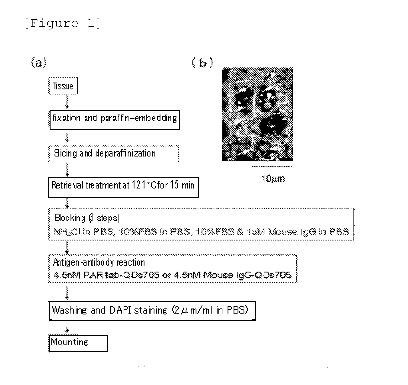
[0044][Method for Preparing an Immunostained Tissue Sample]

[0045]To differentiate from HercepTest (detecting breast cancer tissues using HER2 antibody), all tissue samples used as the human breast cancer pathological tissue were HercepTest negative. On the other hand, there are cancer cases which develop with no connection to three major breast cancer growth-related factors (ER, PgR, HER2) and this “triple negative” breast cancers are generally considered to have poor prognosis and to be difficult to treat. Under the circumstances, the “triple negative” breast cancer tissue samples were also studied. More specifically, the total of 16 samples consisting of 4 samples (one of which is the “triple negatives” [since 20 to 25% of the recurrent patients are the “triple negatives”, 1 sample out of 4 samples was a randomly added “triple negative” sample]) of breast cancer tissues from recurrent patients (recurred within about 1 year and died within 4 years), 8 samples (including the above 1...

example 2

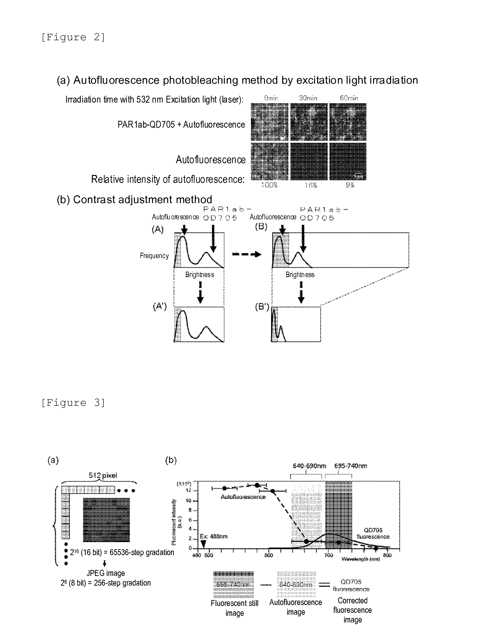
[0046][Problems in Fluorescent Immunohistostaining Method]

[0047]The immunostained tissue samples produced in the above Example 1 were irradiated with 488 nm excitation light using a combination device of a confocal unit (a product of Yokogawa Electric Corporation), a fluorescence microscope (a product of OLYMPUS CORPORATION) and Electron-Multiplier CCD (EM-CCD) Camera (Andor Co., Ltd.), and then a fluorescence image (fluorescent still image) of quantum dot fluorescent particle (detected using PAR1ab-QD705) was acquired using a 695 to 740 nm band pass filter. FIG. 1(b) shows the fluorescent still images of the tissue samples from recurrent breast cancer patients as an example, but the autofluorescence was so intense that the fluorescence of quantum dot fluorescent particle (detected using PAR1ab-QD705) was not identified on the first inspection. When the fluorescent brightnesses of autofluorescence and quantum dot fluorescent particles (detected using PAR1 antibody) were compared, on...

example 3

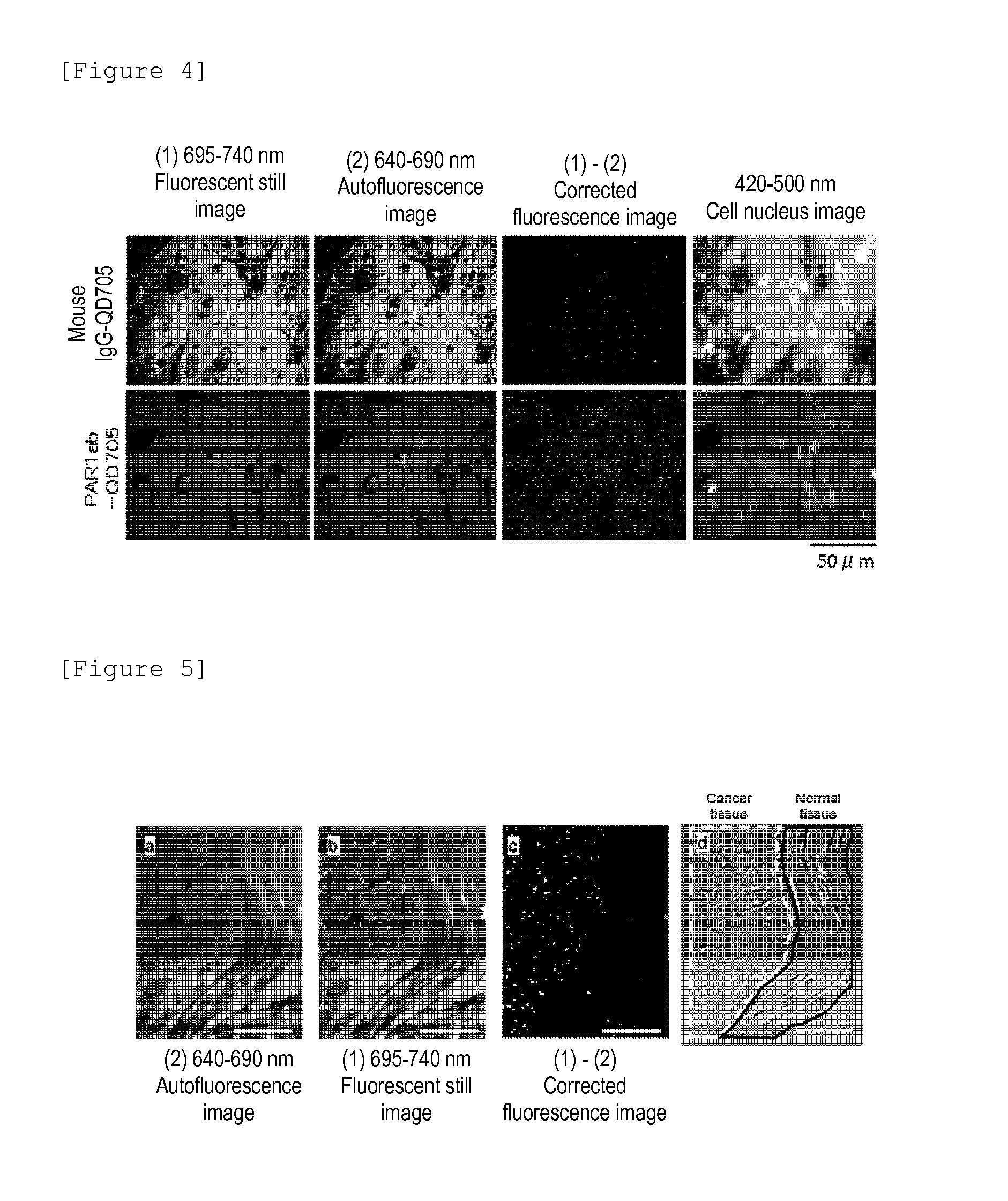
[0049][Production of Corrected Fluorescence Image from Which Autofluorescence is Eliminated]

[0050]A fluorescent still image with a zero (0) fluorescent intensity of the background autofluorescence need to be acquired. Then, the total fluorescence of fluorescent still image can be calculated as the fluorescence derived from quantum dot fluorescent particles. The contrast adjustment method of the above Example 2 uses the division of fluorescent intensity, thus failing to produce zero (0) by the division, and accordingly an image processing method which uses the subtraction capable of producing zero (0) is required. Under the circumstances, the following method is proposed as the image processing method for eliminating the autofluorescence of a fluorescent still image. In the method, first, a breast cancer tissue sample immunostained with quantum dot fluorescent particles is irradiated with excitation light (laser) having an excitation wavelength of 488 nm, an fluorescence image of qua...


PUM


Abstract

A highly accurate and quantitative method for determining cancer onset or cancer onset risk by a quantitative tissue staining method in biological tissues using an antibody capable of recognizing a cancer growth regulatory factor or cancer metastasis regulatory factor such as PAR1 antibody, which inhibits the cancer cell mobility and infiltration is provided. Cancer onset or cancer onset risk is determined using the tissue staining method comprising the steps of: labeling an antibody which recognizes a cancer growth regulatory factor or cancer metastasis regulatory factor with a fluorescent material, and contacting the fluorescent-labeled antibody with a tissue sample; irradiating a tissue site in contact with the antibody with excitation light to acquire a fluorescence image; acquiring an autofluorescence image in a vicinity region of a short wavelength side or long wavelength side of an acquisition region of fluorescence wavelength emitted by the fluorescent material, in the same field of vision and in the same focal point as those of the fluorescence image; acquiring a corrected fluorescence image by image processing to eliminate a fluorescent brightness of the autofluorescence image from the fluorescent brightness of the fluorescence image; counting the number of cells at the tissue site in contact with the antibody; measuring a mean fluorescent brightness of a single fluorescent particle; and calculating the number of fluorescent particles per cell.

Description

TECHNICAL FIELD [0001]The present invention relates to a method for determining cancer onset or cancer onset risk which uses a highly accurate, quantitatively analyzable tissue staining method from which the influence of autofluorescence is effectively eliminated.BACKGROUND ART [0002]Cancer is a disease which splits the causes of adult death with vascular diseases represented by myocardial infarction and cerebral infarction. For example, the breast cancer incidence rate in Japanese is lower than those in European and American countries, but it tends to increase in recent years and has ranked No. 1 in the female incidence rate overtaking the incidence rate of stomach cancer in 1998. According to a recent report, the year 2005 statistics projected by Ministry of Health, Labor and Welfare, the annual incidence number of breast cancer exceeds 50000 cases.[0003]Cancer diagnosis commonly uses, in addition to image diagnoses such as X-ray CT and MRI, methods for detecting a cancer marker w...

Claims


Application Information

Patent Timeline
IPC IPC(8): G01N21/64
CPCG01N21/6428G01N33/57415G01N2800/50G01N33/588G01N21/6458B82Y15/00G01N21/6486
Inventor GONDA, KOHSUKEMIYASHITA, MINORUTAKEDA, MOTOHIROOHUCHI, NORIAKI
Owner TOHOKU UNIV
Why Eureka
Try Eureka