In vitro model for pathological or physiologic conditions

Active Publication Date: 2013-11-21
HEMOSHEAR LLC
View PDF1 Cites 18 Cited by
  • Summary
  • Abstract
  • Description
  • Claims
  • Application Information

AI Technical Summary

Benefits of technology

[0005]Another aspect of the present invention is an in vitro method of testing a drug or a compound for an effect on a pathological condition. The method comprises mimicking the pathological condition, adding a drug or a compound to the culture media, and applying the shear force upon the at least one plated cell type exposed to the drug or the compound. A change in the at least one plated cell type, in the presence of the drug or the compound, indicates that the drug or the compound has an effect on the pathological condition. In this in vitro method of testing a drug or compound, the pathological condition can be mimicked by the in vitro method of mimicking a pathological condition as described above.
[0006]The present invention also provides an in vitro method of testing a drug or compound for an effect. The method comprises adding a culture media to a cell culture container, plating at least one cell type on at least one surface within the cell culture container, adding a drug or a compound to the culture media, and applying a shear force upon the at least one plated cell type exposed to the drug or the compound. The concentration of the drug or the compound in the culture media is within the concentration range of the drug or the compound that achieves the effect in vivo. The shear force results from flow of the culture media induced by a flow device. The flow mimics flow to which the at least one cell type is exposed in vivo. A change in the at least one plated cell type, in the presence of the drug or the compound, indicates that the drug or the compound has the effect. The effect can be, for example, an effect on a physiologic condition or an effect on a pathological condition.

Problems solved by technology

Furthermore, the conventional systems often do not exhibit responses to drugs or compounds at concentrations that induce the response in vivo, instead requiring much higher concentrations of the drug or compound to induce the same response.

Method used


Image

  • In vitro model for pathological or physiologic conditions
  • In vitro model for pathological or physiologic conditions
  • In vitro model for pathological or physiologic conditions

Examples


example 1

An In Vitro Model for Arterial and Venous Thrombosis

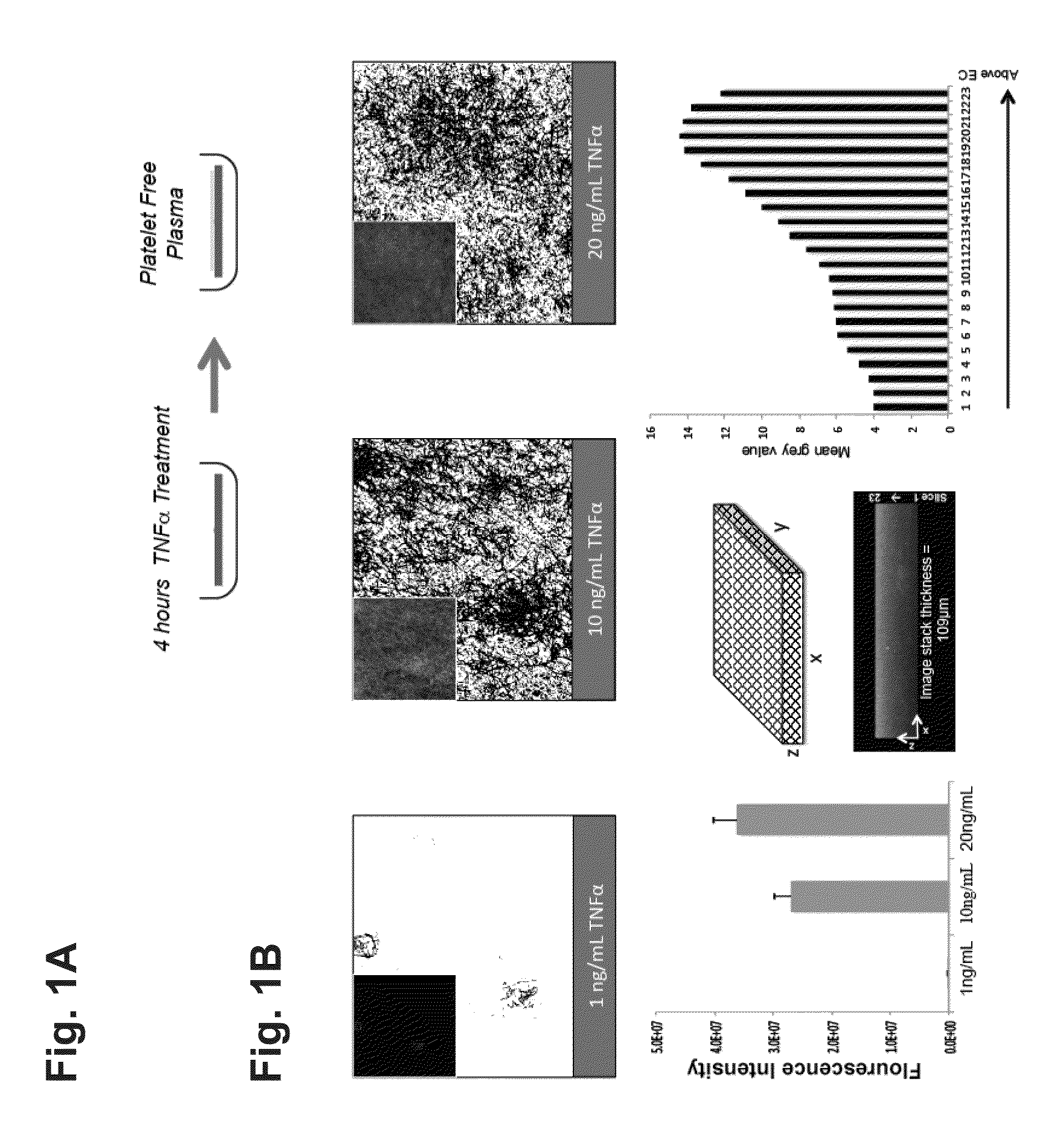
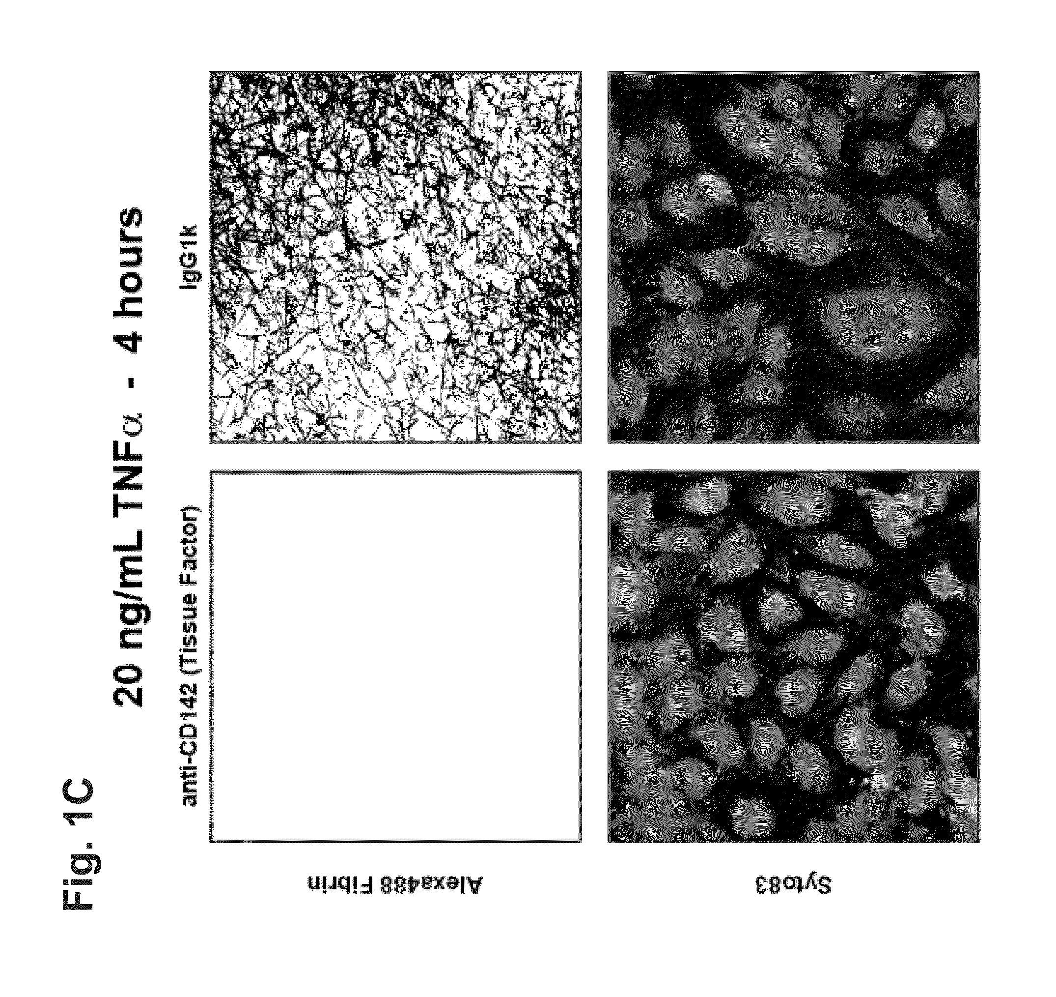
[0296]In the coagulation cascade, thrombin converts fibrinogen to fibrin, which is deposited on the surface of a blood vessel to begin blood clot formation (thrombosis). TNFα is a potent inflammatory cytokine. TNFα and other cytokines have been shown to be potent mediators of endothelial and smooth muscle cell-derived tissue factor in vitro, which mediates fibrin deposition in the vascular wall. Circulating levels of TNFα detected in humans with cardiovascular disease are about 0.01 ng / ml to about 0.1 ng / ml. In healthy individuals, circulating levels of TNFα are much lower or undetectable, for example about 0 ng / ml to about 0.001 ng / ml.

Methods:

[0297]Human endothelial cells were co-cultured with or without smooth muscle cells in the presence or absence of human-derived, region-specific hemodynamics. Endothelial cells were exposed to TNFα at various concentrations and incubated in human, platelet-free plasma supplemented by ALEXA FLU...

example 2

An In Vitro Model for Atherosclerosis

[0320](i) Effects of oxLDL in a Hemodynamic Environment

[0321](a) Methods

[0322]To oxidize LDL, native LDL (nLDL) was dialyzed with PBS for 24 hours to remove EDTA. The LDL was then dialyzed for 3 days in PBS containing 13.8 μM CuSO4. The LDL was then dialyzed with PBS containing 50 μM EDTA for an additional 24 hours. A relative electrophoretic migration number was used to confirm the oxidation level for each batch. Upon completion of oxidation, the oxLDL was stored under nitrogen at 4° C. until use.

[0323]Smooth muscle cells were plated on a first surface of the porous membrane of a TRANSWELL (polycarbonate, 10 μm thickness and 0.4 μm pore diameter, no. 3419, Corning) at a plating density of 20,000 cells / cm2 and allowed to adhere to the membrane for two hours. The TRANSWELL was then inverted and the cells were incubated in reduced serum growth media (M199 supplemented with 2% FBS, 2 mM L-glutamine, and 100 U / ml penicillin-streptomycin) for forty-ei...

example 3

An In Vitro Model for Hypertension

[0364](i) Angiotensin II (ANG2)

[0365]Angiotensin II (ANG2) levels are increased in patients with cardiovascular complications, such as atherosclerosis, diabetes or hypertension. Typical concentrations of ANG2 range from about 1 nM to about 5 nM in healthy patients, and from greater than about 6 nM to about 20 nM in hypertensive patients.

[0366]To assess the effects of ANG2 in the hemodynamic environment, endothelial cells and smooth muscle cells were plated as described above and subjected to 24 hours of shear stress preconditioning with healthy and atheroprone hemodynamic forces. ANG2 at a concentration of 10 nM (10.46 ng / ml) or a DMSO vehicle control (VEH) was added to the upper volume and RNA was collected for gene array analysis.

[0367]Gene array analysis of this condition compared to DMSO vehicle controls revealed many significant inflammatory genes that are upregulated by ANG2. FIG. 13 shows gene expression heat maps for both endothelial cells (...


PUM

PropertyMeasurementUnit
diameteraaaaaaaaaa
angleaaaaaaaaaa
concentrationsaaaaaaaaaa

Abstract

The present invention generally relates to in vitro methods for mimicking in vivo pathological or physiologic conditions. The methods comprise applying shear forces to a cell type or cell type plated on a surface within a cell culture container. Methods for testing drugs or compounds in such systems are also described.

Description

FIELD OF THE INVENTION[0001]The present invention generally relates to in vitro methods for mimicking in vivo pathological or physiologic conditions. The present invention also relates to methods for testing drugs or compounds in such systems.BACKGROUND OF THE INVENTION[0002]Conventional in vitro models of pathological or physiological conditions generally involve culturing one or more cell types under static conditions. However, such models typically require the addition of one or more factors in concentrations much higher than those observed in vivo in the pathological or physiological condition. For example, in order to maintain hepatocytes in static tissue culture, insulin and glucose must be added to the culture media in concentrations significantly higher than the concentrations observed in vivo in healthy individuals (by approximately 2 to 4-fold for glucose, and about 10,000-fold to 40,000-fold for insulin). Similarly, in conventional static monocultures of endothelial cells...

Claims


Application Information

Patent Timeline
Patent Type & Authority Applications(United States)
IPC IPC(8): G01N33/50
CPCG01N33/5067G01N33/5023G01N33/5091G01N2800/7004C12M35/04G01N33/5008G01N2500/10
Inventor BLACKMAN, BRETT R.WAMHOFF, BRIAN R.DASH, AJITSIMMERS, MICHAEL B.FEAVER, RYAN E.
Owner HEMOSHEAR LLC
Why Eureka
Try Eureka