Systems and methods for radiometrically measuring temperature and detecting tissue contact prior to and during tissue ablation

a technology of radiometric measurement and temperature measurement, which is applied in the field of systems and methods for measuring temperature and detecting tissue contact prior to and during tissue ablation, can solve the problems of obscuring any useful information about and clinicians having no useful feedback regarding the temperature of the tissu

Active Publication Date: 2013-12-05
ADVANCED CARDIAC THERAPEUTICS +1
View PDF3 Cites 77 Cited by
  • Summary
  • Abstract
  • Description
  • Claims
  • Application Information

AI Technical Summary

Benefits of technology

[0015]In view of the foregoing, it would be desirable to provide apparatus and methods for treating living tissue that employs a radiometer for temperature measurement and control. In accordance with one aspect of the invention, systems and methods are provided for radiometrically measuring temperature and detecting tissue contact prior to and during RF ablation, i.e., calculating temperature and detecting tissue contact based on signal(s) from a radiometer. Unlike standard thermocouple techniques used in existing commercial ablation systems, a radiometer may provide useful information about tissue temperature at depth—where the tissue ablation occurs—and thus provide feedback to the clinician about the extent of tissue damage as the clinician ablates a selected region of the tissue. Additionally, the radiometer may provide useful information about whether an ablative tip is in contact with tissue, and thus provide feedback to assist the clinician in properly contacting and ablating the tissue.

Problems solved by technology

Although commercially available ablative tips may include thermocouples for providing temperature feedback via a digital display, such thermocouples typically do not provide meaningful temperature feedback during irrigated ablation.
Moreover, for procedures in which the surface of the tissue is cooled with an irrigant, the thermocouple will measure the temperature of the irrigant, thus further obscuring any useful information about the temperature of the tissue, particularly at depth.
As such, the clinician has no useful feedback regarding the temperature of the tissue as it is being ablated or whether the time period of the ablation is sufficient.
If the clinician does not have good tissue contact, ablation energy may heat the blood instead of the tissue, leading to the formation of an edema, e.g., a fluid-filled pocket or blister on the tissue surface.
Such an edema may inhibit adequate destruction of aberrant nerve pathways in the tissue.
For example, edemas may physically interfere with the clinician's ability to contact a desired region of tissue with the ablative tip, and thus may interfere with destruction of a desired nerve pathway.
Additionally, partial lesions or lesions in undesired locations have been found after the clinician completes the procedure and the edema dissipates.
Formation of such partial or undesired lesions are thought to be caused by reduced contact between the ablative tip and the tissue, resulting in a tissue temperature insufficient to cause tissue necrosis.
Edemas and partially formed lesions also may make it more difficult to create an effective lesion in the future, for example during a touch-up ablation within the same procedure or later on during a secondary procedure.
Accordingly, it may only be revealed after the procedure is completed—for example, if the patient continues to experience cardiac arrhythmias—that the targeted aberrant pathway was not adequately interrupted.
In such a circumstance, the clinician may not know whether the procedure failed because the incorrect region of tissue was ablated, because the ablative tip was not actuated for a sufficient period of time to destroy the aberrant pathway, because the ablative tip was not touching or not sufficiently touching the tissue, because the power of the ablative energy was insufficient, or some combination of the above.
Upon repeating the ablation procedure so as to again attempt to treat the arrhythmia, the clinician may have as little feedback as during the first procedure, and thus potentially may again fail to destroy the aberrant pathway.
Additionally, there may be some risk that the clinician would re-treat a previously ablated region of the endocardium and not only ablate the conduction pathway, but damage adjacent tissues.
However, there is again insufficient feedback to assist the clinician in determining whether any of those ablated regions are sufficiently destroyed.
Despite the promise of precise temperature measurement sensitivity and control offered by the use of radiometry, there have been few successful commercial medical applications of this technology.
One drawback of previously-known systems has been an inability to obtain highly reproducible results due to slight variations in the construction of the microwave antenna used in the radiometer, which can lead to significant differences in measured temperature from one catheter to another.
Problems also have arisen with respect to orienting the radiometer antenna on the catheter to adequately capture the radiant energy emitted by the tissue, and with respect to shielding high frequency microwave components in the surgical environment so as to prevent interference between the radiometer components and other devices in the surgical field.
Acceptance of microwave-based hyperthermia treatments and temperature measurement techniques also has been impeded by the capital costs associated with implementing radiometric temperature control schemes.
Radiofrequency ablation techniques have developed a substantial following in the medical community, even though such systems can have severe limitations, such as the inability to accurately measure tissue temperature at depth, e.g., where irrigation is employed.
However, the widespread acceptance of RF ablation systems, extensive knowledge base of the medical community with such systems, and the significant cost required to changeover to, and train for, newer technologies has dramatically retarded the widespread adoption of radiometry.

Method used


Image

  • Systems and methods for radiometrically measuring temperature and detecting tissue contact prior to and during tissue ablation
  • Systems and methods for radiometrically measuring temperature and detecting tissue contact prior to and during tissue ablation
  • Systems and methods for radiometrically measuring temperature and detecting tissue contact prior to and during tissue ablation

Examples


Embodiment Construction

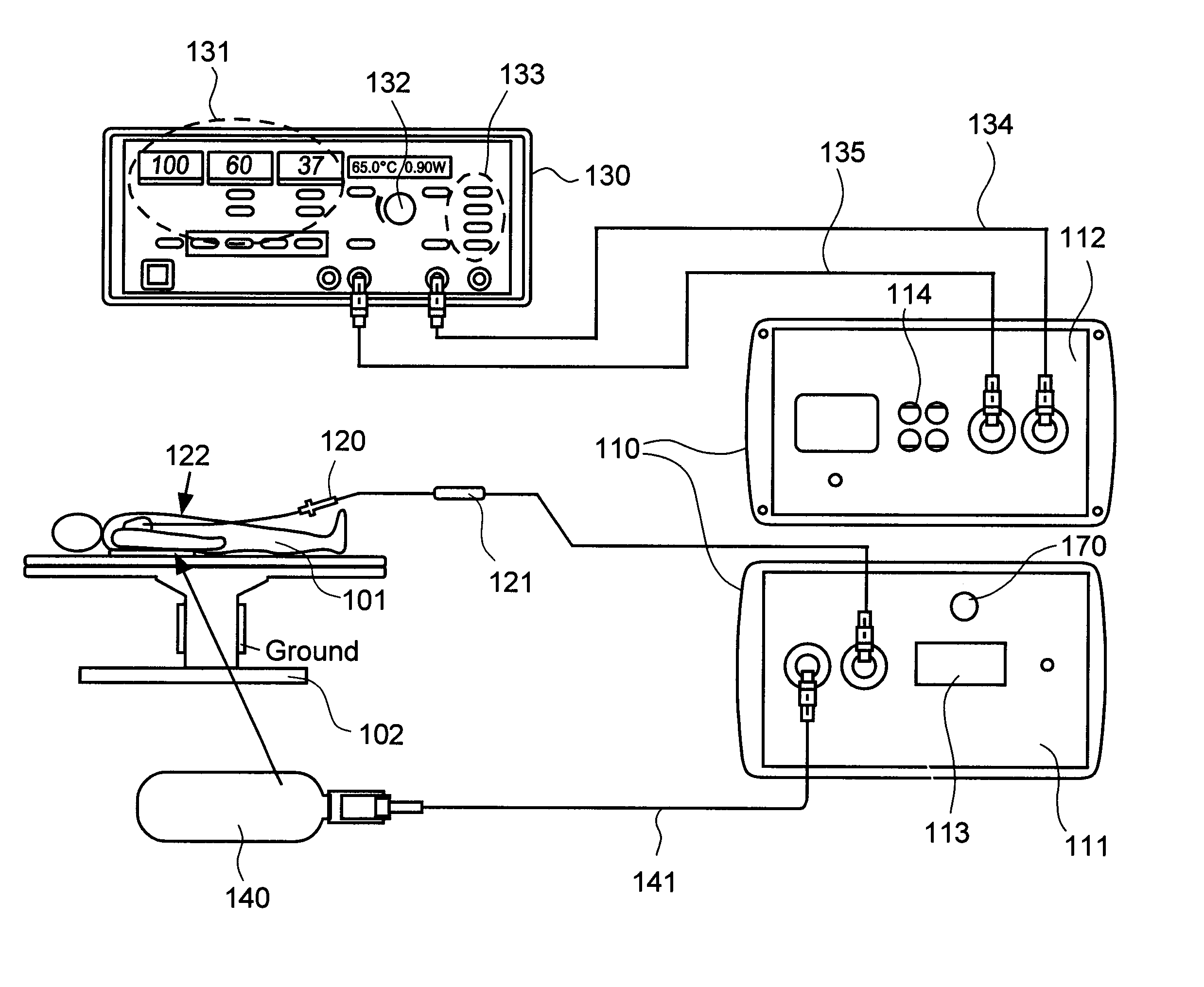
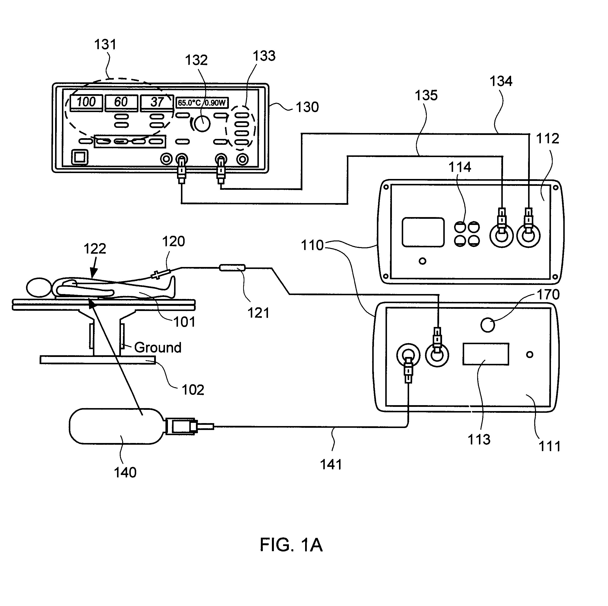
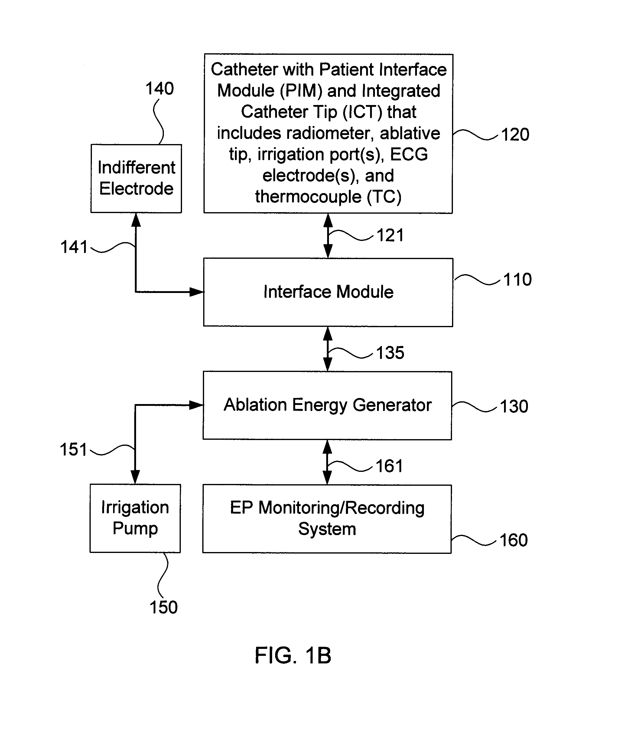
[0032]Embodiments of the present invention provide systems and methods for radiometrically measuring temperature and detecting tissue contact prior to and during ablation, in particular cardiac ablation. As noted above, commercially available systems for cardiac ablation may include thermocouples for measuring temperature, but such thermocouples may not adequately provide the clinician with information about tissue temperature or tissue contact. Thus, the clinician may need to make an “educated guess” about whether an ablative tip is in contact with tissue, as well as whether a given region of tissue has been sufficiently ablated to achieve the desired effect. By comparison, calculating a temperature based on signal(s) from a radiometer is expected to provide accurate information to the clinician about the temperature of tissue at depth, even during an irrigated procedure. Moreover, the signal(s) from the radiometer may be used to determine whether the ablative tip is in sufficient ...


PUM


Abstract

The present invention provides systems and methods for radiometrically measuring temperature and detecting tissue contact during ablation. An interface module includes a first input/output (I/O) port for receiving radiometer and thermocouple signals from an integrated catheter tip (ICT) that includes a radiometer; a second I/O port for receiving ablative energy from an electrosurgical generator; a temperature display; a patient relay; a computer-readable medium storing radiometer and thermocouple parameters and instructions for causing the processor to: calculate a temperature adjacent to the ICT based on the radiometer and thermocouple signals and the parameters; causing the temperature display to display the calculated temperature; closing the patient relay to pass ablative energy from the second to the first I/O port; determining whether the ICT is in contact with tissue based on the radiometer signal. An output device indicates whether the ICT is determined to be in contact with the tissue.

Description

FIELD OF THE INVENTION[0001]This application generally relates to systems and methods for measuring temperature and detecting tissue contact prior to and during tissue ablation.BACKGROUND OF THE INVENTION[0002]Tissue ablation may be used to treat a variety of clinical disorders. For example, tissue ablation may be used to treat cardiac arrhythmias by destroying aberrant pathways that would otherwise conduct abnormal electrical signals to the heart muscle. Several ablation techniques have been developed, including cryoablation, microwave ablation, radio frequency (RF) ablation, and high frequency ultrasound ablation. For cardiac applications, such techniques are typically performed by a clinician who introduces a catheter having an ablative tip to the endocardium via the venous vasculature, positions the ablative tip adjacent to what the clinician believes to be an appropriate region of the endocardium based on tactile feedback, mapping electrocardiogram (ECG) signals, anatomy, and / o...

Claims


Application Information

Patent Timeline
Patent Type & Authority Applications(United States)
IPC IPC(8): A61B18/18
CPCA61B5/01A61B5/150022A61B5/6852A61B5/6886A61B5/7405A61B18/12A61B18/1492A61B2017/00057A61B2018/00351A61B2018/00577A61B2018/00642A61B2018/0066A61B2018/00672A61B2018/00678A61B2018/00708A61B2018/00791A61B2018/00821A61B2018/00898A61B2018/0212A61B2018/1861A61B2090/065A61B5/287A61B5/1405A61B18/1233A61B2017/0007A61B2018/00702A61B2018/1467
Inventor MCCARTHY, JOHNLENIHAN, TIMOTHY J.KANOWSKY, ERIC R.
Owner ADVANCED CARDIAC THERAPEUTICS
Why Eureka
Try Eureka