Method for treating lower extremity varicose vein combined ultrasonic wave and microwave

a combined ultrasonic wave and microwave technology, applied in ultrasonic/sonic/infrasonic image/data processing, tomography, applications, etc., can solve the problems of difficult patient acceptance of the method, pathological changes and clinical manifestations, etc., to reduce the pain of patients, reduce the risk of bleeding, and reduce the effect of surgery

Active Publication Date: 2014-07-03
MENG KAI +1
View PDF9 Cites 2 Cited by
  • Summary
  • Abstract
  • Description
  • Claims
  • Application Information

AI Technical Summary

Benefits of technology

[0011]The present invention uses the ultrasonic imaging microwave treatment technique to treat the lower extremity varicose vein disease, which accurately, firmly and thoroughly closes the varicose blood vessel, has the exact effect, has small trauma and less pain to patients, less bleeding in surgery, quick recovery, is difficult to form

Problems solved by technology

The venous valve insufficiency, the weak venous wall and the continuously increased intravenous pressure are likely pathogenic, which results in pathological changes and clinical manifestations.
Due to large tra

Method used


Image

  • Method for treating lower extremity varicose vein combined ultrasonic wave and microwave
  • Method for treating lower extremity varicose vein combined ultrasonic wave and microwave
  • Method for treating lower extremity varicose vein combined ultrasonic wave and microwave

Examples


example 1

Ultrasonic Imaging Microwave Therapeutic Apparatus

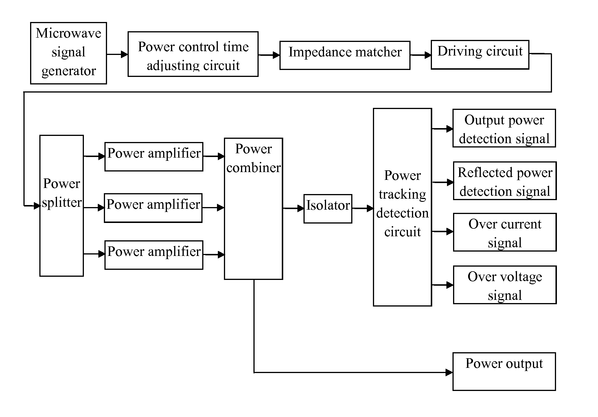
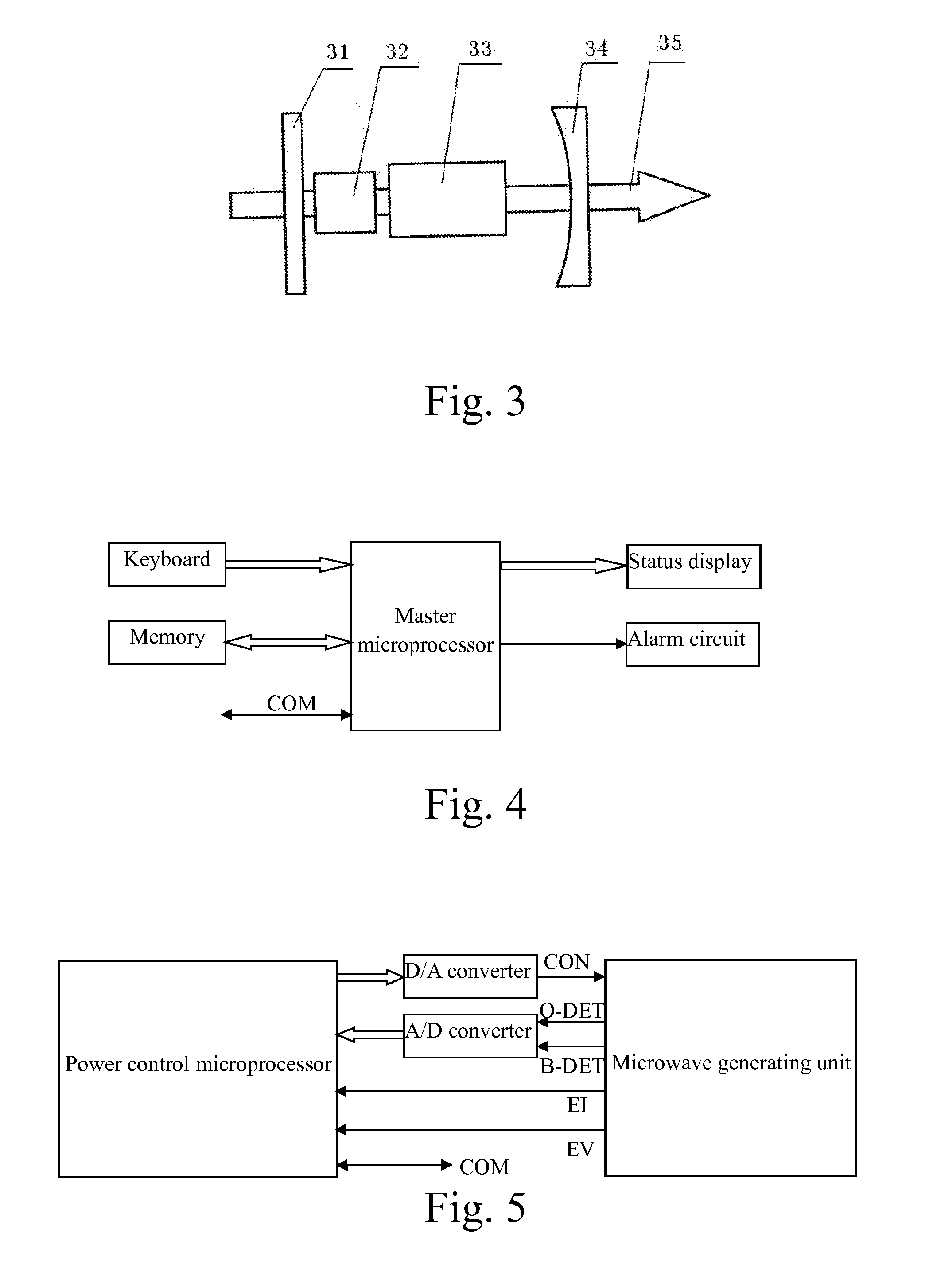
[0023]Referring to FIG. 1 of the drawings, an ultrasonic imaging microwave therapeutic apparatus according to a preferred embodiment of the present invention is illustrated, wherein the ultrasonic imaging microwave therapeutic apparatus comprises a microwave radiation probe (as shown in FIG. 7), a microwave generating unit (as shown in FIG. 2), a laser generating unit (as shown in FIG. 3), a color Doppler ultrasound imaging unit, and a microcomputer control unit (as shown in FIGS. 4-6).

[0024]The microwave radiation probe comprises a front probe part 71, a rear probe part 72 and a probe interface part. A microwave emission hole 73 is provided at the front probe part 71. A laser cursor is provided at the rear probe part 72. The rear probe part 72 of the microwave radiation probe has a hollow structure. A wire for the transmission of microwave and a cable for the transmission of laser are provided within the rear probe part 72 of the mi...

example 2

Method for Treating Lower Extremity Varicose Vein Combined Ultrasonic Wave and Microwave

embodiment 1

[0037]Aiming at treating the lower extremity superficial venous insufficiency, the treatment method of this embodiment comprises steps as follows:

[0038]1. Pre-Operative Preparation:

[0039]1.1 Patient Preparation:

[0040](1) Check the function of the heart, the lung, the liver, the kidney and other major organs;

[0041](2) Check the lower extremity deep vein graft patency and the valvular function, such as color Doppler ultrasound and venography; simultaneously, position and mark the confluence of the femoral saphenous vein;

[0042](3) Prepare skin on the preoperative day whose upper portion and the belly are flat and lower portion reaches the foot (including the perineum), wherein be careful not to involve in operating the surface skin of the varicose vein while shaving the hair, otherwise the operation should be delayed;

[0043](4) Mark the behaviors of the varicose great saphenous vein and its tributaries on the skin by methyl violet (gentian violet) or methylene blue, mark the incision si...


PUM


Abstract

The method for treating the lower extremity varicose vein combined ultrasonic wave and microwave of the present invention includes steps of firstly finding the diseased vein by the ultrasonic imaging equipment; and then directly effecting the microwave treatment catheter/needle on the diseased vein; by the concentric thermal coagulation release effect of the microwave to the tissue, instantly generating high temperature with the certain penetration range in a small range for tissue coagulation; and then gradually making the vascular fibrosis; finally making the completely atresia, thus achieving the therapeutic aim. The present invention uses the ultrasonic imaging microwave treatment technique to treat the lower extremity varicose vein disease, which accurately, firmly and thoroughly closes the varicose blood vessel, has the exact effect, small trauma and less pain to patients, less bleeding in surgery, quick recovery, simple operative procedure and no significant complication, is difficult to form the deep venous thrombosis.

Description

CROSS-REFERENCE TO RELATED APPLICATION[0001]This is a Continuation-In-Part application of U.S. patent application Ser. No. 13 / 726,037, filed on Dec. 22, 2012.BACKGROUND OF THE PRESENT INVENTION[0002]1. Field of Invention[0003]The present invention relates to a treatment technique for the body's thermal ablation, and more particularly to a method for treating the lower extremity varicose vein combined ultrasonic wave and microwave.[0004]2. Description of Related Arts[0005]In the medical vascular surgery field, the lower extremity varicose vein is also called as the lower extremity vein insufficiency, and is a common seen and frequently occurring disease with high incidence. Its main clinical manifestations are soreness, numbness, itching, sense of heaviness and tiredness from the affected limb. While the patient standing, the superficial vein of the affected limb swells, expands, detours and even curls into a group. The severe patient will be complicated with thrombophlebitis; and th...

Claims


Application Information

Patent Timeline
IPC IPC(8): A61B18/18A61B8/08
CPCA61B8/488A61B18/1815A61B8/13A61B8/463A61B8/54A61B8/546A61B2018/00404A61B2018/00589A61B2018/00982A61B2018/00994A61N5/04
Inventor MENG, KAIMENG, QINGYIHUANG, JINXIUSUN, JIEWANG, XIAOPING
Owner MENG KAI
Why Eureka
Try Eureka