
Combination/adjuvant therapy for wt-1-positive disease

a technology for wt-1 and wt-1, which is applied in the field of treatment of wt1positive diseases, can solve the problems of inaccessible classical antibody therapy, limited clinical efficacy of some tkis, for example, imatinib and sunitinib, and achieve the effects of improving anti-tumor response, early inhibition of tumor growth, and maintaining the therapeutic efficacy of tki

Inactive Publication Date: 2014-09-18
MEMORIAL SLOAN KETTERING CANCER CENT
View PDF2 Cites 22 Cited by
  • Summary
  • Abstract
  • Description
  • Claims
  • Application Information

AI Technical Summary

Benefits of technology

The present patent provides a method for treating WT-1 positive diseases by combining a tyrosine kinase inhibitor (TKI) and an anti-WT-1 antibody. This approach offers improved efficacy and reduced side effects compared to using either TKI or anti-WT-1 antibody alone. The combination therapy can be used against various WT-1 positive cancers such as chronic myelogenous leukemia, multiple myeloma, acute lymphoblastic leukemia, acute myeloid / myelogenous leukemia, myelodysplastic syndrome, ovarian cancer, gastrointestinal cancers, breast cancer, prostate cancer, glioblastoma, gastrointestinal stromals tumors, and others. The anti-WT-1 antibody can be fully human and specifically binds to a WT-1 peptide in an HLA restricted manner with high affinity. The combination therapy can also induce antibody dependent cellular cytotoxicity (ADCC) against WT-1-positive cells.

Problems solved by technology

However, the clinical efficacy of some TKIs, for example, imatinib and sunitinib, are limited by rare patient-specific intolerance to the drug or the development of treatment-refractory disease.
WT-1, however, is a nuclear protein and, therefore, is inaccessible to classical antibody therapy.

Method used


Image

  • Combination/adjuvant therapy for wt-1-positive disease
  • Combination/adjuvant therapy for wt-1-positive disease
  • Combination/adjuvant therapy for wt-1-positive disease

Examples


example 1

Materials and Methods

[0104]Cell Samples, Cell Lines and Antibodies.

[0105]After informed consent on Memorial Sloan-Kettering Cancer Center Institutional Review Board approved protocols, peripheral blood mononuclear cells (PBMC) from HLA-typed healthy donors and patients were obtained by Ficoll density centrifugation. All cells were HLA typed by the Department of Cellular Immunology at Memorial Sloan-Kettering Cancer Center. Leukemia cell line, BV173, (WT-1+, A0201+) was kindly provided by Dr. H. J. Stauss (University College London, London, United Kingdom). The cell lines were cultured in RPMI 1640 supplemented with 5% FCS, penicillin, streptomycin, 2 mmol / L glutamine, and 2-mercaptoethanol at 37° C. / 5% CO2.

[0106]Animals.

[0107]Six to eight week-old male NOD.Cg-Prkdc scid IL2rgtm1WjI / SzJ mice, known as NOD / SCID gamma (NSG), were purchased from the Jackson Laboratory (Bar Harbor, Me.) or obtained from MSKCC animal breeding facility.

[0108]Transduction and Selection of Luciferase / GFP Pos...

example 2

Antibody-Dependent Cellular Cytotoxicity (ADCC)

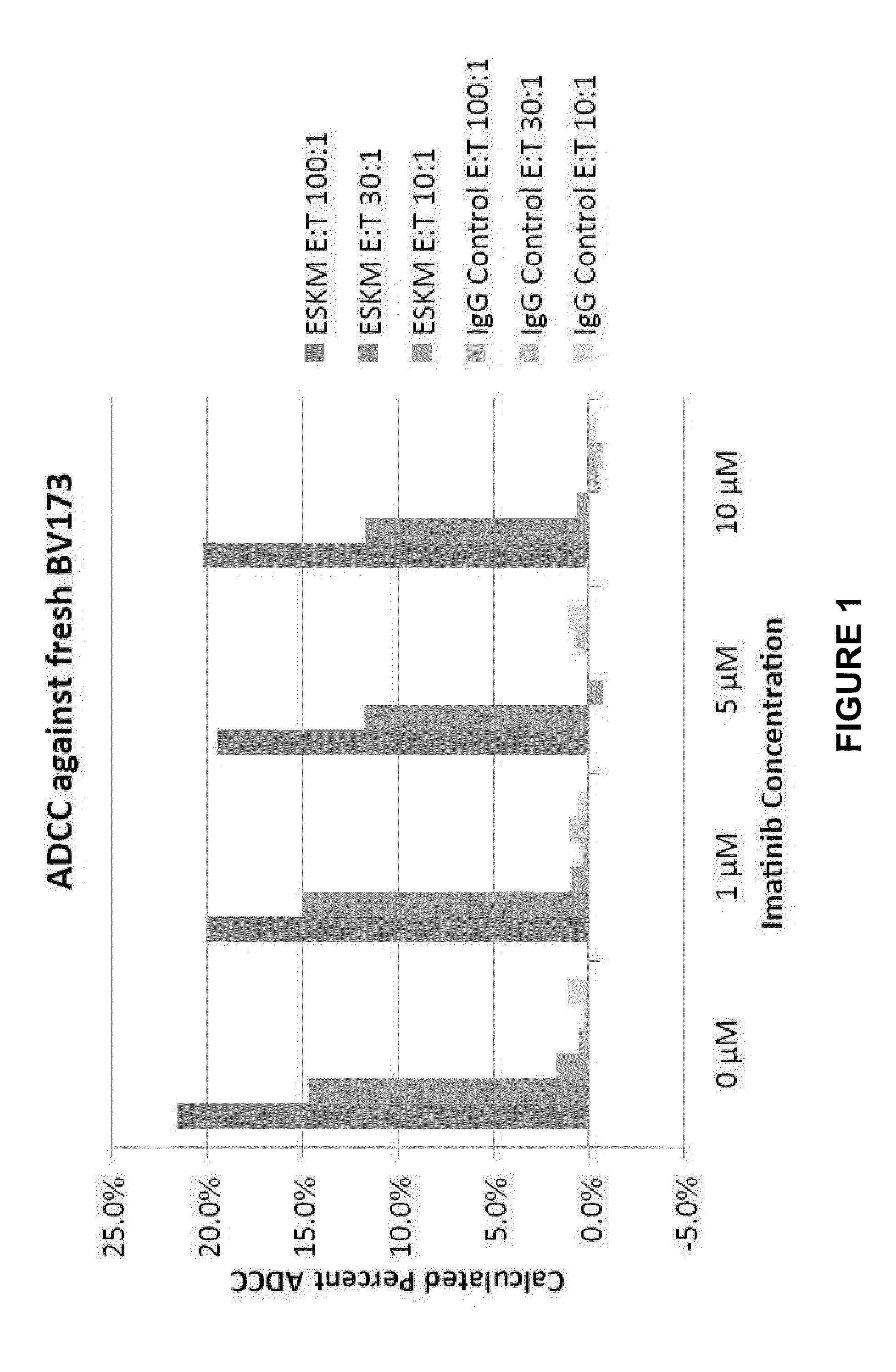
[0110]ADCC is considered to be one of the major effector mechanisms of therapeutic mAb in humans. Evaluation of efficacy, therefore, begins with in vitro experiments measuring ADCC against BV173 cell line, derived from CML in blastic crisis. Fresh BV173 cells were used for ADCC target cells. WT-1 antibody or its isotype control human IgG1 was incubated at 750 ng / ml with target cells and fresh PBMCs at different effector:target (E:T) ratio for 6 hrs. Imatinib was added at concentrations of 0, 1, 5, and 10 μM. The supernatants were harvested and the cytotoxicity was measured by standard chromium 51 release assay.

[0111]In the presence of human PBMC, WT-1 antibody mediated dose-dependent PBMC ADCC against naturally presented RMF epitope by HLA-A0201 molecule on tumor cells, the leukemia cell line BV173. Importantly, WT-1 antibody was able to mediate ADCC in the presence of various doses of imatinib. The killing was consistently observed at ...

example 3

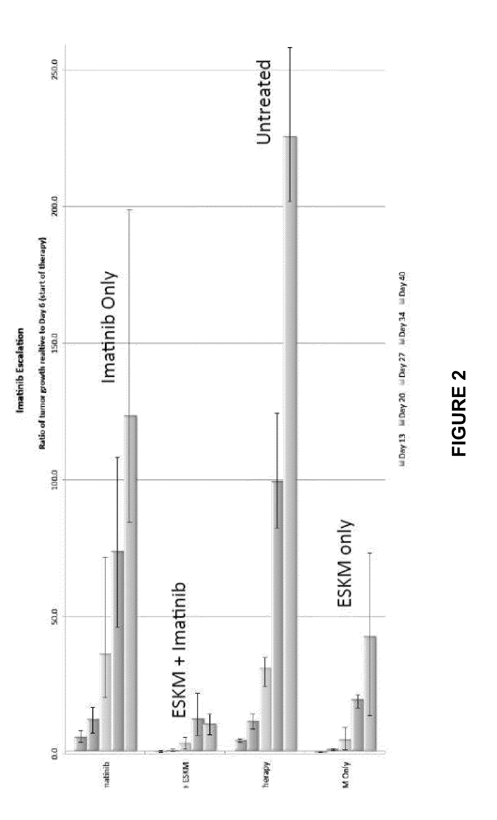
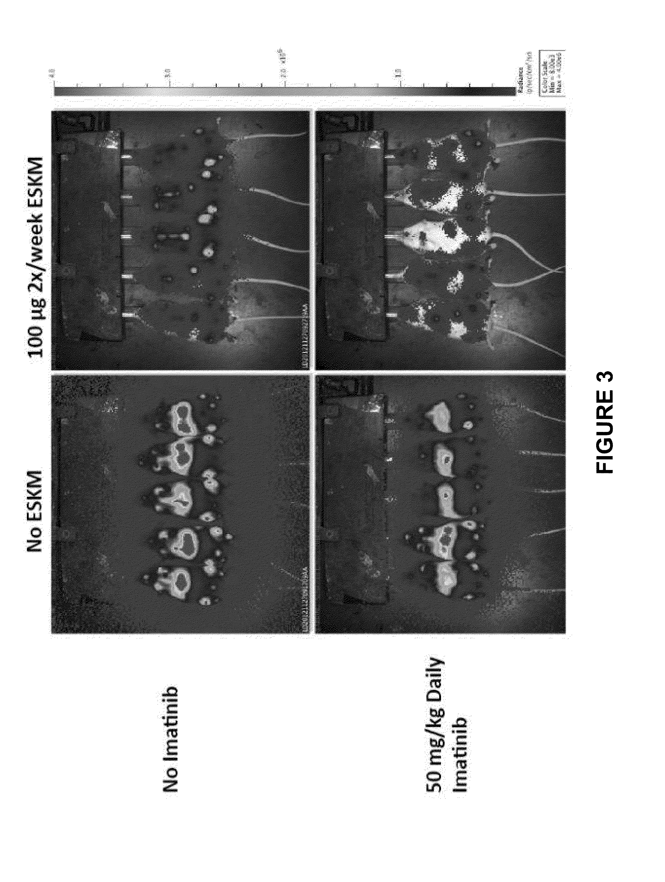
[0112]In vivo efficacy of ESKM with TKIs was evaluated using NSG mice injected with HLA-A0201+ leukemic cell line BV173. The protocol used for imatinib and dasatinib therapy in combination with ESKM consisted of injecting 3×10⁶ cells per mouse via tail vein, luciferin imaging 6 days after injection to assess tumor engraftment, and initiation of therapy immediately after imaging on day 6. Luciferin imaging was used weekly to monitor tumor growth. The TKI is injected intraperitoneally daily (50 mg / kg for imatinib and 20-40 mg / kg for dasatinib.) The antibody is injected intravenously twice per week.


PUM

PropertyMeasurementUnit
Cytotoxicityaaaaaaaaaa

Abstract

In an attempt to improve primary disease responsiveness and / or to overcome resistant disease, the present disclosure provides a method for treating or inhibiting the proliferation of a WT-1-dependent cancer comprising providing to a subject in need thereof a therapeutically effective amount of a tyrosine kinase inhibitor along with an anti-WT-1 / HLA antibody, that is, an antibody that specifically binds to a peptide of Wilms' tumor protein (WT-1) presented on the surface of the cancer cells in an HLA-restricted fashion.

Description

CROSS-REFERENCE TO RELATED APPLICATIONS[0001]This application contains subject matter that is related to the subject matter of commonly-assigned PCT international application serial no. PCT / US2012 / 031892 filed on Apr. 2, 2012 entitled “Antibodies to Cytosolic Peptides” (Docket No. 3314.013AWO), and commonly assigned, co-filed U.S. provisional application No. ______, entitled “Antibodies to Cytosolic Peptides” (Docket No. 3314.030P); the contents of each are hereby incorporated herein by reference in their entirety.STATEMENT OF RIGHTS UNDER FEDERALLY-SPONSORED RESEARCH[0002]This invention was made with government support under grant NIH R01 CA 55349 and P01 CA 23766 awarded by the U.S. National Institutes of Health. The government has certain rights in the invention.SEQUENCE LISTING[0003]This application contains a Sequence Listing, created on Mar. 14, 2012; the file, in ASCII format, is designated 3314031P_Sequence Listing_ST25.txt and is 177 KB. The file is hereby incorporated by r...

Claims


Application Information

Patent Timeline
IPC IPC(8): A61K39/395A61K31/506
CPCA61K31/506A61K39/39558A61K45/06A61K2039/505C07K16/2833C07K16/30C07K2317/32C07K2317/732A61K2300/00
Inventor SCHEINBERG, DAVIDDUBROVSKY, LEONID
Owner MEMORIAL SLOAN KETTERING CANCER CENT
Learn More