Methods and apparatus for stimulating and recording neural activity

a neural activity and neural recording technology, applied in the field of neural activity stimulation and recording methods and apparatuses, can solve the problems of neuronal death, bleeding, neuronal scars, etc., and achieve the effect of reducing the signal-to-noise ratio (snr) and reducing the quality of neural recording

Active Publication Date: 2014-12-18
MASSACHUSETTS INST OF TECH
View PDF13 Cites 34 Cited by
  • Summary
  • Abstract
  • Description
  • Claims
  • Application Information

AI Technical Summary

Benefits of technology

[0003]The Inventors have recognized that conventional technologies for in vivo neural recording and stimulation suffer from several drawbacks. More specifically, conventional devices for electrophysiological recording on the single cell level have elastic moduli that are much higher than the elastic modulus of neural tissue (e.g., tens to hundreds of gigapascals versus kilopascals to megapascals). In other words, conventional electrophysiological recording devices tend to be much stiffer than neural tissue. Because they are stiffer than neural tissue, conventional electrophysiological recording devices may damage tissue during and after implantation in neural tissue. The tissue damage causes the formation of glial scars, neuronal death, and bleeding, all of which contribute to a progressive decrease in the quality of the neural recording (e.g., lower signal-to-noise ratio (SNR), fewer monitored cells). As a result, with a conventional electrophysiological recording devices, it may become impossible to record single neuron activity reliably within several months of implantation.

Problems solved by technology

Because they are stiffer than neural tissue, conventional electrophysiological recording devices may damage tissue during and after implantation in neural tissue.
The tissue damage causes the formation of glial scars, neuronal death, and bleeding, all of which contribute to a progressive decrease in the quality of the neural recording (e.g., lower signal-to-noise ratio (SNR), fewer monitored cells).
As a result, with a conventional electrophysiological recording devices, it may become impossible to record single neuron activity reliably within several months of implantation.
It is also difficult to integrate other devices with conventional electrophysiological recording devices.
For example, conventional electrophysiological recording devices do not allow for straightforward incorporation of other elements that may facilitate neural stimulation functions, such as optical waveguides (e.g., for optogenetic experiments or imaging) and hollow channels (e.g., for drug delivery and fluid sampling).
Thus, drawbacks including excessive stiffness and limited engineering design options limit the usefulness of conventional neural recording device for treating disease and conducting basic scientific studies of neural circuits.

Method used


Image

  • Methods and apparatus for stimulating and recording neural activity
  • Methods and apparatus for stimulating and recording neural activity
  • Methods and apparatus for stimulating and recording neural activity

Examples


Embodiment Construction

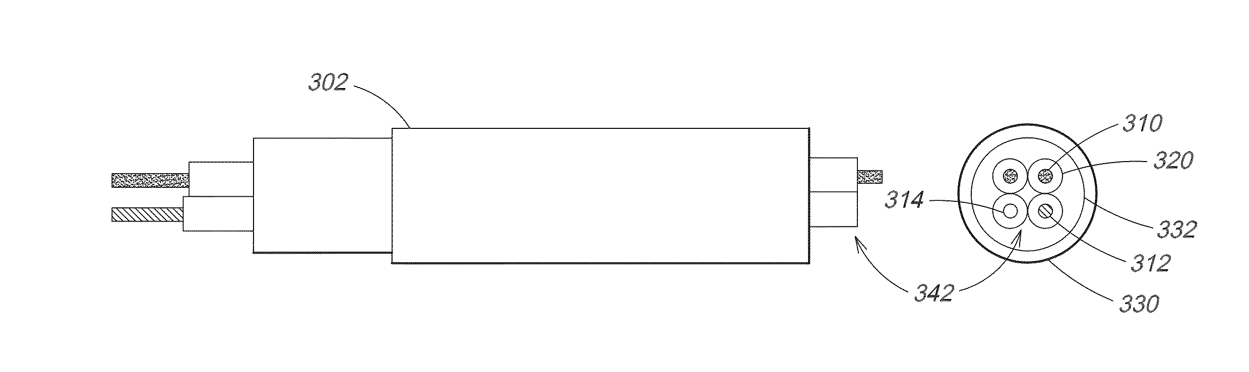
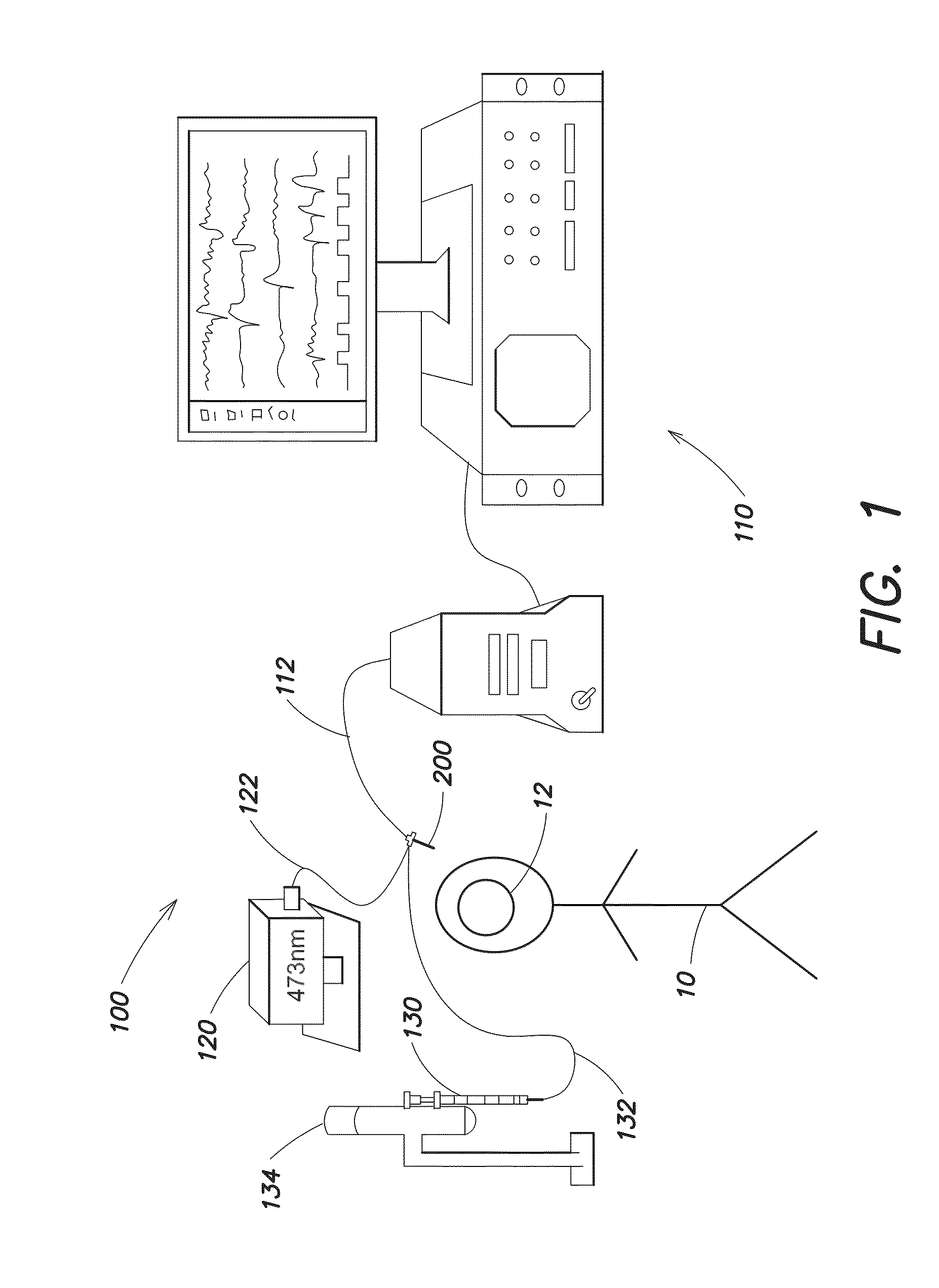
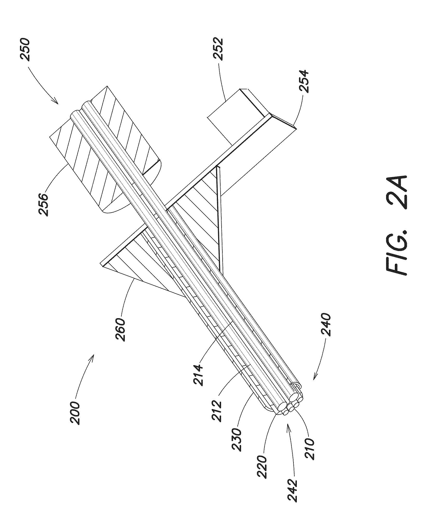
[0051]Embodiments of the present invention include fiber-inspired probes for stimulating and recording neural activity using electrical, optical, and pharmacological interrogation. Exemplary neural probes can be implanted in human patients and animal models to assess and modulate neural activity in the context of diseases such as epilepsy, Parkinson's, Alzheimer's, major depressive disorder, autism spectrum disorders, substance addiction, post-traumatic stress disorder, and traumatic brain injury. They can also be used to research basic principles of brain function, including but not limited to learning and memory processes. Furthermore, an exemplary neural probe can be used in a brain-machine interface to restore motor and cognitive function lost due to limb amputation, locked-in syndrome, paraplegia, or quadriplegia resulting from spinal cord or peripheral nerve injury.

[0052]An exemplary neural probe can be made using a thermal drawing process similar to the drawing processes used...


PUM


Abstract

Thermal drawing processes can be used to make multifunctional, high-resolution neural probes for neural recording and stimulation. An exemplary neural probe may include one or more conductive fibers or microelectrodes coated with two or more layers of insulating material, at least one of which is partially etched to expose a tip at the neural probe's distal end. The conductive fibers conduct electrical signals (e.g., neural spikes or electrical stimulation) between the tip and the neural probe's proximal end. Optional optical and fluidic waveguides may guide light and fluid, respectively, between the tip and the proximal end. A neural probe may be flexible enough for long-term (chronic) implantation in neural tissue (e.g., the brain) without excessive tissue damage, even during movement of the brain in the skull. The probe may be made from biocompatible materials, such as insulating and conductive polymers, that have negligible (insignificant) interaction with the surrounding tissue.

Description

GOVERNMENT SUPPORT[0001]This invention was made with government support under Grant No. DMR-0819762 awarded by the National Science Foundation. The government has certain rights in this invention.BACKGROUND[0002]Neural probes are used to record and stimulate neural activity in vivo in neural tissue, including the brain. As understood by those of skill in the art, stimulating a neuron causes a brief change in the neuron's electrical membrane potential. The change in membrane potential, also called a nerve impulse or spike, plays an important role in cell-to-cell communication and can be detected using electrodes inserted or implanted in the neural tissue. Relatively low-impedance (e.g., less than about 150 kΩ) electrodes can be used to record the local field potential (LFP), which is the low-frequency component of the extracellular voltage detected in the cortex. Electrodes with higher impedance (e.g., about 150 kΩ to about 10 MΩ) can be used to record the unit potential, which is th...

Claims


Application Information

Patent Timeline
Patent Type & Authority Applications(United States)
IPC IPC(8): A61N1/05A61M37/00A61B5/04A61N5/06
CPCA61N1/0551A61N5/0601A61M37/00A61M2202/09A61N2005/063A61M2207/00A61B5/04001A61B5/0084A61B5/4064A61N1/0529A61N5/0622A61N2005/0662A61B5/24A61B5/6868
Inventor ANIKEEVA, POLINA OLEGOVNAJIA, XIAOTINGLU, CHICANALES, ANDRESFRORIEP, ULRICH PAULTRINGIDES, CHRISTINA MYRAFINK, YOEL
Owner MASSACHUSETTS INST OF TECH
Why Eureka
Try Eureka