
Cationic Liposomal Drug Delivery System for Specific Targeting of Human CD14+ Monocytes in Whole Blood
a cd14+ monocyte and cationic liposome technology, applied in the field of liposomes, can solve problems such as adverse side effects of anti-inflammatory drugs given systemically
- Summary
- Abstract
- Description
- Claims
- Application Information
AI Technical Summary
Benefits of technology
Problems solved by technology
Method used
Image

Examples
example 1
Liposome Preparation
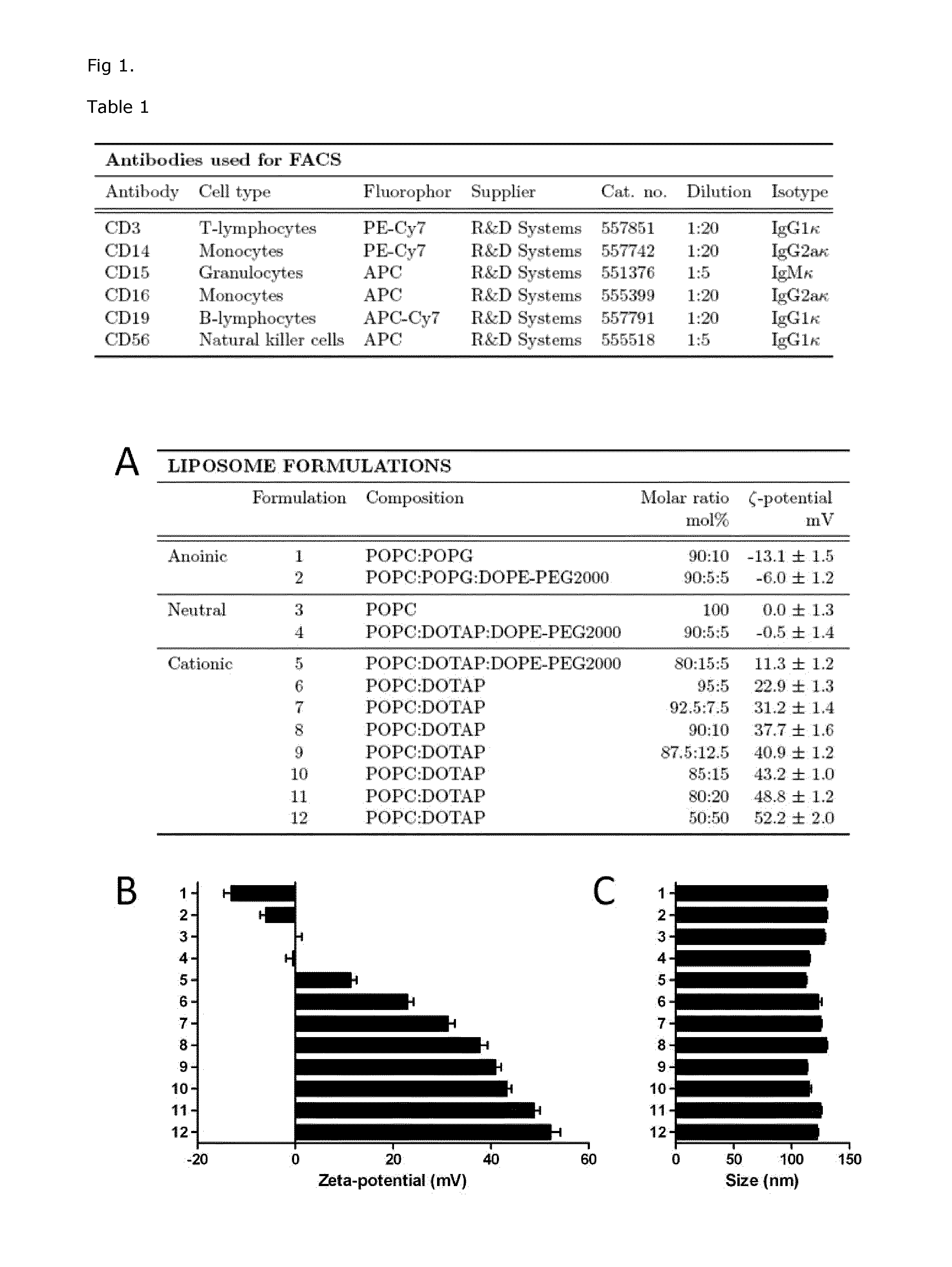
[0133]Unilamellar fully hydrated liposomes were made from mixtures of 1-palmitoyl-2-oleoyl-sn-glycero-3-phosphocholine (POPC), 1-palmitoyl-2-oleoyl-sn-glycero-3-phosphoglycerol (POPG), 1,2-dioleoyl-3-trimethylammonium-propane (DOTAP) and 1,2-dioleoyl-sn-glycero-3-phosphoethanolamine-N-[methoxy(polyethylene glycol)-2000] (DOPE-PEG2000). As a fluorescence marker to measure presence of liposomes in biological systems, 0.5% 1,2-dioleoyl-sn-glycero-3-phosphoethanolamine-N-rhodamine (DOPE-RhB) was mixed with the lipids as a tracer. The molar ratios of each lipid in the liposomes are outlined in FIG. 1A. All lipids were all obtained from Avanti Polar lipids. Briefly, appropriate weighed amounts of POPC, POPG, DOTAP and DOPE-PEG2000 were dissolved in chloroform. The solvent was removed by a gentle stream of N2 and the lipid films were dried overnight under low pressure to remove trace amounts of solvent. Multilamellar vesicles were prepared by dispersing the dried lipids...
example 2
Characterization of Liposome Size and Surface Charge Dependent on Composition
[0134]Liposomes prepared as outlined in FIG. 1A were prepared with the attempt to design liposomes with ability to be recognized and taken up by monocytes but not other cells in the blood. We designed liposomes with 0-50% net positive charge (formulation 6-12 in FIG. 1A), together with control liposomes with negative or nearly neutral charge (formulation 1-5 in FIG. 1A). The liposomes were prepared as described in example 1, and their size measured in nanometer (nm) by dynamic light scattering on a ZetaPALS zeta potential analyzer from Brookhaven Instruments in a buffer consisting of 300 mM glucose, 10 mM HEPES, 1 mM CaCl2 in MilliQ water, pH 7.4. The liposomes showed sizes between 110±20 nm in diameter (FIG. 1B). The surface charge (Zeta potential) of the liposomes was measured in mV and showed surface charge dependent on lipid composition (FIG. 1C). Addition of the negatively charged POPG (10 molar percen...
example 3
Liposome Targeting to Monocytes Dependent on Liposome Composition
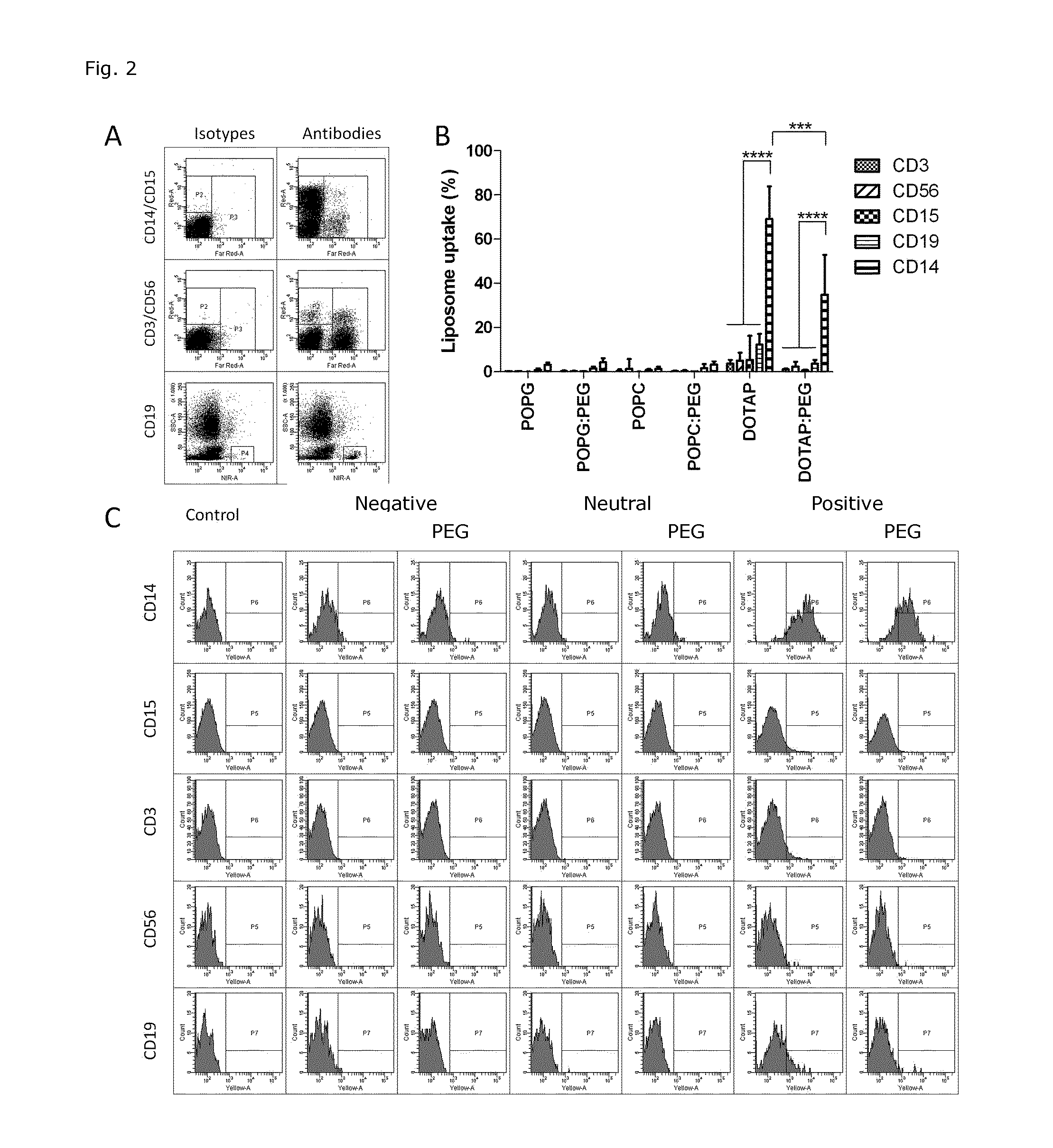
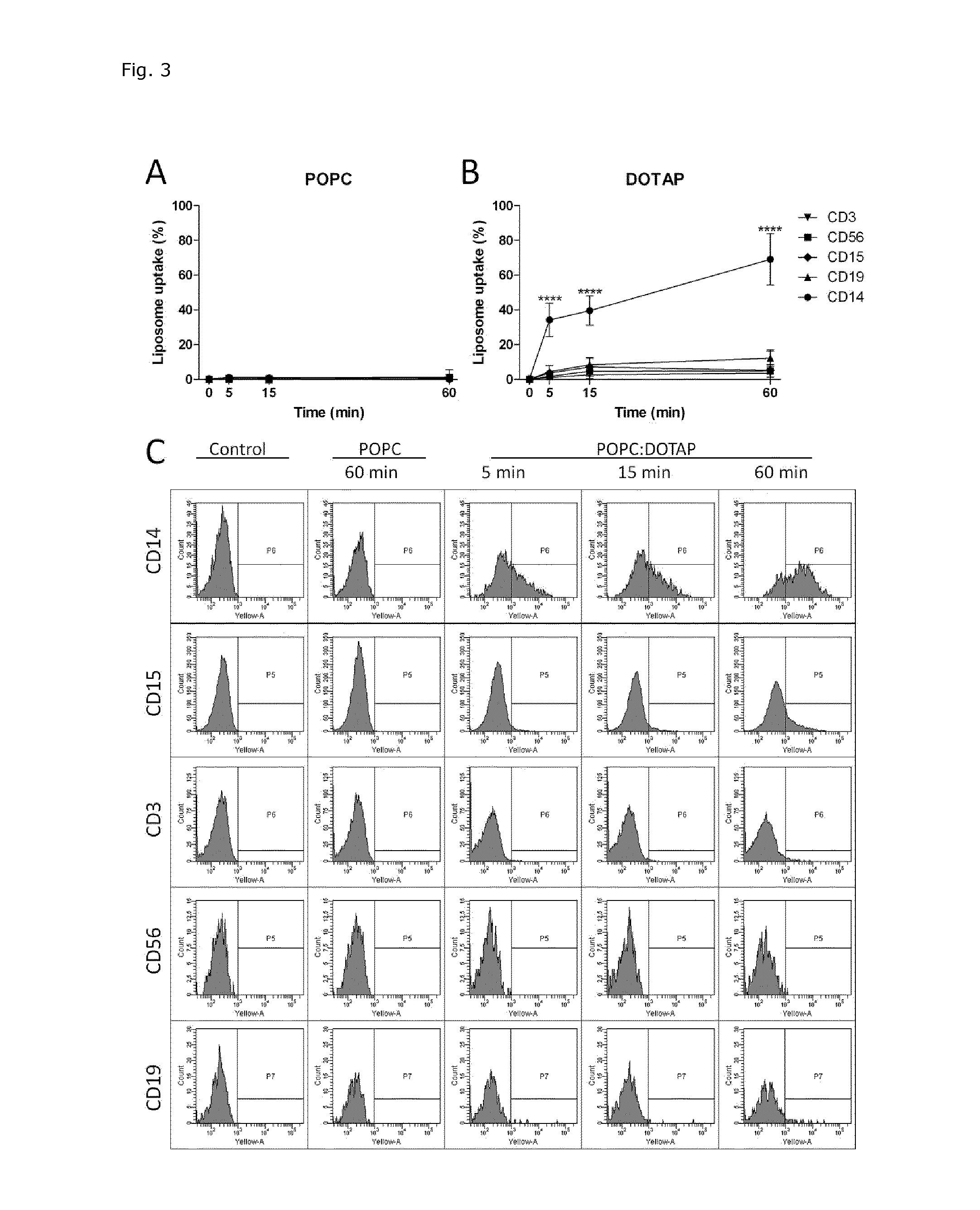
[0135]The cellular uptake of modified POPC liposome formulations was determined based on fluorescence of RhB incorporated into the liposomal membrane. The total amount of liposome associated with cells (indicated as ‘uptake’ and include cell membrane bound liposomes and liposomes already internalized) was estimated using excitation at 532 nm and emission at 564-606 nm. Liposomal uptake in five different cell populations in whole blood was analyzed.
[0136]The following markers were used to distinguish the different populations: CD14 (monocytic marker), CD15 (granulocytic marker), CD3 (T-lymphocytic marker), CD19 (B-lymphocytic marker), and CD56 (natural killer cell marker). Whole blood was obtained from healthy volunteers by standard methods in BD Vacutainer containing ethylenediaminetetraacetic acid (EDTA, 366450). Briefly, 10 μl liposome preparations (formulation 1-5 and 8 in FIG. 1A) were added to 200 μl fresh whole b...
PUM
| Property | Measurement | Unit |
|---|---|---|
| diameter | aaaaa | aaaaa |
| diameter | aaaaa | aaaaa |
| diameter | aaaaa | aaaaa |
Abstract
Description
Claims
Application Information