Contrast-Enhancing Agent for Nuclear Magnetic Resonance Imaging Comprising Melanin Nanoparticles Stably Dispersed in Water

Active Publication Date: 2015-05-21
MELANIS CO LTD
View PDF1 Cites 4 Cited by
  • Summary
  • Abstract
  • Description
  • Claims
  • Application Information

AI Technical Summary

Benefits of technology

[0043]The contrast agent for nuclear magnetic resonance imaging according to the present invention contains melanin nanoparticles having a uniform. shape and size, thereby providing good dispersibility i

Problems solved by technology
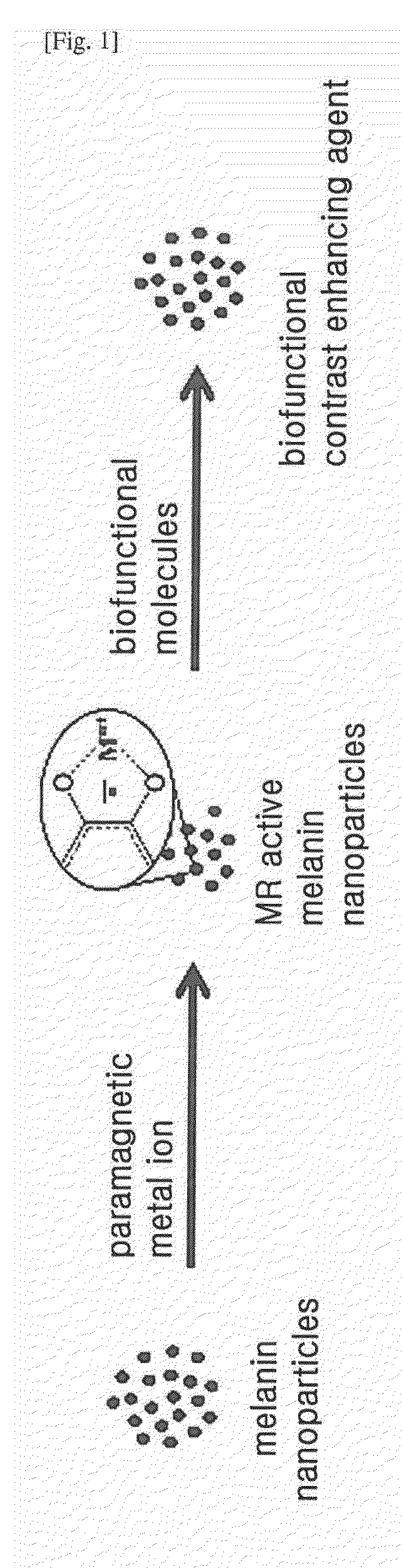
However, the gadolinium (Gd) agent itself is highly toxic, and therefore, its safety must be improved it the form of chelates.
However, it is difficult to obtain pure melanins, because many biological materials such as proteins other than melanins are also obtained when obtained from natural sources.
In addition, even though the biological materials are removed by purification, melanin particles themselves ar

Method used


Image

  • Contrast-Enhancing Agent for Nuclear Magnetic Resonance Imaging Comprising Melanin Nanoparticles Stably Dispersed in Water
  • Contrast-Enhancing Agent for Nuclear Magnetic Resonance Imaging Comprising Melanin Nanoparticles Stably Dispersed in Water
  • Contrast-Enhancing Agent for Nuclear Magnetic Resonance Imaging Comprising Melanin Nanoparticles Stably Dispersed in Water

Examples


example 1

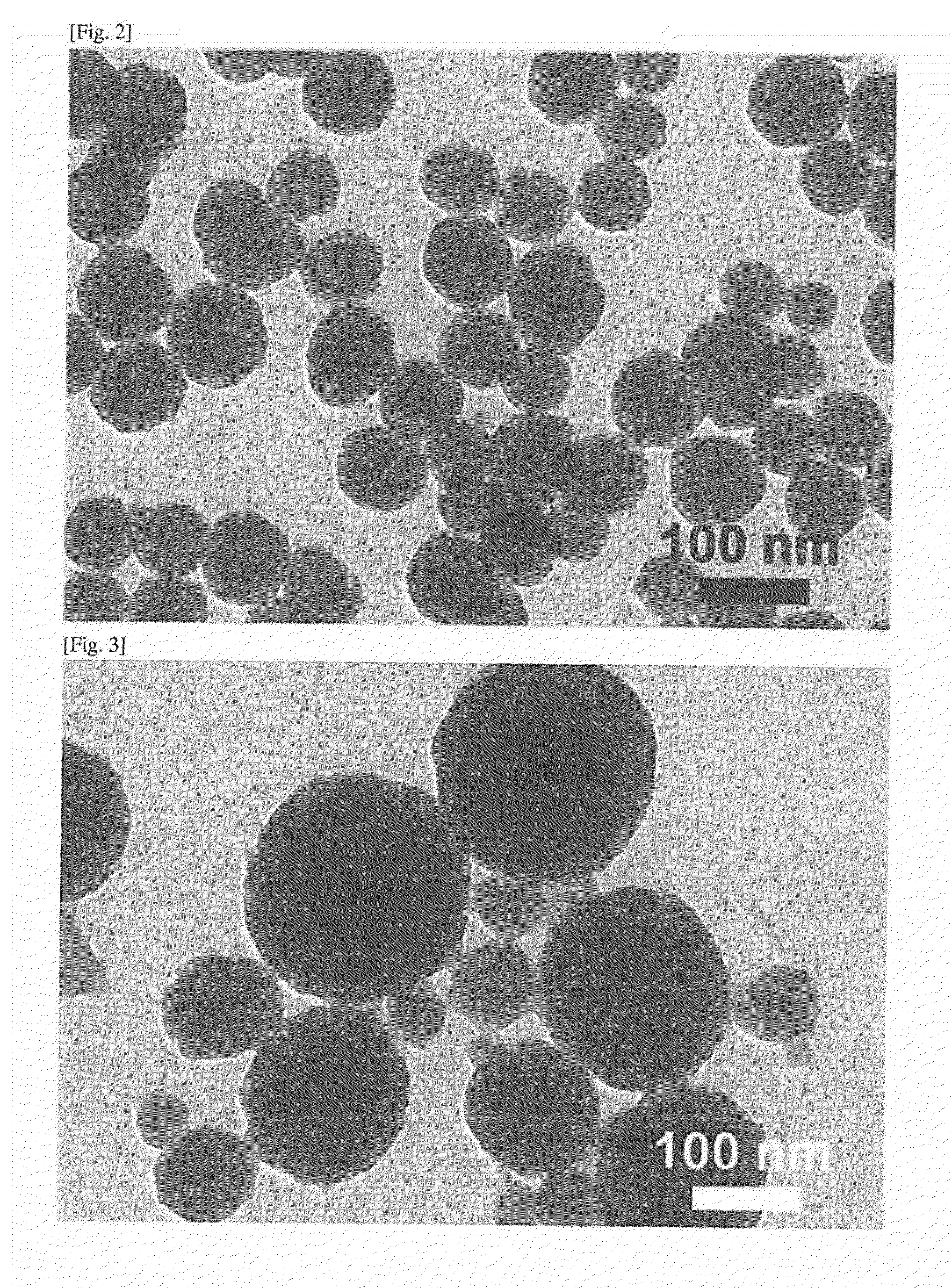
Preparation of Melanin Nanoparticles

[0058]1) Example 1-1 (MelNPs)

[0059]180 mg of dopamine hydrochloride (Aldrich Chemical) was dissolved in 90 mL of deionized water. 760 μl of 1 N NaOH solution was added to the dopamine hydrochloride solution at 50° C. with vigorous stirring. When NaOH was added, the solution immediately changed to light yellow, and gradually to deep brown. After reaction for 5 hours, melanin nanoparticles were recovered by centrifugation (18,000 rpm), and washed with deionized water several times. Precipitates were removed after low speed centrifugation (4000 rpm), and then melanin nanoparticles were stored as a dispersibility solution.

[0060]2) Example 1-2 (MelNPs)

[0061]Melanin nanoparticles were prepared in the same manner as in Example 1-1, except that the addition amount, of NaOH was from 400 to 950 μl, the amount of deionized water was from 45 to 180 mL, and the reaction temperature was from 20 to 70° C.

[0062]In detail, 180 mg of dopamine hydrochloride was diss...

example 2

Preparation of Paramagnetic Metal Ion-Coordinated Melanin Nanoparticles

[0069]1) Example 2-1 (Fe3+-MelNPs)

[0070]100 μl of Fe3+ solution (1 mg / mL) was added to 10 ml of melanin nanoparticles solution (1 mg / mL) prepared in Example 1-1 with vigorous stirring. After 3 hours, Fe3+-coordinated melanin nanoparticles were recovered by centrifugation (19,000 rpm), and the supernatant was fettered using a membrane filter (0.45μm pore size) and Fe3+ concentration was measured by ICP-AES to calculate the amount of Fe3+ bound to the melanin nanoparticles.

[0071]The Fe3+-coordinated melanin nanoparticles thus recovered were washed with deionized water several times, and diluted and stored.

[0072]2) Example 2-2 (Fe3+-MalNPs)

[0073]Fe3+-coordinated melanin nanoparticles were prepared in the same manner as in Example 2-1, except that the melanin nanoparticles prepared in Example 1-2 were used instead of the melanin, nanoparticles prepared in Example 1-1.

[0074]2) Example 2-3 (Fe3+-Sepia Melanin)

[0075]Fe3...

example 3

Preparation of PEGylated Paramagnetic Metal Ion-Coordinated Melanin Nanoparticles (PEGylated Fe3+-MelNPs)

[0079]150 mg of methoxy-poly(ethylene glycol)thiol (mPEG-SH; 2 kDa; SunBio (Korea)) was added, to 10 mL of Fe3+-coordinated melanin nanoparticles solution (1 mg / mL) prepared in Example 2-1, and NH4OH solution (28 wt %) was added to adjust the pH of the solution to approximately 10.3. After stirring for 1 hour. surface-modified melanin nanoparticles were recovered by centrifugation (18,000 rpm), and washed with deionized water several times using redispersibility / centrifugation processes so as to prepare PEGylated Fe3+-coordinated melanin nanoparticles.

[0080]The TEM images and FT-IR spectra of the prepared melanin nanoparticles were measured and shown in FIGS. 9A and 9B, respectively. Further, the dispersibility of the prepared melanin nanoparticles was observed with the naked eye (FIG. 9C), and plots of T1 excitation time vs Fe3+ concentration were obtained (FIG. 9D).


PUM

PropertyMeasurementUnit
Diameteraaaaaaaaaa
Diameteraaaaaaaaaa
Sizeaaaaaaaaaa

Abstract

The present invention relates to a contrast agent for nuclear magnetic resonance imaging, and more particularly, to a contrast agent for nuclear magnetic resonance imaging containing melanin nanoparticles having a uniform shape and size, thereby providing good dispersibility in water, no cell toxicity, and a long retention time in vivo.

Description

TECHNICAL FIELD[0001]The present invention relates to a contrast agent for nuclear magnetic resonance imaging, which has good dispersibility in water, no cytotoxicity, and a long retention time in vivo, thereby being usefully applied as an MRI contrast agent.BACKGROUND ART[0002]Magnetic resonance imaging (MRI) is a rapidly developing diagnostic imaging modality, which detects magnetic resonance signals emitted from water molecules after irradiation of low electromagnetic energy into a sample. Magnetic resonance imaging allows repeatable acquisitions of non-invasive magnetic resonance images to study tissue anatomy with high resolution for a short time, and therefore, it is known as the most suitable method for diagnosing a patient's disease and monitoring drug treatment. Signal intensity of magnetic resonance imaging is determined by two time parameters, which are T1 and T2 relaxation time and by proton density of water molecule from spin density. With regard to the contrast of magn...

Claims


Application Information

Patent Timeline
IPC IPC(8): A61K49/18A61K49/12A61K49/16A61K49/10
CPCA61K49/1818A61K49/16A61K49/126A61K49/106A61K49/103A61K49/186
Inventor LEE, JIN KYUJU, KUK YOUNLEE, JUNG HEE
Owner MELANIS CO LTD
Why Eureka
Try Eureka