Methods and kits for reversible adhesion of implants to an eye sclera

a technology of implant and eye sclera, which is applied in the direction of radiation therapy, medical devices, enzymes, etc., can solve the problems of affecting the effect of sclera, and difficulty in finding and removing sutures, so as to achieve accurate placement of implants, no tilting, and easy removal of implants

Inactive Publication Date: 2015-07-02
TEL HASHOMER MEDICAL RES INFRASTRUCTURE & SERVICES
View PDF3 Cites 4 Cited by
  • Summary
  • Abstract
  • Description
  • Claims
  • Application Information

AI Technical Summary

Benefits of technology

[0014]According to some embodiments, the methods presented herein enable accurately placing an implant on a subject's sclera, strongly affixing the implant to the sclera for a desired time period and easily removing the implant at the end of treatment.
[0015]Advantageously, affixing an implant to the sclera of a subject according to methods presented herein is safe with regard to ocular surface tissue reaction and causes no elevation in intraocular pressure, as exemplified herein below. In addition, the implants show negligible horizontal movement and no tilting, thus facilitating treatment success.
[0016]When reducing the invention to practice the inventors found that, according to some embodiments, it is preferable to apply the fibrin-based tissue adhesive on top of the implant after the implant is placed on the sclera. As used herein, the “top surface” of the implant is the surface that is not facing the sclera. According to these embodiments the fibrin-based tissue adhesive is not applied to the space between the implant and sclera. According to these embodiments a layer of tissue adhesive is formed that preferably covers the top surface of the implant and a portion of the sclera in the immediate surroundings of the implant. The layer is preferably not formed beneath the implant, in the space between the implant and sclera. According to some embodiments, applying the fibrin-based tissue adhesive on top of the implant results in a layer of fibrin glue which covers the implant while binding both to the sclera and the conjunctiva. Thus, according to some embodiments, applying the fibrin-based tissue adhesive on top of the implant results in affixing the implant both to the sclera and the conjunctiva. According to some embodiments, applying the fibrin-based tissue adhesive on top of an implant enables firmly affixing implants which are inert to the fibrin-glue, such as a gold plaque, to the sclera and conjunctiva.

Problems solved by technology

Furthermore, affixing the plaque using sutures may result in movement of the plaque from the exact area to be treated, resulting in treatment failure (Almony A. et al., 2008, Arch. Opthalmol., 126(1):65-70).
Moreover, the radiation may induce inflammation of the conjunctiva, which makes finding and removing the sutures difficult (Gunduz K et al., 2010, Clin. Ophthalmo., 24(4):159-161).
However, several studies indicated that adhesive strength using fibrin glue is lower than that achieved using sutures.
Other limitations of the related art will become apparent to those of skill in the art upon a reading of the specification and a study of the figures.

Method used


Image

  • Methods and kits for reversible adhesion of implants to an eye sclera
  • Methods and kits for reversible adhesion of implants to an eye sclera
  • Methods and kits for reversible adhesion of implants to an eye sclera

Examples


example 1

Fibrin Glue is Able to Strongly Affix a Plaque to the Sclera of an Eye in-vitro

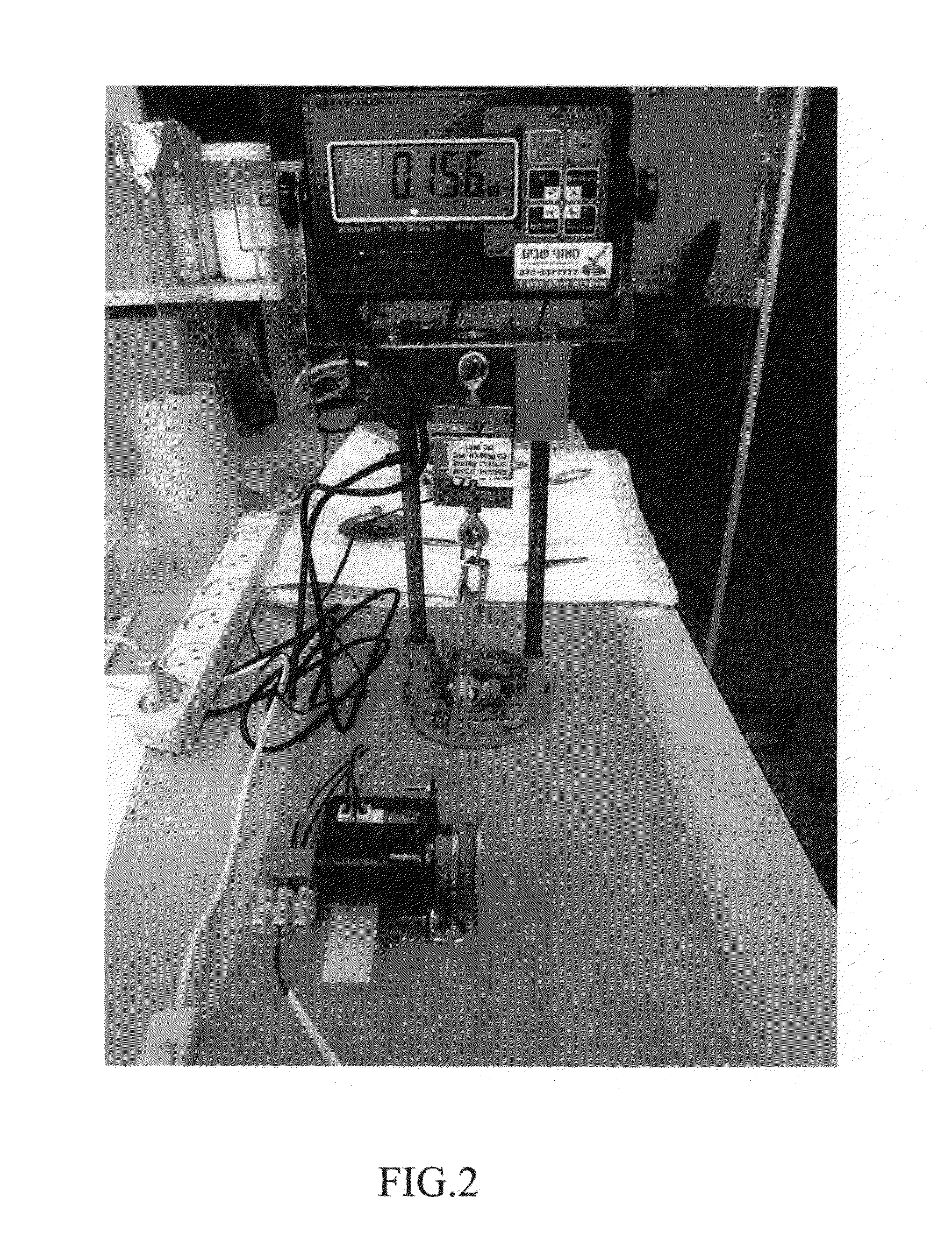
[0134]In order to test whether fibrin glue may be used for affixing a plaque to the sclera of an eye, extracted porcine eyes were used. The porcine eyes were enucleated from 6 month old (weight ˜90 Kg) domesticated pigs. The eyes were eviscerated through an 180°-270° corneo-limbal incision, filled with glass beads and sutured with a 5 / 0 prolen suture in order to maintain a constant intraocular pressure. The eyes were then anchored to a stainless steel perforated cover using 3 / 0 silk sutures (full thickness through the conjunctiva and sclera). The cover was attached to a vertical stand to simulate a supine lying patient.

[0135]In five of the examined eyes, 12 mm COMS-style gold plaques without radioactive seeds (Eckert & Ziegler BEBIG, Berlin, Germany) were affixed to the sclera using 5 / 0 nylon sutures through eyelets 1, 3 and 5 followed by closure of the conjunctiva using vicryl 7 / 0 sutures, as known in th...

example 2

Plasminogen Activator Facilitates Detachment of a Plaque Affixed to a Sclera of an Eye through Fibrinogen Glue

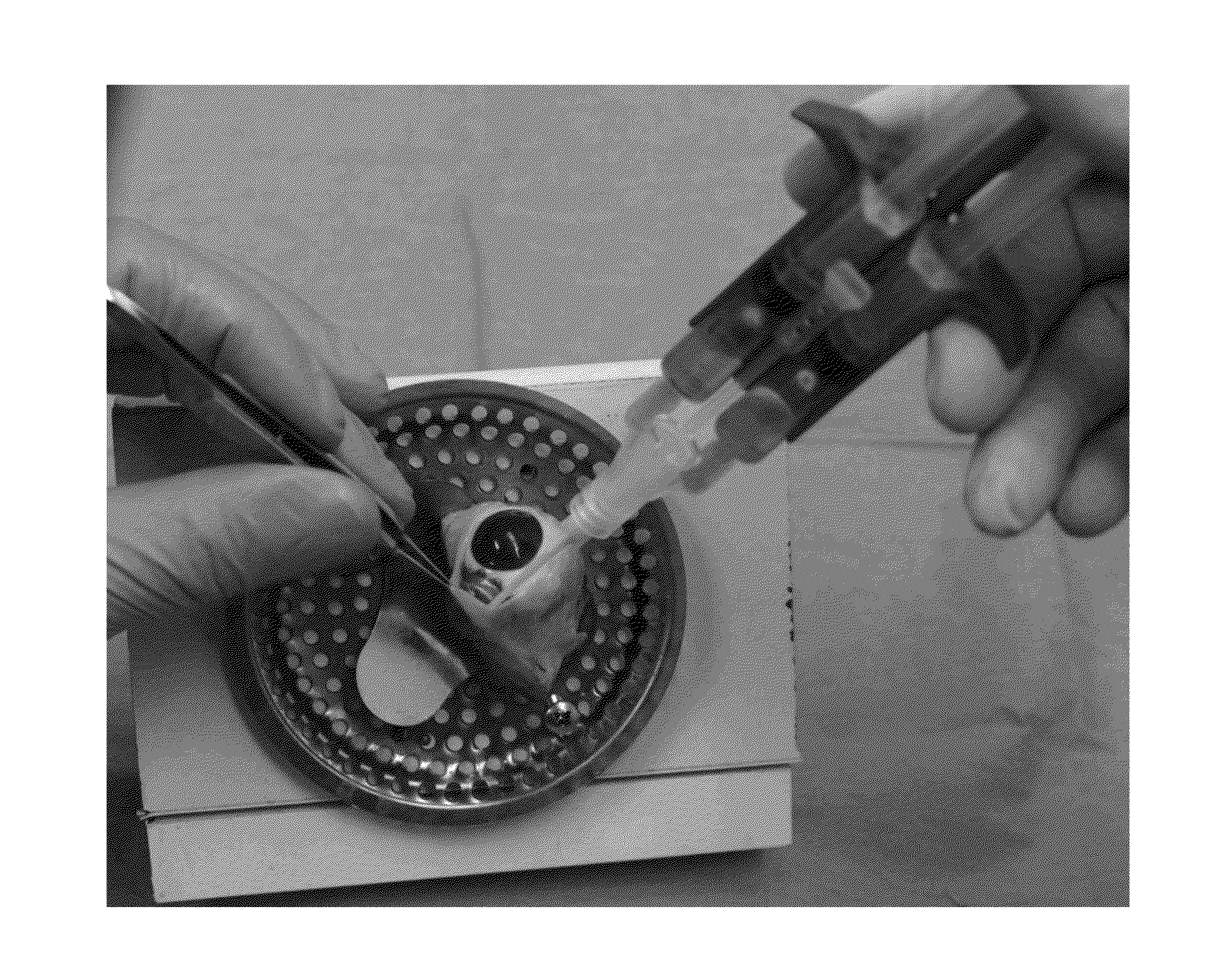
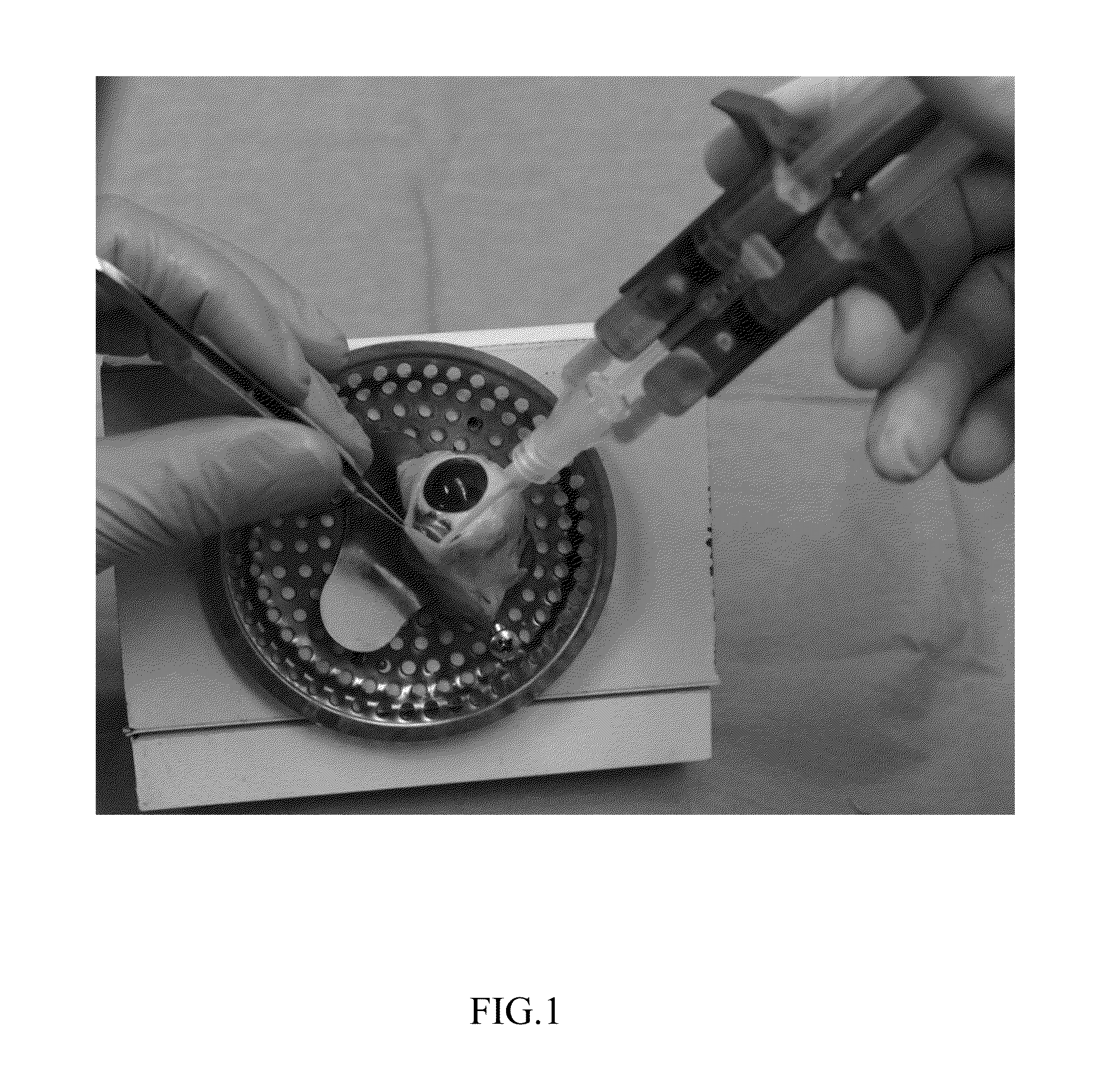
[0141]In order to examine whether a plasminogen activator is able to facilitate detachment of a plaque affixed to a sclera through fibrinogen glue, six extracted porcine eyes were used. The eyes were prepared as described in Example 1, followed by affixing 12 mm COMS-style gold plaques (Eckert & Ziegler BEBIG, Berlin, Germany) to the eyes as described for the group treated with fibrin glue and conjunctiva in Example 1. Administration of the fibrin glue on the gold plaques is demonstrated in FIG. 1. The eyes with the affixed plaques were then incubated in plasma (MDAIS, Ramat Gan, Israel) for 3 days (72 hours) at room temperature to simulate a fibrinolytic environment (the plasma was replaced every 36 hours).

[0142]In order to examine whether a composition containing a plasminogen activator is able to detach the affixed plaques, three eyes were treated with 10 ml of Normal Sal...

example 3

Fibrin Glue is Able to Affix a Plaque to the Sclera of a Human Eye in-vitro

[0144]A section of a human sclera, derived from a donor, was placed in a petri dish and a 16 mm gold plaque (without radioactive seeds) has been placed on the sclera. Next, 2 ml of fibrin-glue components (Evicel, Johnson&Johnson) were dripped on the plaque such that the excess glue spilled on the sclera and petri dish. After approximately 1 minute, once the fibrin glue has set, the sclera-plaque-glue complex was covered in plasma to simulate a fibrinolytic environment, and incubated for three days as described in Example 2. The plaque remained strongly bound to the sclera after three days of incubation. Next, 10 ml of Urokinase solution, as described above, was applied on the hardened fibrin glue. The fibrin glue broke down within 2-3 minutes, enabling extraction of the plaque from the eye.


PUM

PropertyMeasurementUnit
timeaaaaaaaaaa
diameteraaaaaaaaaa
time-periodaaaaaaaaaa

Abstract

Methods and kits are provided for reversibly affixing an implant to a sclera of an eye of a subject. The methods and kits are based on applying a composition comprising at least one fibrin-based tissue adhesive to at least one of said implant and said sclera, thereby affixing said implant to the sclera, and applying a composition comprising at least one plasminogen activator to at least one of said implant and said sclera following a desired time-period, thereby detaching said implant from said sclera, and removing said implant from said eye.

Description

FIELD OF THE INVENTION[0001]The present invention relates, according to some embodiments, to methods and kits for affixing eye implants to the sclera of an eye via a fibrin-based tissue adhesive and for breaking down said fibrin-based tissue adhesive.BACKGROUND OF THE INVENTION[0002]Brachytherapy, also known as sealed source radiotherapy, is a form of radiotherapy where a radiation source is placed inside or next to the area requiring treatment. Brachytherapy may be used in the eyes of patients suffering from eye neoplasms in order to prevent tumor growth. Brachytherapy may further be used for treatment of disorders such as choroidal neovascularization (CNV) in which arrest of neovascularization is desired.[0003]Eye neoplasms may be primary neoplasms, which originate from within the eye, or metastatic neoplasms which spread to the eye from another organ. The two most common cancers that spread to the eye from another organ are breast cancer and lung cancer. The most common primary i...

Claims


Application Information

Patent Timeline
Patent Type & Authority Applications(United States)
IPC IPC(8): A61L24/10A61N5/10A61F9/00A61K38/48A61F9/007
CPCA61L24/106A61K38/482A61F9/007A61F9/0017A61F2220/005C12Y304/21031A61N2005/1024A61F2250/0067A61N5/1027A61K45/06A61L24/0015A61L2300/406A61L2300/41A61L2300/414A61L2300/416A61N5/1017
Inventor FABIAN, IDO DIDI
Owner TEL HASHOMER MEDICAL RES INFRASTRUCTURE & SERVICES
Why Eureka
Try Eureka