
Methods of Detecting Conjugation Site-Specific and Hidden Epitope/Antigen

a conjugation site and antigen technology, applied in the field of methods, antibodies, reagents, immunoassays and kits for designing and detecting hidden antigens, can solve the problems of many antigens, many antigens, and most antibodies that don't work as users, so as to reduce non-specific binding

Active Publication Date: 2015-08-27
LIU CHUNLI +1
View PDF1 Cites 13 Cited by
  • Summary
  • Abstract
  • Description
  • Claims
  • Application Information

AI Technical Summary

Benefits of technology

The patent is about a method called ACE that can be used to improve the accuracy and reduce non-specific binding in antibody-based applications. This method uses a chemical bond-specific hydrolysis to create and expose the specific structures that the antibody needs to bind, while breaking up other structures that might bind to it non-specifically. These techniques can improve the reliability and accuracy of antibody-based assays and make them more reliable for use in research and medical settings.

Problems solved by technology

Furthermore, most antibodies don't work as the users intend, and about 75% of antibodies do not work in all antibody-based applications (http: / / www.sdix.com / Products / Custom-Antibody-Services / Engagement / Antibodies.aspx?Q=2).
A key reason of much fewer antibodies than “antibodiable” antigens is that many antigens are hidden in samples or sample preparations and thus poorly accessible to antibodies.
Disease-specific macromolecule-to-macromolecule conjugates are present in body fluids such as blood serum or cerebrospinal fluid (CSF), but few reliable methods are currently available to detect them.
Therefore, antibodies to macromolecule-to-macromolecule conjugation sites are difficult to make and are not currently available for assays of macromolecule-to-macromolecule conjugation sites in all antibody-based applications.
In comparison to monomeric modification site-specific antibodies, there is no effective method currently available for making polymeric macromolecular conjugation site-specific antibodies.
However, these efforts have been so far proven futile (Matsumoto et al., 2008).
As a result, there are few conjugation site-specific antibodies currently available.
However, this antibody does not recognize isopeptide bond-branched ubiquitin-to-ubiquitin conjugation, which are dominant forms of cellular ubiquitin conjugates.
However, the glycine-glycine-to-(lysine) antibodies recognize only the glycine-glycine-to-(lysine), rather than the lysine surrounding sequence of a specific ubiquitinated protein, and thus they are also not conjugation site-specific antibodies, and cannot be used to detect individual ubiquitin-to-protein conjugation sites.
In addition, the glycine-glycine-to-(lysine) antibodies cannot be used in regular antibody-based applications, rather they were developed for the pre-LC-MS / MS profiling applications (Peng et al., 2003; Denis et al., 2007; Xu et al., 2010).
Also, the phage display approach usually has technical challenges associated with it.
For instance, it is acknowledged that the affinity and specificity of binding partners generated by phage display are often suboptimal, relative to conventional antibody, and the loss of the original heavy-and light-chain pairings is also a challenge.
However, these methods cannot be used to design and detect hidden antigens, and they are also principally and profoundly different with the methods of the present invention.
All currently available cleavage-specific antibodies were developed to detect the naturally occurring cleavage sites in vivo, and cannot be used to detect hidden antigens such as macromolecule-to-macromolecule conjugation sites.
However, polysaccharide-to-protein and lipid-to-protein conjugation site-specific antibodies are not currently available, most probably because they are hidden antigen and no reliable methods can successfully make them.

Method used


Image

  • Methods of Detecting Conjugation Site-Specific and Hidden Epitope/Antigen
  • Methods of Detecting Conjugation Site-Specific and Hidden Epitope/Antigen
  • Methods of Detecting Conjugation Site-Specific and Hidden Epitope/Antigen

Examples


example 1

A SUMO-To-Protein Conjugation Site-Specific ACE Structure Design

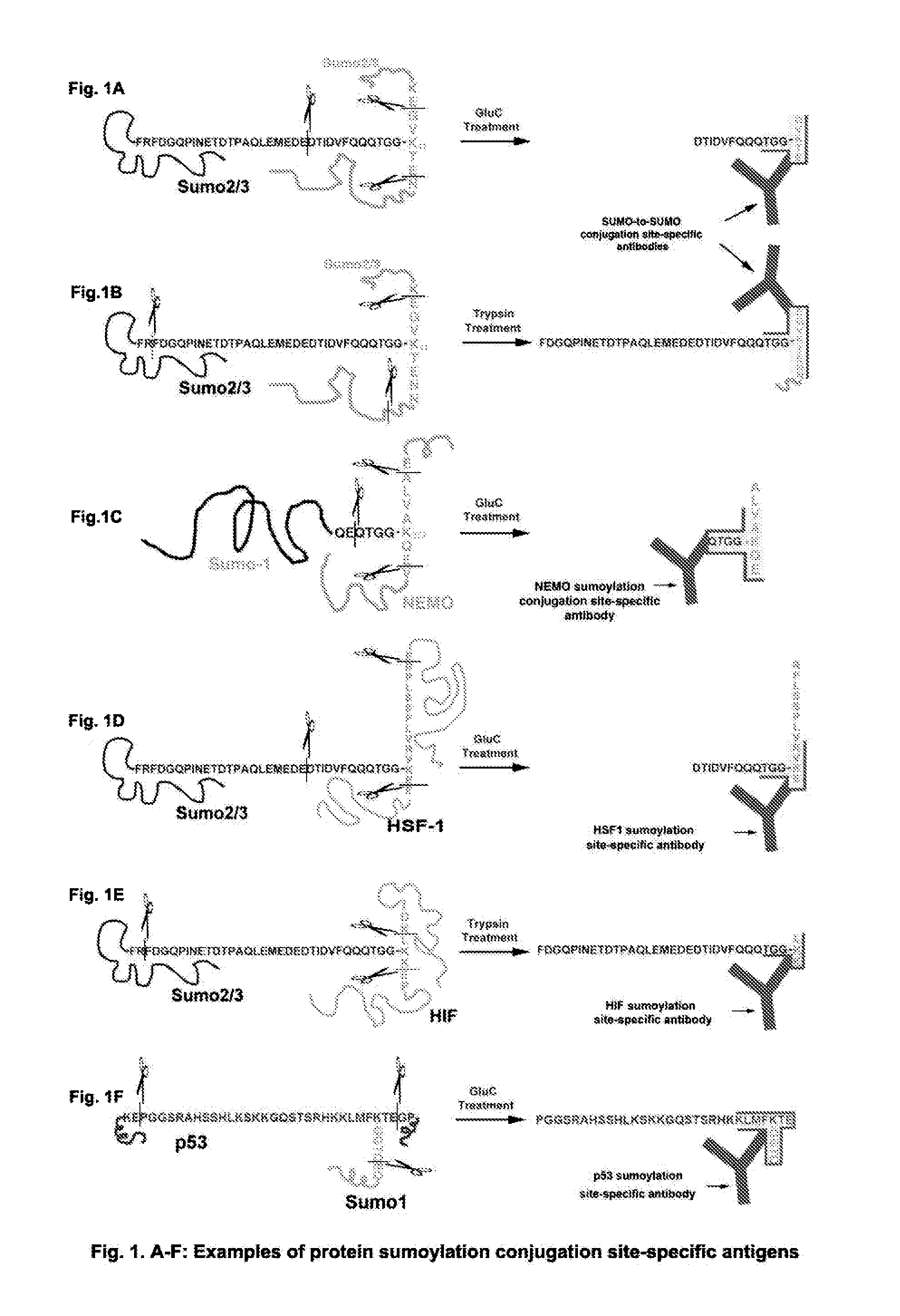
[0140]A SUMO can conjugate to many proteins including itself by an isopeptide bond. As demonstrated in FIG. 1A, a SUMO-to-SUMO conjugation site is between one SUMO C-terminal end glycine (G) and another SUMO internal lysine K11. Based on the ACE methods, a GluC-cleaved SUMO-to-SUMO conjugation site-specific hapten can be designed as GVK(GGTQQQFVDITDC)TE, wherein “C” is added to the hapten and conjugated to an immunogenic carrier, including, but not limited to, KLH or BSA to make the hapten a complete antigen. The conjugation site-specific antibody to GVK(GGTQQQFVDITDC)TE can be made with the complete antigen and purified with the hapten-conjugated resin or other supportive materials [e.g., GVK(GGTQQQFVDITDC-resin) TE]. The non-conjugation site antibodies can be removed by non-conjugated linear peptide-linked resin(s), e.g., (resin-GVKTE) and (GGTQQQFVDITDC-resin). After treatment with a protease or agent such as the des...

example 5

The fatty Acid-To-Protein Conjugation Site-Specific ACE Design

[0162]Similar to lipidated proteins described above in Example 2, a wide variety of cellular or viral proteins are covalently conjugated with a 14-carbon fatty acid myristate(s) and / or a 16-carbon fatty acid palmitate(s), as well as other fatty acids or lipid-related molecules. These types of protein lipidation are for associating the proteins to cell membranes from the cytoplasmic sides. Examples of making fatty acid-to-protein conjugation site-specific antibodies and their use thereof are shown in FIG. 6. Caveolin-1 is palmitoylated at three cysteine residues C133, C143 and C156 (Homo sapiens). A C133 palmitoylation site-specific ACE contains a base sequence set forth by SEQ ID NO: 3 and can be designed as CGGGAVVPC133(palmitate)IK, wherein the N-terminal C is added for conjugation to an immunogenic carrier; wherein “GGG” is a spacer; and wherein palmitate is linked covalently to the C133. The conjugation site-specific ...

example 6

GPI-to-Protein Conjugation Site-Specific ACE Design

[0165]GPI is post-translationally conjugated to a group of structurally and functionally diversified proteins via an ethanolamine linker. This group of proteins is anchored in the outer leaflet of the cell membrane. FIG. 7 shows two examples of the ACE antigen design for making conjugation site-specific antibodies to GPI-anchored proteins and their use thereof. One is the prion protein (PrP) (Homo sapiens) that is conjugated by a GPI-anchor at the cysteine residue C230. A conjugation site-specific ACE hapten contains a base sequence set forth by SEQ ID NO: 8 and can be designed as GC-phosphoethanolamine-SGGGC, wherein “SGGG” is added as a spacer and a linker that, via the serine hydroxyl side chain, links to phosphoethanolamine; wherein C is added for conjugation to KLH or BSA for making the hapten a complete antigen. The conjugation site-specific antibody to GC-phosphoethanolamine can be made with the complete antigen and purified ...


PUM


Abstract

This invention discloses “Artificially Cleaved Epitope (ACE)” methods, antibodies, reagents, immunoassays, and kits for designing and detecting hidden epitopes / antigens. The ACE methods can detect epitopes that are either absent or poorly accessible naturally to antibodies, and thus must be specifically and artificially created (free terminals) and / or exposed in samples and sample preparations for antibody detection. The ACE structures include, but are not limited to, macromolecule-to-macromolecule conjugation sites, and any types of linear hidden epitopes. The ACE methods comprise ACE antigen design and ACE antigen detection. The ACE methods, antibodies, reagents, immunoassays, and kits are useful in research and discovery, diagnostic, and therapeutic applications. In another aspect, the ACE methods can artificially and specifically expose hidden antigens while reducing the antibody non-specific bindings in all antibody-based applications.

Description

CROSS-REFERENCE TO RELATED APPLICATIONS[0001]This application is a continuation of U.S. patent application Ser. No. 13 / 312,285, filed Dec. 6, 2011, which claims priority to Ser. No. 61 / 420,354 filed on Dec. 7, 2010, both incorporated herein by reference in their entirety.TECHNICAL FIELD[0002]The present description relates to methods, antibodies, reagents, immunoassays and kits for designing and detecting hidden antigens, including, but not limited to macromolecule-to-macromolecule conjugation sites or any types of hidden antigens, either in situ or ex situ in sample preparations, collectively defined as the ACE methods, and their use thereof.BACKGROUND OF THE INVENTION[0003]It is estimated that about 50,000 -100,000 antibodies are currently available worldwide representing about 5,000 -10,000 different genes / proteins. This number is significantly below 400,000 proteins in the proteome, and millions of other “antibodiable” antigens. Furthermore, most antibodies don't work as the use...

Claims


Application Information

Patent Timeline
IPC IPC(8): G01N33/68
CPCG01N33/6878G01N2400/02G01N2440/38G01N2440/10C07K16/18C07K2317/21C12Q1/34C12Q1/37C12Q1/61G01N33/68
Inventor LIU, CHUNLIHU, BINGREN
Owner LIU CHUNLI
Learn More