
Mri-safe robot for transrectal prostate biopsy

a robot and prostate cancer technology, applied in the field of medical devices, can solve the problems of limiting the lifespan of patients, affecting the normal tissue of the prostate, and the unreliable use of standard gray-scale ultrasound to differentiate the prostate from the normal tissue,

Active Publication Date: 2015-09-24
THE JOHN HOPKINS UNIV SCHOOL OF MEDICINE
View PDF0 Cites 10 Cited by
  • Summary
  • Abstract
  • Description
  • Claims
  • Application Information

AI Technical Summary

Benefits of technology

This patent was made with support from the government through a specific program. The government has certain rights in the invention. The technical effect of this patent is modifying a certain system to improve its efficiency and reliability.

Problems solved by technology

A large number of these represent indolent tumors unlikely to limit the lifespan of the patient.
Still, many PCa are aggressive, causing an estimated 28,170 mortalities this year.
But, standard gray-scale ultrasound is unreliable in differentiating PCa from normal tissues.
But, standard gray-scale ultrasound provides minimal PCa specific information being unreliable in differentiating PCa from normal gland tissues.
Yet biopsies do not sample the entire gland because the number of cores is limited (typically 12).
For an exhaustive, saturation biopsy the number of biopsy cores is very high making them impractical for most patients and the common transrectal biopsy path.
Moreover, due to limited technology, the common manually-operated TRUS-guided biopsy is difficult and quality control is subjective.
Studies have shown the biopsy schema is difficult to define and follow, resulting in biopsy samples that are clustered and miss regions of the gland.
While these errors have less impact on the detection of small tumors, the detection rate of clinically significant lesions is worsened.
Systematic TRUS biopsies have typically low sensitivity and low negative predictive value.
These imaging approaches and PCa image biomarkers still require further validation and have recognized limitations, including the potential for false positive and false negative results.
Even though no current imaging method can absolutely differentiate benign from malignant lesions, imaging could point to abnormalities that should be biopsied.
Technically, the accuracy of these systems relies on challenging cross-modality image-to-image registration and deformable registration methods.
These differences contribute to targeting errors that are difficult to quantify.
Also, the means of verifying the registration are very limited, especially after the initial alignment.
But, its operation is time consuming and it can't be used with an endorectal coil, which limits the quality of prostrate imaging.
But commonly, the manual devices were difficult to operate due to the limited access within the scanner and numerous table moves were required to access the patient.
However, making a robot that can operate safely and accurately in the MRI scanner without being influenced by and without interfering with the functionality of the imager has been a very challenging engineering task.
The use of electricity may cause interference leading to signal to noise attenuation, signal distortions, and image artifacts.
As such, most of the components commonly used in robotics may not be used in close proximity of the MRI.
For example, the ubiquitous electromagnetic motor is clearly MRI unsafe because it functions based on magnetism.
Field conditions that define the specified MR environment include field strength, spatial gradient, dB / dt (time rate of change of the magnetic field), radio frequency (RF) fields, and specific absorption rate (SAR).MRI-Unsafe Is an item that is known to pose hazards in all MR environments.
Interestingly, carbon fiber is not MRI-Safe because it is a good electrical conductor.
These are magnetism free but use metallic components and electricity which typically affect the quality of MR images.
Even without power, it was shown that the wiring may debase the signal-to-noise-ratio by as much as 50%.
Moreover, piezoelectric robots may only be MRI-Conditional but not MRI-Safe.
However, fluid leakage is difficult to control, especially when nonmetallic components are used.
For medical applications this raises contamination and sterility concerns.
But, the major limitation of classic pneumatic actuators is their notoriously difficult motion control.
Pneumatic servo control is a very delicate problem, because the compressibility of the air and the stiction of the piston make the system highly nonlinear and hardly manageable.
Pneumatic servo control in the MRI is even more intricate because long hoses are needed to connect them to pneumatic valves, which are typically located distal from the scanner.
Moreover, pneumatic cylinders have another major problem for medical applications: they are direct drive actuators.
If they malfunction, direct drives may swiftly spring off, fully and quickly unwinding and potentially hurting the patient or personnel.
But unfortunately, due to all the additional complexity, the robots still included metallic components and were shown to debase the signal to noise ratio of the MRI.
Unfortunately, this cylinder remained highly sensitive to disturbances such as small temperature changes.
But apparently the actuators of device are not encoded yet, or the feedback is not being used for control so that the device is not a robot but remotely controlled by the physician observing the MRI.
This is an especially difficult task because the MRI has relatively long acquisition times.
Hitherto, very few interventional MRI-guided prostate biopsy devices have been tested clinically in very few patients.
Yet, these have shown the feasibility of interventional MRI and CRS biopsy targeting.

Method used


Image

  • Mri-safe robot for transrectal prostate biopsy
  • Mri-safe robot for transrectal prostate biopsy
  • Mri-safe robot for transrectal prostate biopsy

Examples


examples

[0085]The following Examples have been included to provide guidance to one of ordinary skill in the art for practicing representative embodiments of the presently disclosed subject matter. In light of the present disclosure and the general level of skill in the art, those of skill can appreciate that the following Examples are intended to be exemplary only and that numerous changes, modifications, and alterations can be employed without departing from the scope of the presently disclosed subject matter. The following Examples are offered by way of illustration and not by way of limitation.

[0086]Bench Tests of Robot Precision and Accuracy: A set of experiments was conducted to determine the targeting performance of the robotic device independent of the imaging components. A 30×20×20 mm region within the workspace of the robot was divided in 10 mm interval, creating a target set of 36 target locations. A Polaris optical tracker (NDI, Canada) was used to measure the actual location of ...


PUM


Abstract

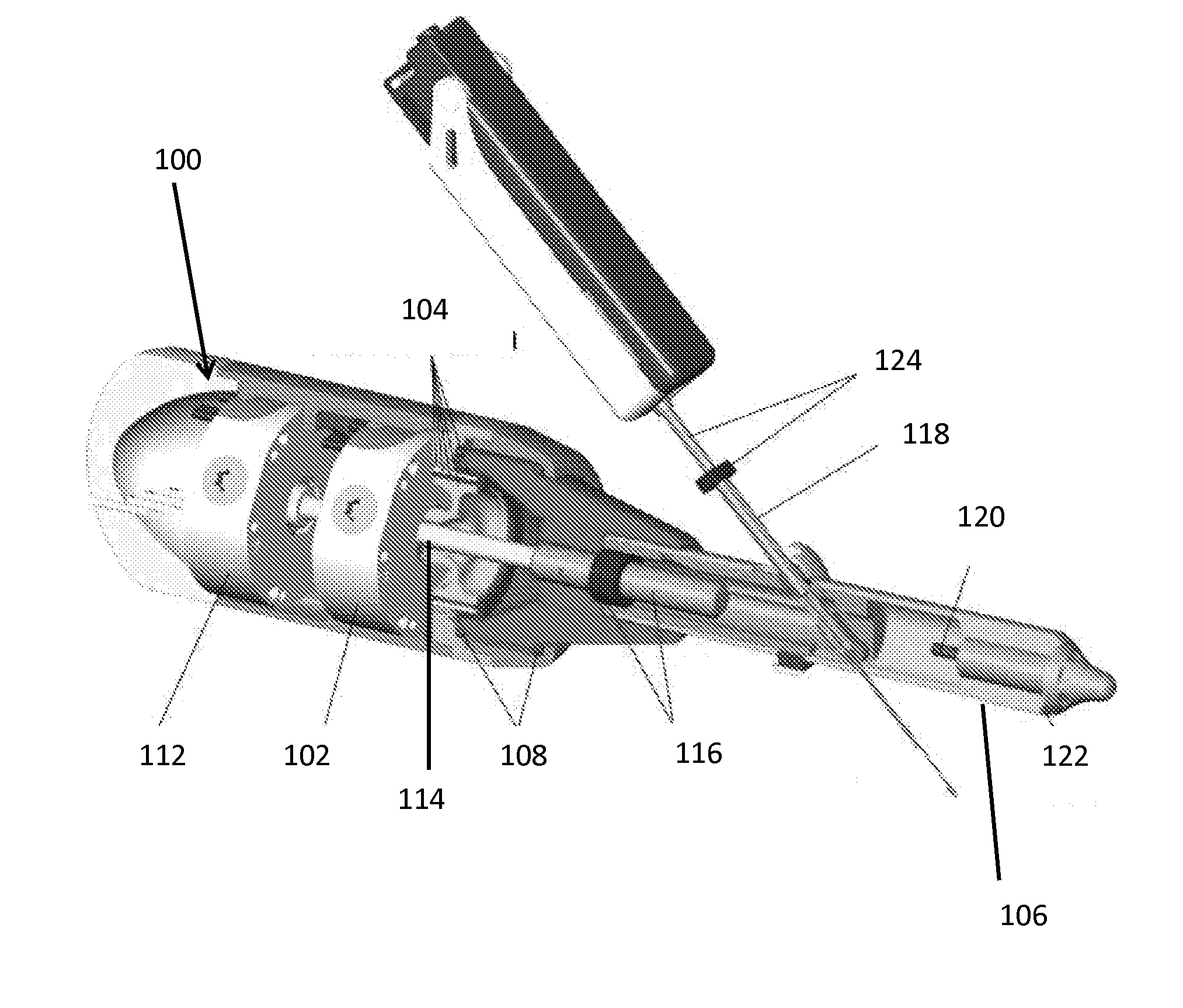
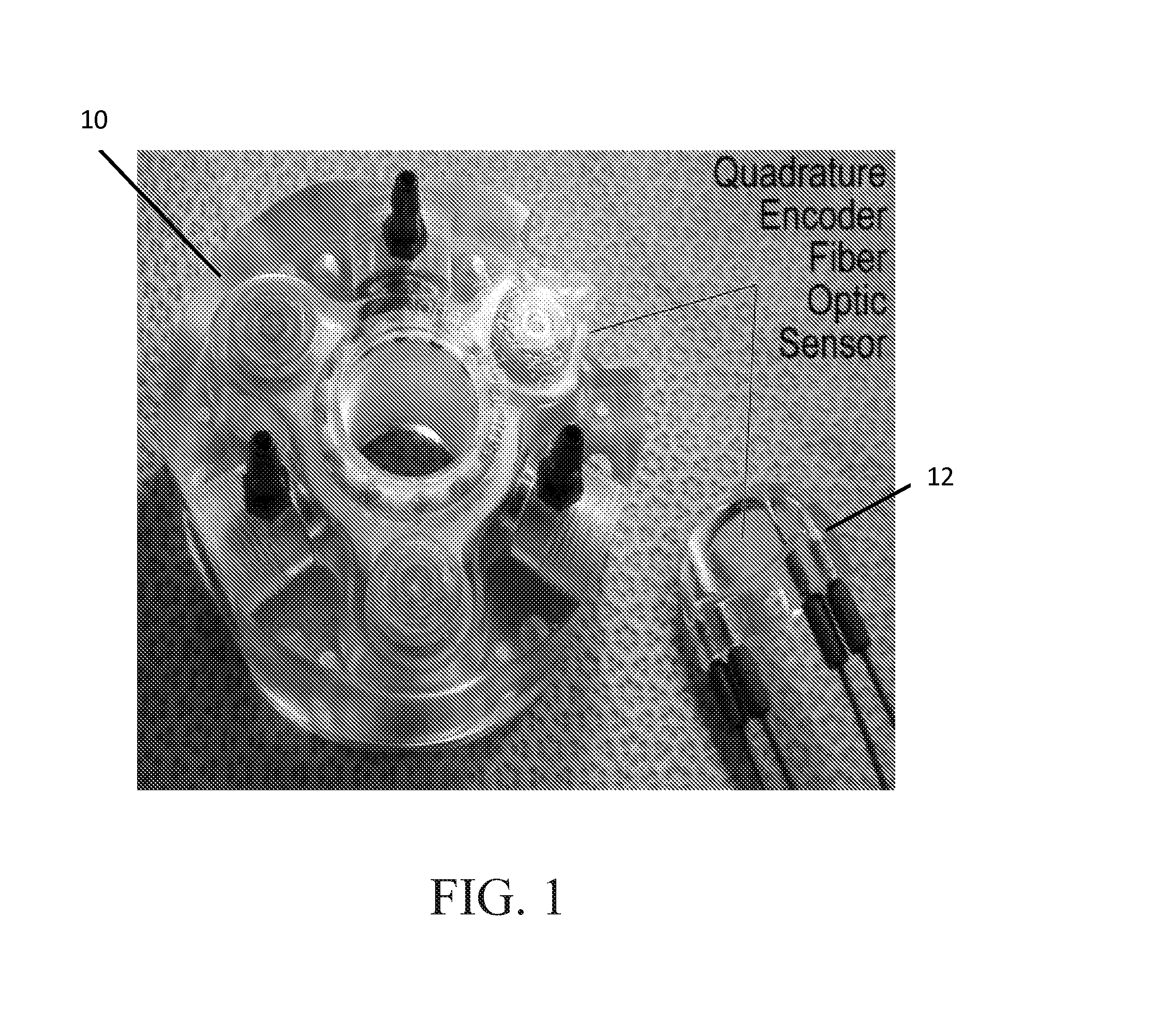
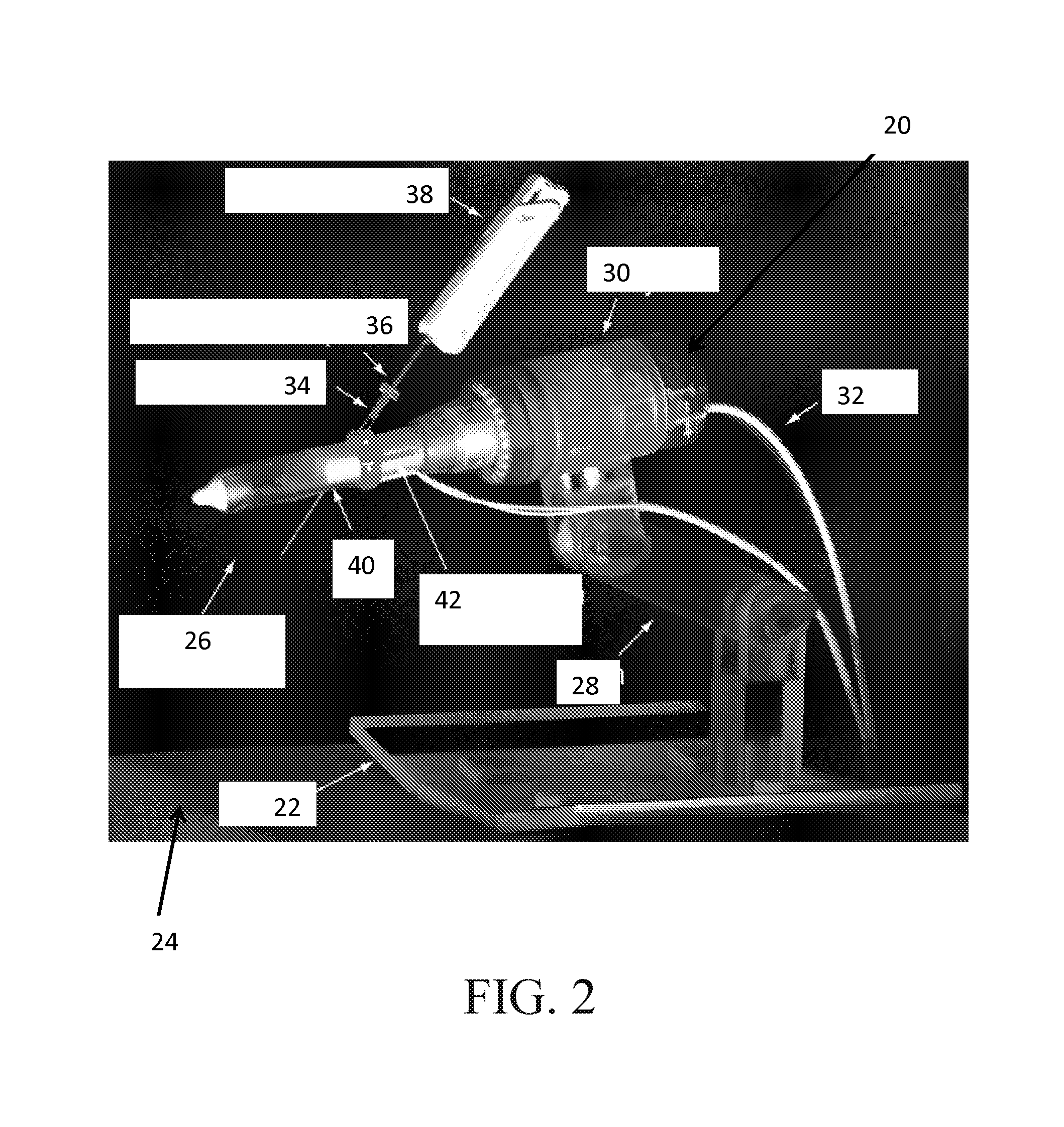
An MRI safe robot for guiding transrectal prostate biopsy comprises a support arm, a robot body operatively connected to the support arm and a transrectal biopsy device operatively connected to the robot body. The transrectal biopsy device includes an endorectal extension and a biopsy needle device, the endorectal extension including an MRI coil for MRI imaging of the prostate. The robot body includes a first driver module for generating rotational motion of the endorectal extension and a second driver module for angulating the biopsy needle device toward a target area of the prostate for biopsy. The biopsy needle device is rotatable relative to the endorectal extension about a fixed axis and translatable through the endorectal extension. Each of the first and second driver modules include at least one pneumatic motor, wherein the MRI images are used by a physician to determine the target area for biopsy.

Description

CROSS REFERENCE TO RELATED APPLICATIONS[0001]This application claims the benefit of U.S. Provisional Patent Application No. 61 / 710,909 filed on Oct. 8, 2012, which is incorporated by reference, herein, in its entirety.GOVERNMENT SUPPORT[0002]This invention was made with government support under W81XWH-08-1-0221 awarded by the Department of Defense. The government has certain rights in the invention.FIELD OF THE INVENTION[0003]The present invention relates to medical devices. More particularly, the present invention is related to a MRI-safe robot for transrectal prostate biopsy.BACKGROUND OF THE INVENTION[0004]In 2012, an estimated 241,740 new prostate cancer (PCa) cases will be diagnosed in the US alone. A large number of these represent indolent tumors unlikely to limit the lifespan of the patient. A recent study has shown that it is necessary to treat 48 men to prevent one death from PCa, suggesting that significant overtreatment exists. Still, many PCa are aggressive, causing an ...

Claims


Application Information

Patent Timeline
Patent Type & Authority Applications(United States)
IPC IPC(8): A61B19/00A61B5/055A61B10/04G01R33/28G01R33/48A61B10/02A61B5/00
CPCA61B19/2203A61B2019/5454A61B10/0275A61B5/055A61B5/4381G01R33/287G01R33/48A61B10/04A61B19/201A61B19/54A61B5/742A61B5/748A61B2010/045A61B2010/0208A61B10/0241A61B34/30A61B90/11A61B90/39A61B2017/00544A61B2090/062A61B2090/374A61B2090/3954
Inventor STOIANOVICI, DANPETRISOR, DORUKIM, CHUNWOOSEBRECHTS, PETER
Owner THE JOHN HOPKINS UNIV SCHOOL OF MEDICINE
Learn More