Biological sample collection and preservation

a biomarker and sample technology, applied in the field of biomarker collection and preservation, can solve the problems of limiting the analysis of biomarkers, affecting the detection accuracy of biomarkers, etc., and achieving the effects of reducing the difficulty of biomarker extraction, and reducing the number of samples

Active Publication Date: 2016-02-04
SPOT BIOSCI LLC
View PDF2 Cites 15 Cited by
  • Summary
  • Abstract
  • Description
  • Claims
  • Application Information

AI Technical Summary

Problems solved by technology

Current tissue preservation methods such as formalin-fixed, paraffin embedded (FFPE) are suitable for histopathology studies but not amenable to biomarker analysis due to poor protein and nucleic acid recovery.
Even though recent reports describe some success with genetic analysis, the poor quality of DNA and RNA restricts analysis by common techniques such as RT-PCR, microarrays and sequencing.
Extraction of full-length, non-degraded, immunoreactive proteins from FFPE tissue has also proved challenging, with limited detection by common methods such as ELISA and bead-based multiplexed immunoassays.
While this method may be suitable for limited sampling, it is not practical for wide-scale use due to high costs and infrastructure requirements as well as logistical issues in collecting, maintaining and shipping samples at sub-freezing temperatures.
A major limitation for disease and biomarker research is a lack of robust and relevant biological samples.
Small collections of biological samples are spread throughout research institutions, but sample collection and storage is not uniform and samples are often compromised, which can lead to faulty data.
As a key recent example, NCI attempted to form a cancer biobank through the Cancer Genome Atlas program but found that up to 99% of stored blood and tissue samples were unacceptable for research.
The development of alternative methods for simplified tissue sample storage has proved challenging.

Method used


Image

  • Biological sample collection and preservation
  • Biological sample collection and preservation
  • Biological sample collection and preservation

Examples


working examples

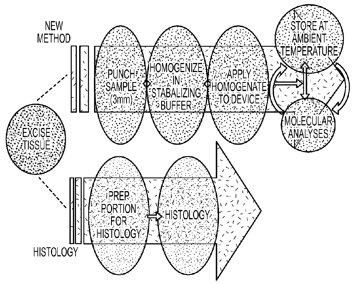
[0035]Rat liver tissue (approx. 50 mg) was homogenized in two separate reagent mixtures, A: phosphate buffer saline with 0.5% Triton and B: 10 mM Tris, 1 mM EDTA with 0.5% Triton-X100. Enzymatic activity was compared for homogenate samples that remained wet and equivalent samples that were dried on filter paper (FIG. 5). Activities of alpha-galactosidase (alpha-GAL) and beta-galactosidase (beta-GAL) were measured by incubating at 37° C. for 2 hours with 4-methylumbelliferyl-alpha-D-galactopyranoside and 4-methylumbelliferyl-beta-D-galactopyranoside, respectively. 4-methylumbelliferone (4-MU) formation was measured by fluorescence. Activity levels were similar between wet and dried homogenization samples.

[0036]Three separate reagent mixtures were used to homogenize rat liver tissue, A: nuclease-free water B: nuclease-free water with 0.5% CHAPS and C: nuclease-free water with 0.5% Triton-X100. Portions of the homogenates were frozen while separate portions were applied to HemaSpot Dev...


PUM

PropertyMeasurementUnit
sizeaaaaaaaaaa
temperatureaaaaaaaaaa
sub-freezing temperaturesaaaaaaaaaa

Abstract

An embodiment of the claimed invention is directed to a method that greatly streamlines and reduces costs for tissue preparation, preservation, long-term storage and sample retrieval for molecular analysis using a method based on dried blood spot (DBS) technology. In this method, a small needle punch sample of freshly excised tissue will be homogenized in stabilizing reagent and inserted into a device containing absorbent material and drying agent. This device is suitable for long-term sample storage at ambient temperature and allows for easy removal of sections for biomarker analysis.

Description

CROSS-REFERENCES TO RELATED APPLICATIONS[0001]This application claims the benefit under 35 U.S.C. §119(e) of U.S. Provisional Patent Application No. 61 / 782,393 filed Mar. 14, 2013 which is incorporated herein by reference in its entirety as if fully set forth herein.BACKGROUND OF THE INVENTION[0002]The collection, preservation and storage of tissue samples for molecular analysis is essential for cancer treatment and for research and development of tissue-based biomarkers for disease pathophysiology. Much effort is currently focused on determining markers based on nucleotides (i.e. DNA, mRNA, miRNA), proteins and metabolites for cancer staging, prognosis and treatment selection. Detection of these biomarkers would more efficiently direct patients to treatments with the highest potential for benefit.[0003]Current tissue preservation methods such as formalin-fixed, paraffin embedded (FFPE) are suitable for histopathology studies but not amenable to biomarker analysis due to poor protei...

Claims


Application Information

Patent Timeline
Patent Type & Authority Applications(United States)
IPC IPC(8): A61B10/00G01N1/30A61B10/02G01N1/08G01N1/31
CPCG01N1/30A61B10/0266G01N1/08G01N1/31A61B10/0096A61B10/0038A61B10/0233A61B10/0241B01L3/0231B01L3/5023B01L3/5055B01L2200/141B01L2300/0803B01L2300/105G01N2001/4027
Inventor HILL, JAMESHILL, JEANETTE
Owner SPOT BIOSCI LLC
Why Eureka
Try Eureka