However,
mammography requires compression of the breast which is often uncomfortable for the patient.
Mammography also exposes the patient to
ionizing radiation and may fail to detect malignant tumors in some patients, especially younger individuals, e.g., those under fifty years old.
Most guidelines do not recommend routine screening for these younger patients because of concerns regarding the effects of
radiation exposure and false positive identification rates.
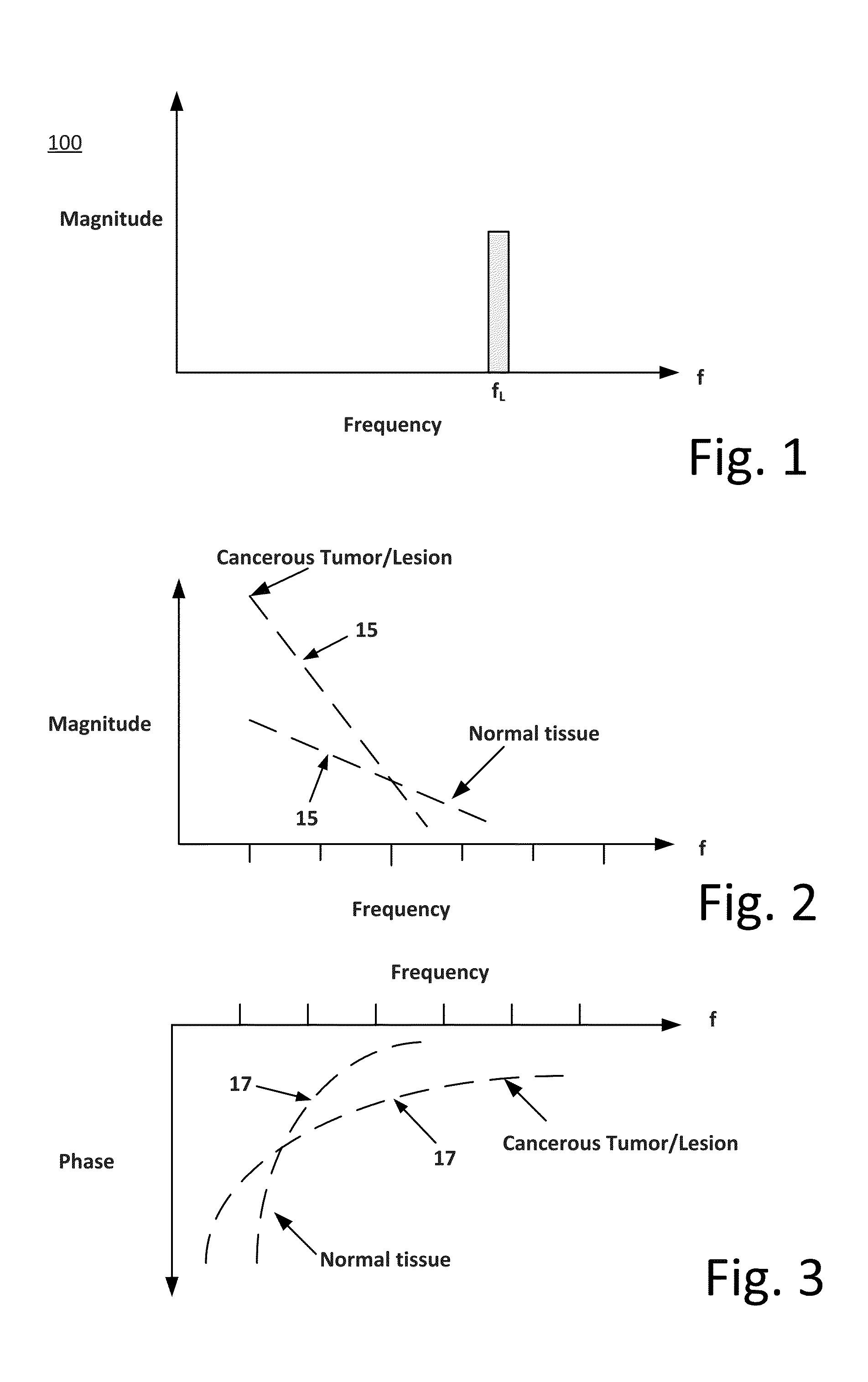
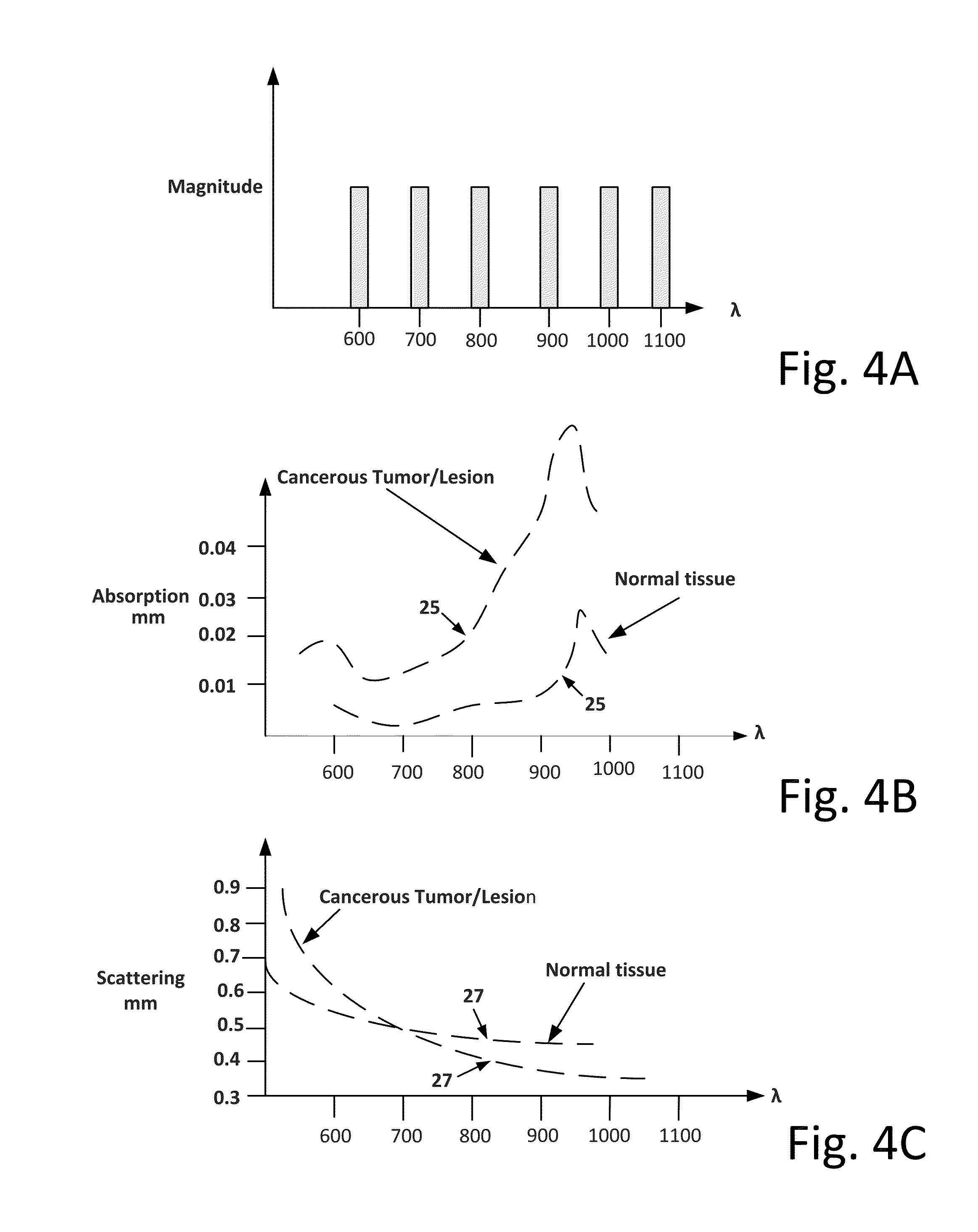
First, referring to FIG. 4A, while a number of different modulated lasers have been used in previous efforts, the
demodulation processing has only provided partial spectral information for use in response determination due to the limitation of the bandwidth of the modulating waveform. This is illustrated in FIGS. 2 and 3.
Partial Fourier spectral information, i.e. discontinuous Fourier spectral information with gaps 15 / 17, leads to gaps 25 / 27 in corresponding measurements of the optical parameters, Absorption and Scattering, as shown in FIGS. 4B and 4C. The reason for the gaps 15 / 17 is that the incident modulated
laser beams can only generate Fourier spectral information in the
modulation bandwidth around the
laser carrier. The gaps 15 / 17 in the Fourier spectral information collected are significant because the resulting gaps 25 / 27 in the measurements of optical parameters can affect the sensitivity and specificity, and thus accuracy, of the information provided. Specifically, the gaps 25 / 27 can lead to distortions in the signals that will affect the
interpretability with respect to tissue discrimination and thus the accuracy of diagnosis which can result in both false positive and negative diagnoses, both of which can seriously and deleteriously
impact patient care. Traditional DOS
processing assumes that these gaps contain no information and therefore ignores the gaps. However, this is a very narrow
view based upon speculation and a desire to simplify the
processing, and it is not necessarily true.
Second, the computation of all of the Fourier spectral information has to be carried out with extreme accuracy. The greater the accuracy in Fourier spectral information, the greater the reliability in the computation of the optical parameters, Absorption and Scattering. This enhances the discrimination of the measured Absorption and
Scattering parameters of the
normal tissue from the absorption and
scattering parameters of the suspected cancerous tissues, which in turn enhances both the reliability of detection of cancerous tissue and determination of the success of the therapeutic program. However, traditional DOS processing ignores the effects of
noise, interference, inaccuracies in
digitization, biasing in sampling and other deleterious effects.
Third, there is the need to make the device or system small enough to be portable so that it can be employed in the physician's office, rather than sending the patient to a series of appointments at centralized laboratories. To date, attention has not been directed at this concern.
Fourth, the optical parameters, the Absorption and Scattering, should to be computationally processed in such a way as to give the physician a transparent and understandable indication of the detection of
cancer, and if so detected, an indication of the patient's response to a subsequent course of therapy. Presenting the optical parameters by themselves to the physician is not worthwhile. To date, attention has not been directed at this concern.
However, this is not absolutely certain and there are probabilities associated with the resulting physician diagnosis as to whether a patient is responding to the therapy.

