Combined agent for cell therapy of corneal endothelial cell

a corneal endothelial cell and cell therapy technology, which is applied in the direction of medical preparations, pharmaceutical delivery mechanisms, unknown materials, etc., can solve the problems of hypertrophy of the corneal guttae and descemet's membrane, corneal endothelial cells have very limited regeneration ability, and the corneal endothelial cells have very limited ability to regenerate, so as to improve the establishment of the transplantation of the corneal endothelial cells, the effect of effectiv

Active Publication Date: 2016-11-03
KYOTO PREFECTURAL PUBLIC UNIV CORP +2
View PDF1 Cites 3 Cited by
  • Summary
  • Abstract
  • Description
  • Claims
  • Application Information

AI Technical Summary

Benefits of technology

[0036]The present invention provides a medicament that can treat or prevent a disease associated with the corneal endothelium by using a myosin II-specific inhibitor, where the technique can also be materialized as eye drops or the like. The present invention has drawn attention particularly as an invention that markedly improves the establishment of transplantation of a corneal endothelium.
[0037]The present invention has verified that therapy using a myosin II-specific inhibitor achieves the same level of effect as the ...

Problems solved by technology

Once damaged, human corneal endothelial cells have very limited ability to regenerate.
For example, Fuchs' endothelial corneal dystrophy is a disease causing abnormality in endothelial cells inside the cornea, resulting in edema of the cornea.
In Fuchs' endothelial corneal dystrophy, extracellular matrix such as collagen is deposited on a part of the back surface of a Descemet's membrane at the back of the cornea, resulting in hypertrophy of a corneal guttae and Descemet's membrane.
Hypertrophy of the corneal guttae and Descemet's membrane is a cause of photophobia or blurred vision in Fuchs' endothe...

Method used


Image

  • Combined agent for cell therapy of corneal endothelial cell
  • Combined agent for cell therapy of corneal endothelial cell
  • Combined agent for cell therapy of corneal endothelial cell

Examples


reference example 1

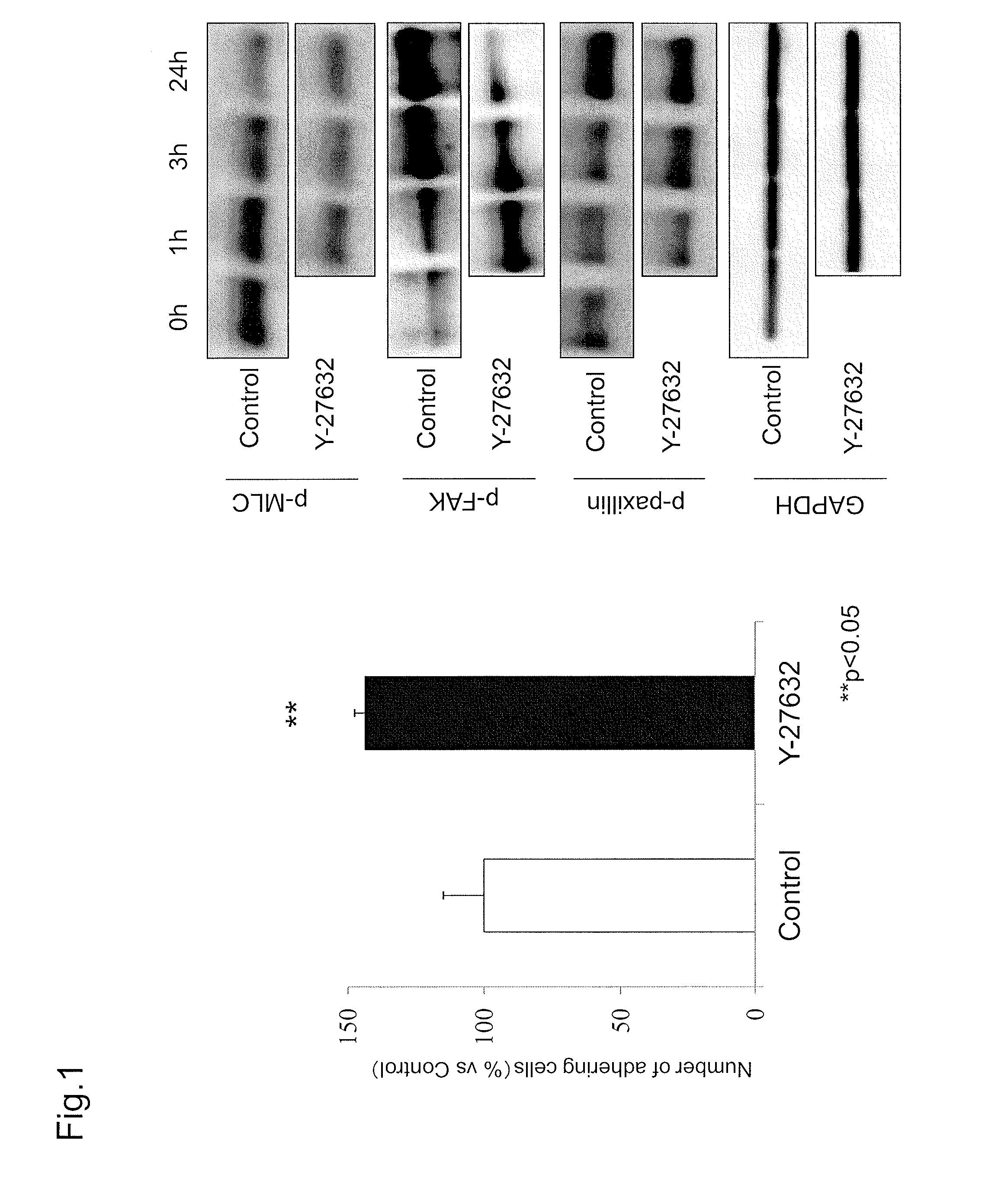
Examination of Cell Adhesion Due to ROCK Inhibitor and Effect Thereof on Adhesion-Related Molecule

[0128]Reference Example 1 added a ROCK inhibitor to study the effect on cell adhesion due to inhibiting the Rho-ROCK pathway.

[0129](Materials and Methods)

[0130](Corneal Tissue)

[0131]All monkey corneal tissues used in this experiment were corneas of a cynomolgus monkey euthanized for other research purposes (Nissei Bilis Co., Ltd., Ohtsu, Japan, or Keari Co., Ltd., Wakayama, Japan). All corneas were preserved at 4° C. in a preservation medium (Optisol; Chiron Vision Corporation, Irvine, Calif.).

[0132](Cell Culture)

[0133]In primary culture of monkey corneal endothelial cells, a Descemet's membrane including an endothelial cell layer was detached from a corneal tissue, and placed in 2 mg / ml Collagenase A (catalog No.: 70164923; Roche Applied Science, Penzberg, Germany) dissolved in DMEM (Gibco-Invitrogen) and incubated at 37° C. After 12 hours, the sample was centrifuged at 1000 rpm for 5 ...

reference example 2

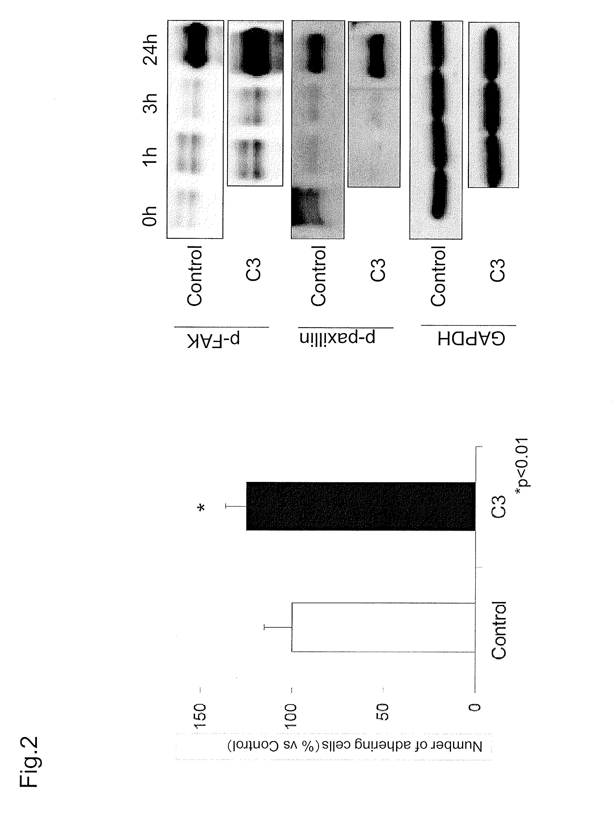
Examination of Cell Adhesion Due to RhoA Inhibitor and Effect Thereof on Adhesion-Related Molecule

[0142]Reference Example 2 used a RhoA inhibitor (botulinum C3 enzyme) to study the effect on cell adhesion.

[0143](Materials and Methods)

[0144]In short, cell adhesion due to RhoA inhibitor was examined by assessing the number of adhering cells with CellTiter-Glo®. Further, the activity of adhesion-related molecules after the addition of a RhoA inhibitor was measured by Western blot.

[0145](Examination of Number of Adhering Cells).

[0146]The number of adhering cells was analyzed by using CellTiter-Glo®. First, cultured monkey corneal endothelial cells were seeded onto a 96 well plate at 5000 cells / well in DMEM (Gibco-Invitrogen) to which C3 botulinum toxin (CALBIOCHEM, catalog number: 341208) was added such that the final concentration would be 300 ng / ml. DMEM (Gibco-Invitrogen) was used as a control. The cells were washed with PBS(−) after three hours from seeding. PBS(−) was removed by ta...

example 1

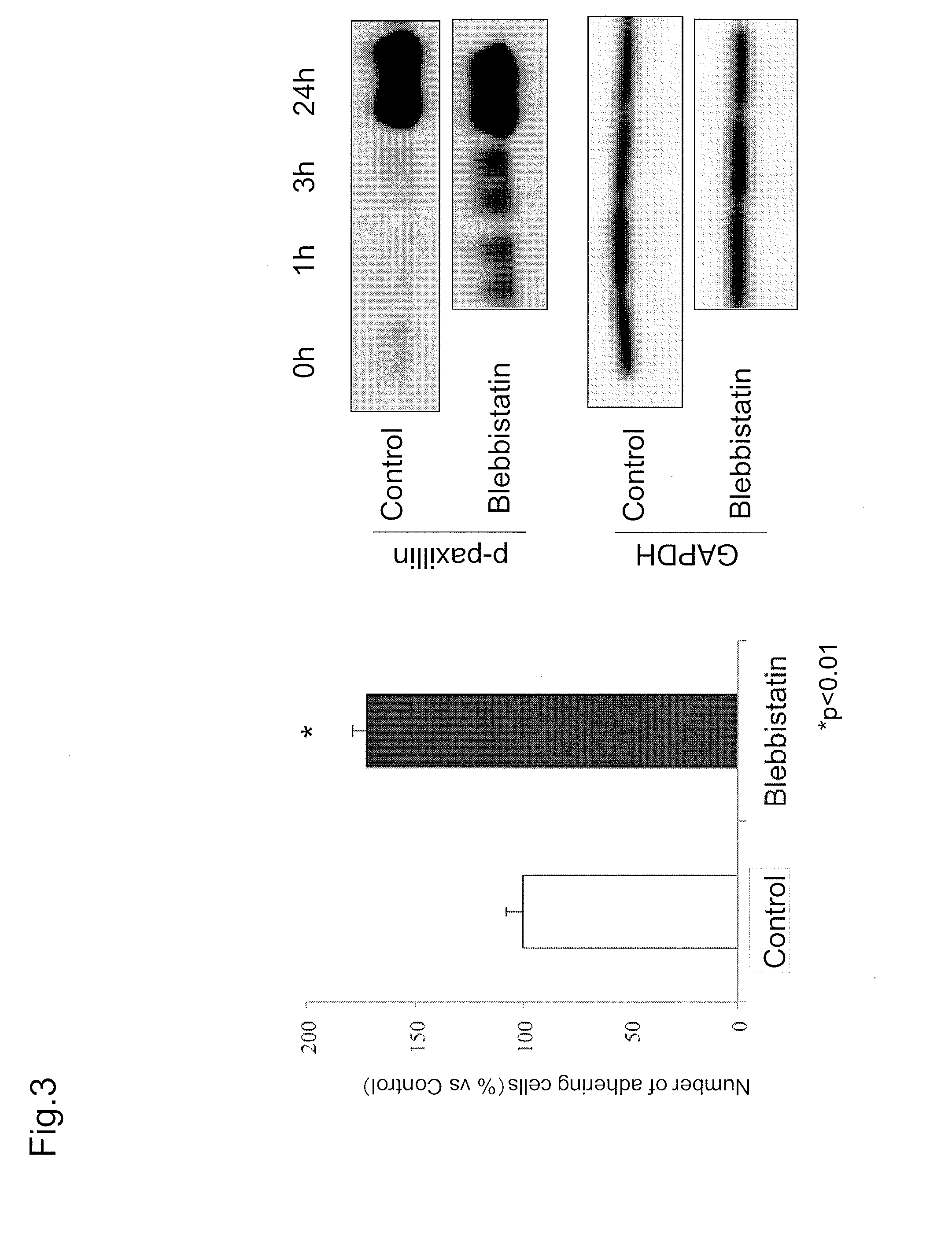
Examination of Cell Adhesion Due to MLC Phosphorylation Inhibitor

[0151]The present Example examined the involvement of MLC, which is downstream of a Rho-ROCK pathway and phosphorylated by the activation thereof, in cell adhesion.

[0152](Materials and Method)

[0153]In short, cell adhesion due to a p-MLC inhibitor was examined by assessing the number of adhering cells with CellTiter-Glo®. Further, the activity of adhesion-related molecules after the addition of an RhoA inhibitor was measured by Western blot.

[0154](Examination of Number of Adhering Cells).

[0155]The number of adhering cells was analyzed by using CellTiter-Glo®. First, cultured monkey corneal endothelial cells were seeded onto a 96 well plate at 5000 cells / well in DMEM (Gibco-Invitrogen) to which blebbistatin (MILLIPORE, catalog number: 203391) was added such that the final concentration would be 100 μM. DMEM (Gibco-Invitrogen) was used as a control. The cells were washed with PBS(−) after three hours from seeding. PBS(−) ...


PUM


Abstract

The present invention provides an agent for treating or preventing a disease, a disorder or a condition of a corneal endothelium, said agent comprising a corneal endothelial cell and a myosin II-specific inhibitor. More specifically, the myosin II-specific inhibitor is blebbistatin. The disease, the disorder or the condition is typically a disorder associated with Fuchs endothelial corneal dystrophy or corneal endothelial dysfunction (bullous keratopathy). Also provided is a method for treating or preventing a corneal endothelial disease, said method comprising a step of administering an effective amount of a myosin II-specific inhibitor together with a corneal endothelial cell to a subject.

Description

TECHNICAL FIELD[0001]The present invention relates to a technique, a method, an agent and the like for treating or preventing a disease, disorder or condition of a corneal endothelium, and more specifically to a combined agent for treating corneal endothelial cells.BACKGROUND ART[0002]Visual information is recognized when light transmitted into the cornea, which is a transparent tissue at the front-most part of an eyeball, reaches the retina and excites nerve cells of the retina, and a generated electric signal is transmitted through the optic nerve to the visual cortex of the cerebrum. To attain good vision, it is necessary that the cornea is transparent. The transparency of the cornea is maintained by holding the water content constant with pumping and barrier functions of corneal endothelial cells.[0003]Human corneal endothelial cells are present at a density of about 3000 cells per 1 mm2 at birth. Once damaged, human corneal endothelial cells have very limited ability to regener...

Claims


Application Information

Patent Timeline
IPC IPC(8): A61K31/4745A61K9/00A61K35/44A61K35/30
CPCA61K31/4745A61K35/44A61K9/0048A61K35/30A61K45/06A61P27/02
Inventor KOIZUMI, NORIKOOKUMURA, NAOKIKINOSHITA, SHIGERU
Owner KYOTO PREFECTURAL PUBLIC UNIV CORP
Why Eureka
Try Eureka