

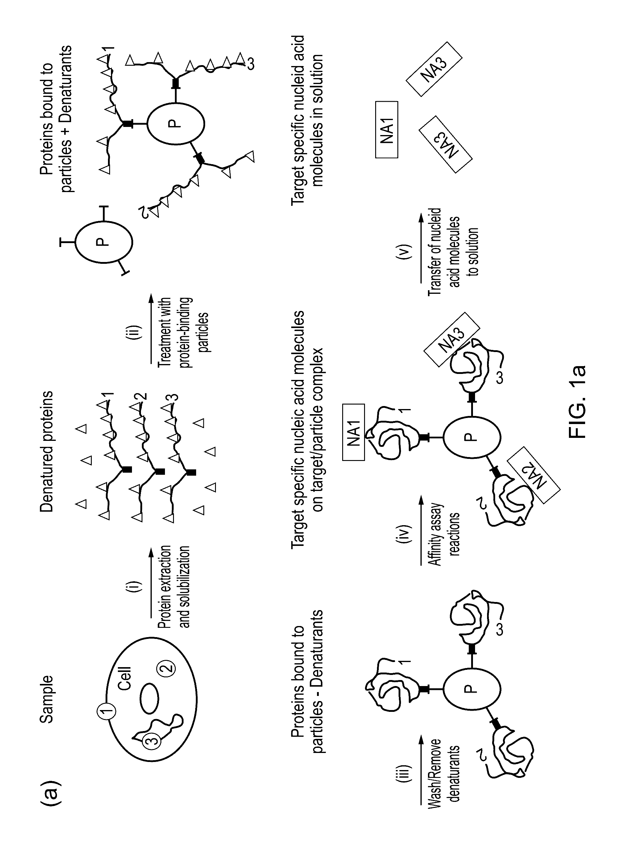
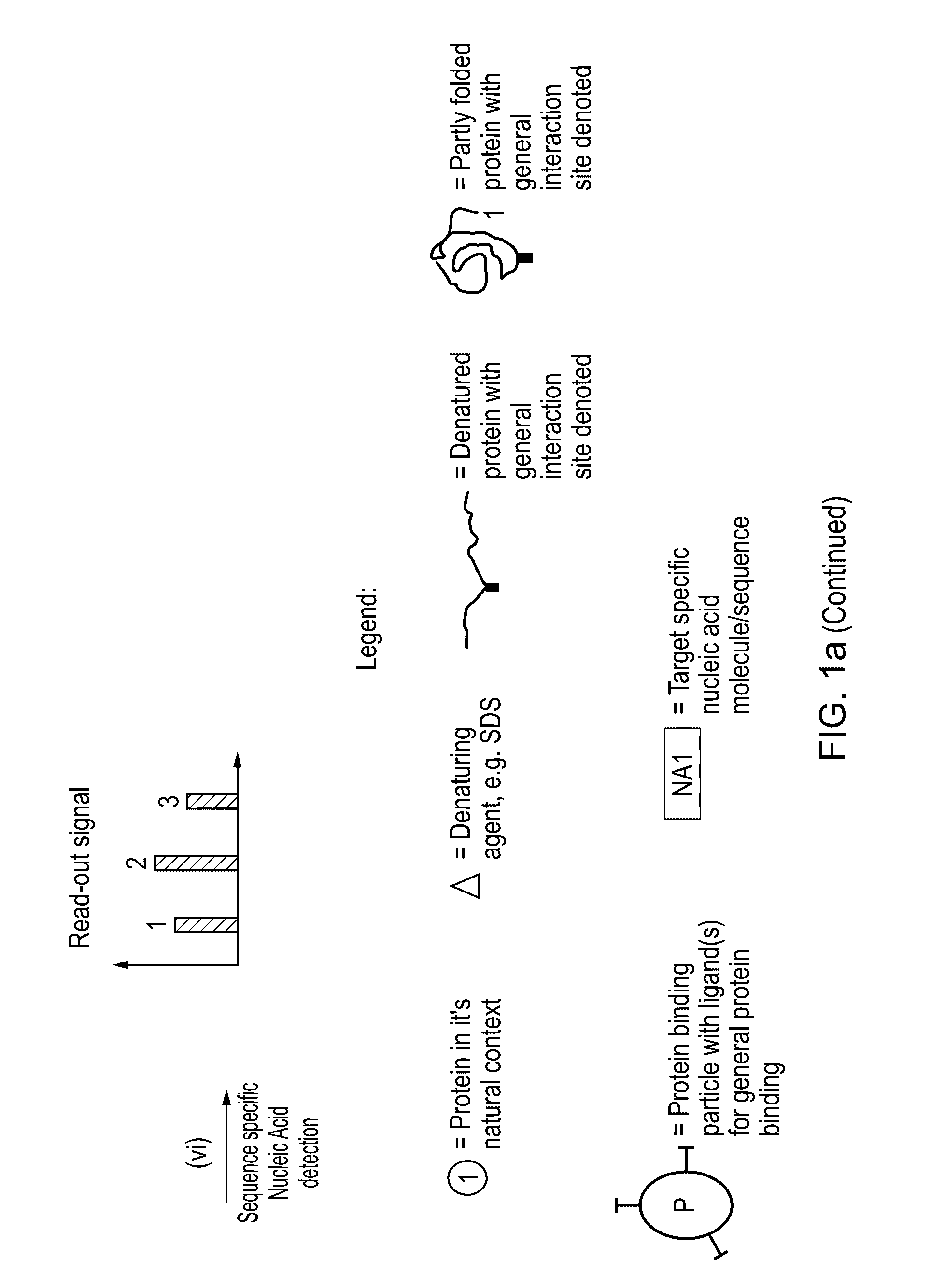
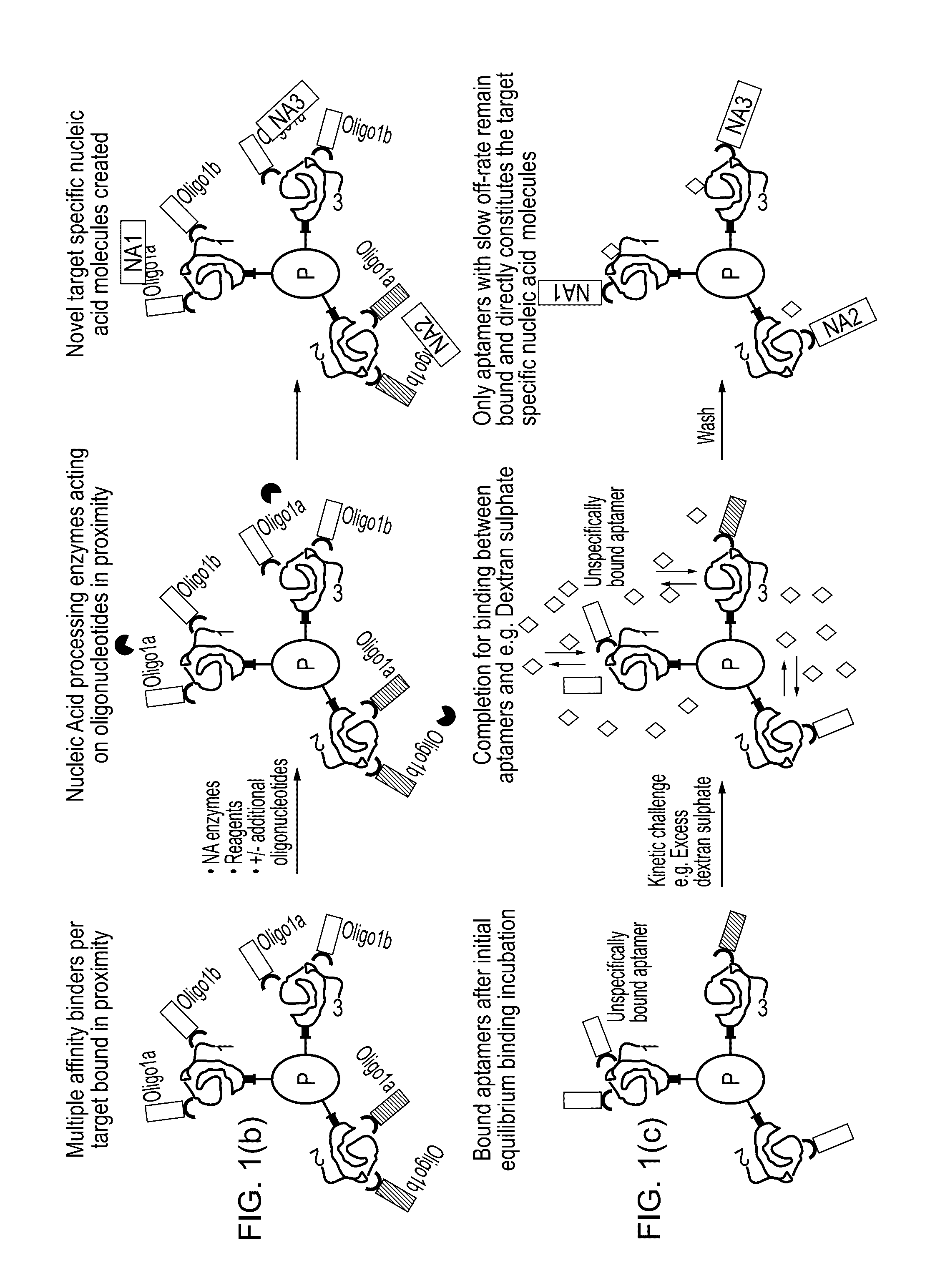
Method for protein analysis
a protein analysis and protein technology, applied in the field of protein analysis, can solve the problems of no example available, best knowledge, prior art, etc., and achieve the effects of high versatility, increased detection sensitivity and ability, and accurate multiplexing results in complex solution based protein samples
- Summary
- Abstract
- Description
- Claims
- Application Information
AI Technical Summary
Benefits of technology
Problems solved by technology
Method used
Image

Examples
example 1
f Complex Protein Samples to NHS Activated Paramagnetic Beads in the Presence of SDS
Experimental
[0048]Total protein from sonicated clarified Vero cells, was used for the investigation. Samples containing 1 mg / ml protein and 0-0.5% SDS in 150 mM triethanolamine, 500 mM sodium chloride, pH 8.3 were prepared. The samples containing SDS were heated at 95° C. for 5 minutes prior to coupling.
Coupling to NHS MagSepharose
[0049]32 μl NHS MagSepharose beads (i.e. 160 μl of 20% slurry, GEHC Bio-Sciences AB) were equilibrated with 1mM HCl and incubated with 32 μl sample with slow end-over-end mixing for 20 min at room temperature. The sample was removed and analyzed by SDS-PAGE [11] (reducing conditions) using Deep Purple Total Protein Stain (fluorescence) and imaging with Ettan DIGE imager (GEHC Bio-Sciences AB).
Results and Discussion
[0050]To investigate how efficiently a complex protein mixture can be coupled to magnetic beads after treatment with SDS at 95° C., total protei...
example 2
ting Beads Assay Implementation Using Paramagnetic NHS Beads and Proximity Extension Assay Reactions
Experimental
[0051]Transferrin (Calbiochem) was used as a target protein and was serial diluted in a Vero cell lysate made by sonication in PBS (Lonza). Final samples were conditioned with coupling buffer [12] prior to coupling to beads and contained transferrin concentrations in the range of 0.001 ng / ml to 10000 ng / ml in 100 μg / ml total cell lysate, either without SDS or including 0.1% SDS (Merck). Samples for full workflow implementation were first incubated with 2% SDS for 5 min at 95° C. (at 20× the above protein concentrations) before being 20-fold diluted in coupling buffer [12].
Blotting Beads Assay
[0052]Polyclonal anti-transferrin antibody (GEHC, internal) was split into two aliquots and conjugated to either probe A or probe B (Olink Bioscience, www.olink.com) according to the manufacturer protocol [13]. NHS MagSepharose beads (10 μl of 20% slurry, GEHC) were e...
example 3
ty of Blotting Beads Assay Implementation Using NHS Based Biotinylation and Binding to Paramagnetic Beads with Streptavidin Ligands
[0056]All protein samples (GFP-His, transferrin, Vero cell lysate) were biotinylated using EZ-Sulfo-NHS-LC-Biotin reagent (Thermo Scientific, art. no. 21217) according to the manufacturer. For tests with GFP-His a recombinant histidine-tagged Green Fluorescent Protein) 21 mg / ml (745 μM) in pH 7.4 was used without or in the presence of 3% SDS (0.5% SDS and 7.4 mg / ml protein in actual biotinylation reaction).
Estimation of Biotinylation Degree
[0057]Residual excess of the biotinylation reagent in the GFP-His samples was removed by purification of the histidine-tagged proteins by IMAC (Immobilized Metal-ion Affinity Chromatography) using His MagSepharose Ni magnetic beads (GEHC, art. no. 28-9673-88). Sample load was about 14.8 mg GFP-His / ml beads. Samples were incubated for 15 minutes at room temperature. The beads were then was...
PUM
| Property | Measurement | Unit |
|---|---|---|
| concentrations | aaaaa | aaaaa |
| pH | aaaaa | aaaaa |
| concentrations | aaaaa | aaaaa |
Abstract
Description
Claims
Application Information