Apparatus and Method for Characterization of Acute Otitis Media

a technology of otitis media and appendix, which is applied in the field of devices for the detection of middle ear effusion, can solve the problems of delayed speech and language skills development, no one can achieve the diagnostic accuracy of invasive myringotomy and tympanocentisis, and the overall likelihood of obtaining an accurate diagnosis using any of the non-invasive methods is no better than 50%, so as to achieve the effect of determining the viscosity

Active Publication Date: 2017-01-19
OTONEXUS MEDICAL TECHNOLIGIES INC
View PDF11 Cites 28 Cited by
  • Summary
  • Abstract
  • Description
  • Claims
  • Application Information

AI Technical Summary

Benefits of technology

[0027]A speculum tip includes an ultrasound transducer for sending and receiving ultrasound energy through an ear canal and a comparatively low frequency tympanic membrane excitation source. The tympanic membrane excitation source generates a subtle movement of the tympanic membrane during an interval coincident with an ultrasound transmitter delivering acoustic wave ultrasound energy to the tympanic membrane either in CW form or in pulsed form. A receiver for ultrasound reflected from the tympanic membrane measures displacement of the tympanic membrane as a phase change in the received signal when compared to the transmit frequency, thereby indicating a temporal displacement of the tympanic membrane. An analysis of the temporal displacement of the tympanic membrane, as measured by the phase shifts of the reflected ultrasound in response to the pneumatic excitation coupled to the tympanic membrane, in combination with comparison to the temporal displacement or from templates or metrics associated with the delay in and amplitude of response between the excitation stimulus to and ultrasound response from the tympanic membrane, is used to determine the viscosity of the fluid behind the tympanic membrane. Measurement of the viscosity of the fluid behind the tympanic membrane is thereafter used to characterize the type of effusion fluid present in the middle ear as one of: no fluid, serous fluid, or purulent fluid.

Problems solved by technology

Left untreated in children, recurrent AOM can also lead to delays in the development of speech and language skills.
Because they are invasive and have significant associated risks of complications, myringotomy and tympanocentisis are not used as standard diagnostic methods for AOM except in research settings.
Several other non-invasive diagnostic tests are available for evaluating AOM, including acoustic reflectometry, tympanometry, pneumatic otoscopy, and otoscopy, however, none of these tests achieves the diagnostic accuracy of invasive myringotomy and tympanocentisis; the overall likelihood of obtaining an accurate diagnosis using any of the non-invasive methods is no better than 50%.
More importantly, the various non-invasive methods are useful only in identifying the presence of middle ear effusion; they provide no information regarding the type of effusion.
Because of the risks associated with undiagnosed AOM, and the recognized unreliability of the non-invasive diagnostic tests, patients who are diagnosed with middle ear effusions based on any of these non-invasive tests are often prescribed antibiotics.
In addition to the increased cost burden of unnecessary antibiotic treatment, the patients are exposed to the side effects of antibiotics and the attendant and significant risk of developing antibiotic resistance.
Thus, current diagnostic and treatment methods are not lowering the rate of AOM in the United States.
In spite of decades of research, optimal management of OM remains controversial.
Given the high spontaneous resolution rate of AOM, there are serious questions about the need for antibiotics in most cases.
Thus, physicians and parents are frequently uncertain about proper treatment because there are no clear-cut clinical findings that might reliably predict which cases will resolve spontaneously and which cases would be better treated with an oral antibiotic.
These children do not benefit from antimicrobial therapy, even though many receive it as a precaution.
Detecting MEE is difficult without expensive equipment, such as a tympanometer or an audiometer.
While screening tympanometers are available, they are not widely used in primary care offices where the majority of cases of AOM / OME are first seen.
Although the sensitivity and specificity of acoustic reflectometry is similar to that of tympanometry, neither device will predict which cases may resolve spontaneously and which cases will require treatment.
OME may cause hearing loss without other symptoms.
Some effusions cause substantial hearing loss.
Typically, middle ears that are impacted with the characteristic viscous effusion (glue ear) are associated with substantial hearing loss that may persist for years.
Primary care physicians, unlike ENT specialists, lack a robust clinical method that can distinguish between a mucoid effusion (glue ear) and one that contains a serous (watery) effusion, which is more likely to resolve spontaneously.
One of the major sources of controversy about OM in clinical practice is accuracy of diagnosis.
Although with practice, many physicians become proficient otoscopists, a monocular examination of the TM of a struggling infant through a tiny speculum remains a difficult and challenging maneuver.
However, this expensive equipment is rarely found in primary care practices where the majority of AOM diagnoses are made.
Tympanometry is not widely used in primary care offices because of equipment expense and training requirements.
However, the pressurization may cause mild discomfort in the presence of an acute infection.
Audiometry often reveals a substantial conductive hearing loss in OME.
However, audiometry is expensive and not widely used in primary care practice.
Audiometry is important in surgical planning but is too nonspecific for evaluation of effusion type.
While acoustic reflectometry is indeed simple, safe, and inexpensive, it is too unreliable for making treatment decisions and is used infrequently by physicians.

Method used


Image

  • Apparatus and Method for Characterization of Acute Otitis Media
  • Apparatus and Method for Characterization of Acute Otitis Media
  • Apparatus and Method for Characterization of Acute Otitis Media

Examples


Embodiment Construction

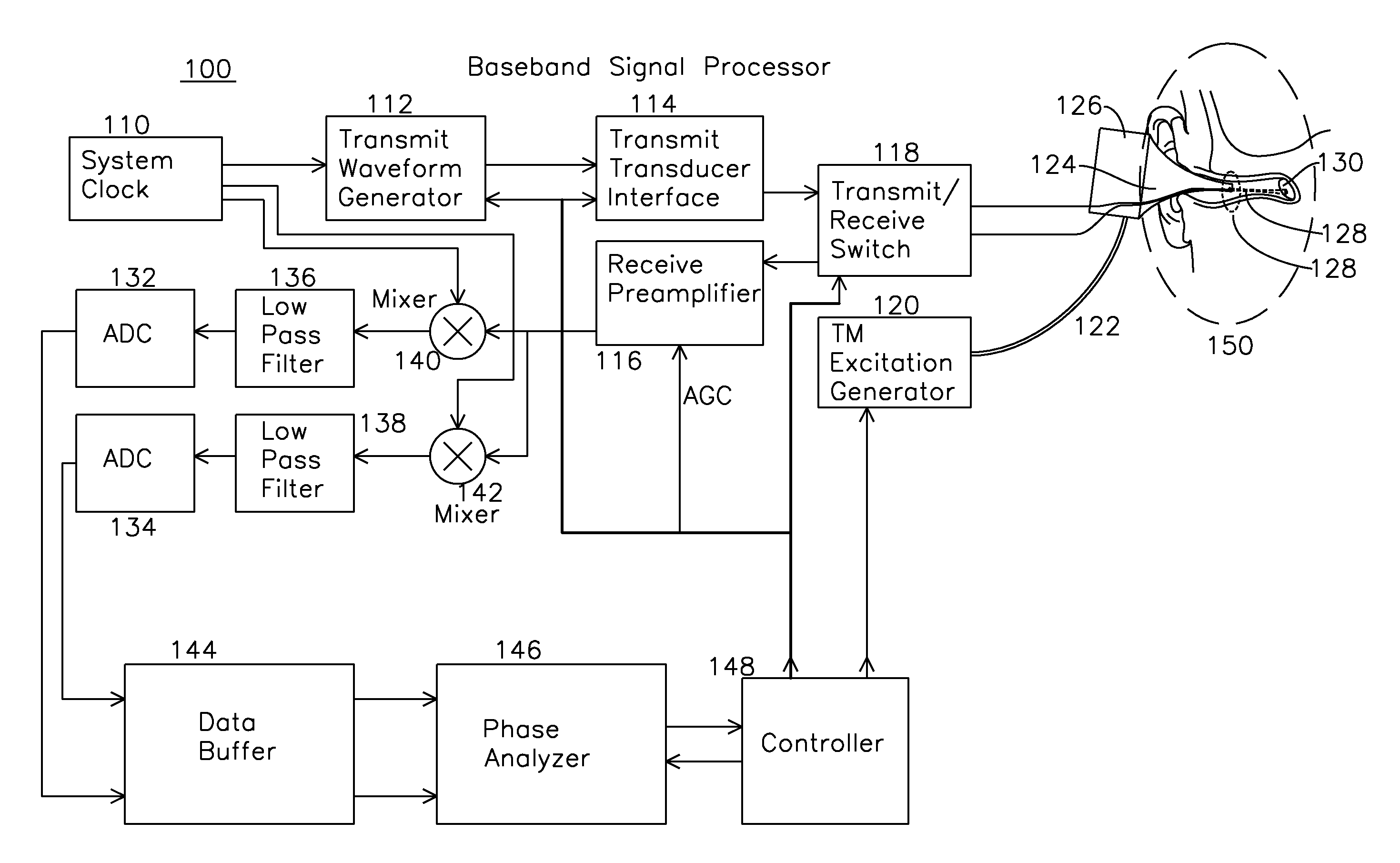
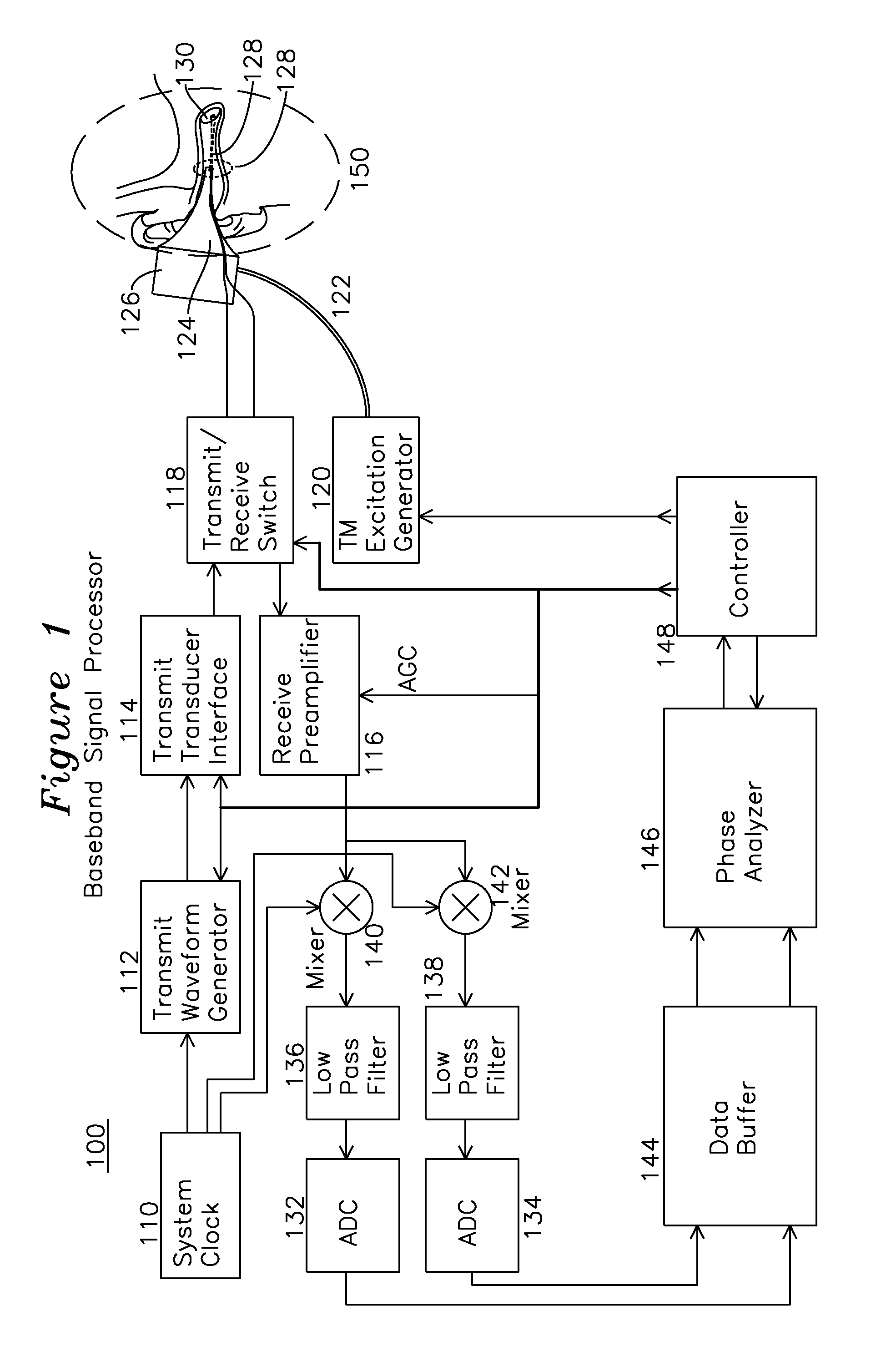
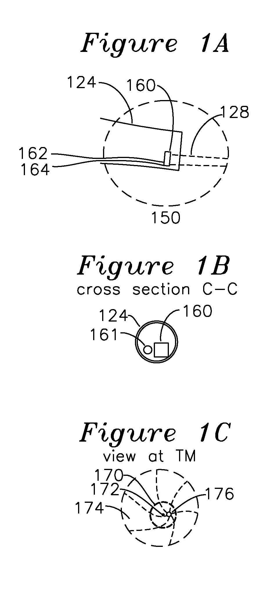
[0044]FIG. 1 shows a signal processor for an example embodiment of a tympanic membrane characterization system. Region 150 (shown in magnified view FIG. 1A) includes a cross section view of a middle ear and tympanic membrane 130 of a subject being examined. The tympanic membrane 130 is interrogated by an ultrasound beam 128 from an ultrasound transducer 160 (shown in FIG. 1A) which is optionally mounted on the inner surface of a speculum tip 124, and is detachable from an otoscope speculum mounting adapter 126. In one embodiment of the invention, an optical source 161 seen in the FIG. 1B cross section view of FIG. 1A, generates a visual indication the region of insonification by the ultrasound by illumination of a target or region of the tympanic membrane within the ear canal, as seen in FIG. 1C. FIG. 1C shows the view of the tympanic membrane as seen through the speculum, including the tympanic membrane 174, “cone of light”176, which is a reflective region of the TM which is normal...


PUM


Abstract

An ultrasound signal processor uses an excitation generator to cause displacement of a tympanic membrane while a series of ultrasound pulses are applied to the tympanic membrane. Phase differences between a transmitted signal and received signal are examined to determine the movement of the tympanic membrane in response to the applied excitation. An examination of the phase response of the tympanic membrane provides a determination as to whether the fluid type behind the tympanic membrane is one of: no fluid, serum fluid, or purulent fluid.

Description

FIELD OF THE INVENTION[0001]The present invention relates to a device for the detection of middle ear effusion with discrimination of fluid type. In particular, the invention relates to the characterization of middle ear effusion behind the tympanic membrane by stimulating the tympanic membrane using a low frequency excitation such as acoustic and measuring the displacement behavior with a comparatively higher frequency excitation such as ultrasound.BACKGROUND OF THE INVENTION[0002]Acute otitis media (AOM) is an inflammatory process in the middle ear and is the most common clinical condition seen by pediatricians in children fifteen years and younger. AOM is generally associated with the presence of a middle ear effusion and is considered a middle ear inflammation. Complications of undiagnosed AOM can include hearing loss. Left untreated in children, recurrent AOM can also lead to delays in the development of speech and language skills.[0003]There are two key factors in the diagnosi...

Claims


Application Information

Patent Timeline
Patent Type & Authority Applications(United States)
IPC IPC(8): A61B5/12A61B1/227A61B1/32A61B1/07A61B1/05A61B8/00A61B1/00A61B1/06A61B8/08A61B8/12
CPCA61B5/12A61B8/08A61B1/227A61B1/32A61B8/12A61B8/5223A61B1/07A61B8/4416A61B8/463A61B1/00105A61B8/4494A61B1/063A61B1/0684A61B1/05A61B1/0661A61B5/126A61B8/085A61B8/4427A61B8/4444A61B8/488
Inventor MOEHRING, MARK A.CHESAVAGE, JAY A.SINGH, RAHULGATES, GEORGEKREINDLER, DANNY
Owner OTONEXUS MEDICAL TECHNOLIGIES INC
Why Eureka
Try Eureka