Automated microscopic cell analysis

a cell analysis and microscopy technology, applied in the field of analytical instruments, can solve the problems of unreliable cell counts with high standard deviations, difficult to accurately predict the cell count of cells, so as to improve sample preparation, eliminate air bubbles, and improve quantitative accuracy

Inactive Publication Date: 2017-11-16
MEDICA CORP
View PDF5 Cites 35 Cited by
  • Summary
  • Abstract
  • Description
  • Claims
  • Application Information

AI Technical Summary

Benefits of technology

[0012]In one embodiment the size of the vessel is of sufficient capacity to provide diluent-stain to dilute several samples with a diluent-stain to sample ratio of 10:1 to about 250:1. The reagent supply module can include a self-priming mechanism for priming the diluent-stain and for eliminating air bubbles. In such an embodiment, the reagent supply module may also include a vacuum chamber and vacuum port which interfaces with, and is connected to, the vacuum ports of the test cartridge when the cartridge is in the cradle to provide a vacuum to the vacuum channel or imaging chamber. The reagent supply module may further include a chamber for collecting waste diluent-stain from the priming process as explained below.
[0013]Systems according to the invention can exhibit better quantitative accuracy than manual microscope analyses, which tend to be limited by variability in sample preparation and limited counting statistics. In the present invention, sample preparation is improved by removing critical operator fluid handling steps and by automation of all dilution steps. A complete set of system controls can be incorporated to ensure that sample dilutions are accurate and repeatable. This can be particularly important in laboratories where metrology standards are not universally followed and skill level is low. Where every...

Problems solved by technology

The hemocytometer with microscope method can be extremely time-consuming and require skilled technicians.
It can also be operator-dependent, and is susceptibl...

Method used


Image

  • Automated microscopic cell analysis
  • Automated microscopic cell analysis
  • Automated microscopic cell analysis

Examples


Embodiment Construction

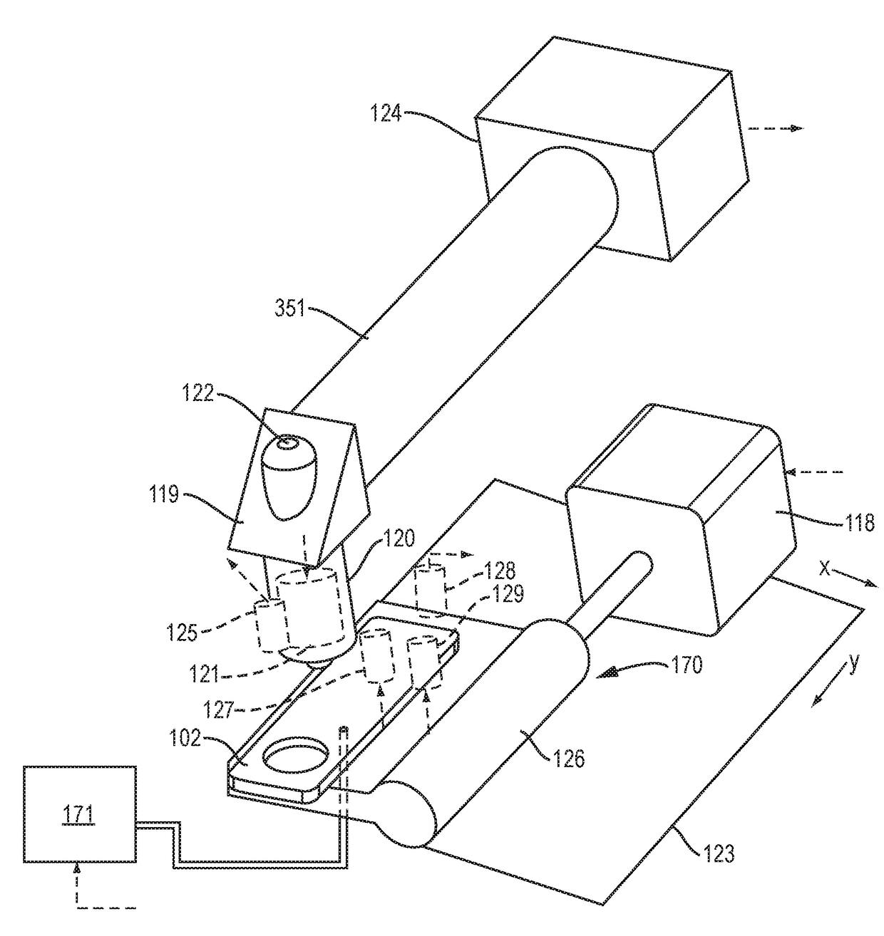
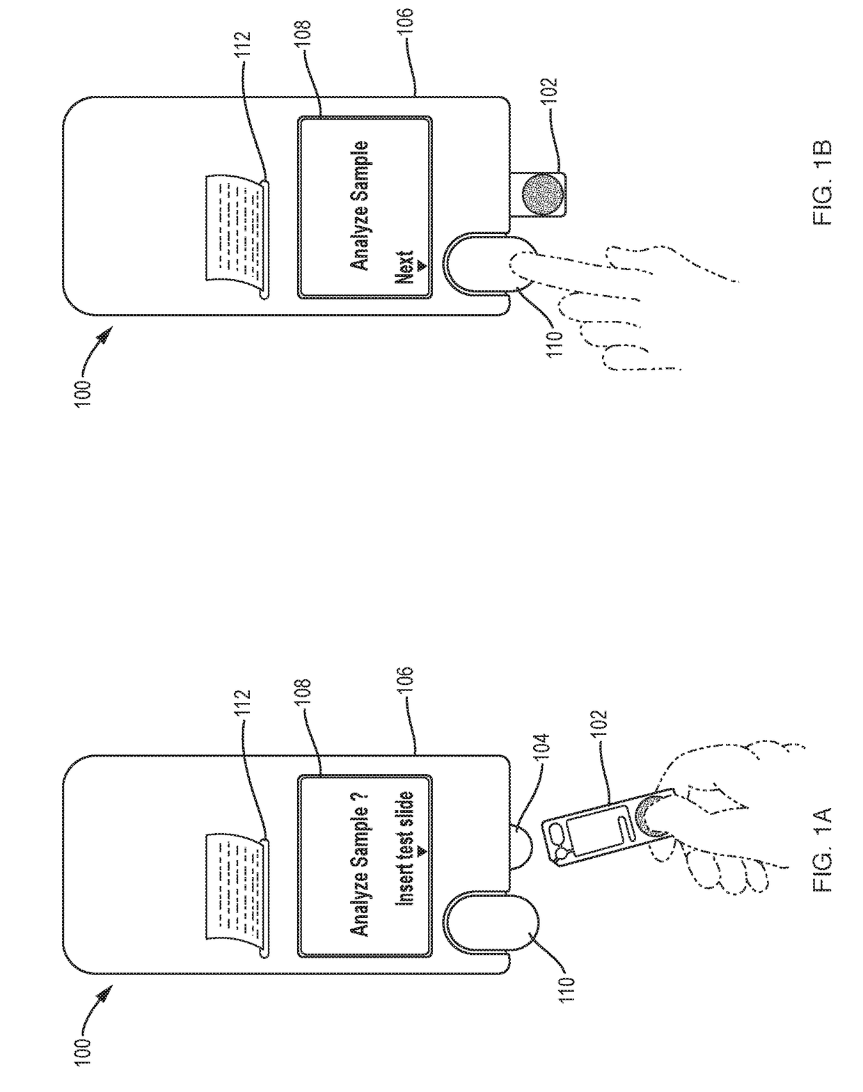
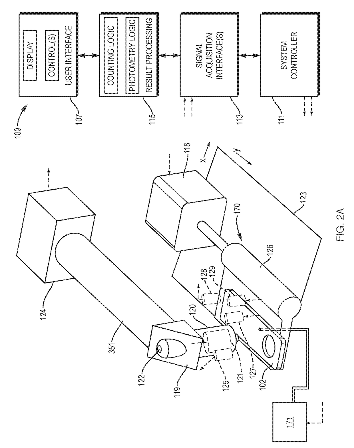
[0046]Referring to FIGS. 1A, 1B, and 2A, an illustrative cell analyzer 100, according to the invention, includes a housing 106 that supports a removable reagent supply module 170 with a cradle 104 into which a test cartridge 102 can be inserted. The housing also supports a “go” button 110, a display screen 108, and a printer 112. Other configurations of the analyzer are of course possible without departing from the invention. The “go” button's function could be triggered by insertion of the test cartridge or be provided through soft prompting on a touchscreen instead of through the use of a discrete button. The printer could be housed separately from the analyzer. And while the cartridges may shaped like microscope slides, a working system could be built around cartridges dimensioned in a variety of other shapes and sizes.

[0047]In operation, a technician or other operator first collects a sample, such as a blood sample from a patient finger stick, heel stick, or by venipuncture in t...


PUM


Abstract

Disclosed in one aspect is a method for performing a complete blood count (CBC) on a sample of whole blood by metering a predetermined amount of the whole blood and mixing it with a predetermined amount of diluent and stain and transferring a portion thereof to an imaging chamber of fixed dimensions and utilizing an automated microscope with digital camera and cell counting and recognition software to count every white blood cell and red blood corpuscle and platelet in the sample diluent/stain mixture to determine the number of red cells, white cells, and platelets per unit volume, and analyzing the white cells with cell recognition software to classify them.

Description

CROSS-REFERENCE TO RELATED APPLICATION[0001]This application claims priority to U.S. provisional applications Nos. 62 / 138,359, filed Mar. 25, 2015, 62 / 084,760, filed Nov. 26, 2014, and 62 / 113,360 filed Feb. 6, 2015, which are all herein incorporated by reference.FIELD OF THE INVENTION[0002]This invention pertains to analyzers and methods for automatically performing microscopic cell analysis tasks, such as counting blood cells in biological samples.BACKGROUND OF THE INVENTION[0003]There are a variety of methods for enumerating particles, such as cells, in biological samples. The most rudimentary method consists of a hemocytometer and a microscope. A hemocytometer is a precision slide device where biological samples are introduced into a chamber by capillary action or by pipette with ruled square markings, such as 1×1 mm squares, and a known depth. These known dimensions are used to compute a unit volume, typically 0.01 uL, so that the concentration of cells can be accurately determi...

Claims


Application Information

Patent Timeline
IPC IPC(8): G01N35/00B01L3/00G02B21/16G01N1/31G01N35/10G02B21/34G01N15/14G01N15/00G01N15/10
CPCG01N35/00029B01L3/502G01N15/1463B01L3/52G01N35/1002G01N35/1016G01N1/312G02B21/34G02B21/16B01L2200/0605B01L2400/0622B01L2400/049B01L2300/0867B01L2300/0654B01L2400/0694B01L2300/123B01L2400/0406G01N2015/0065G01N2015/1486G01N2015/1006B01L2200/16G01N2035/00138G01N2015/0073G01N2015/008G01N1/38G01N35/00069B01L3/502715B01L9/527B01L2200/04B01L2200/0684B01L2200/0689B01L2300/021B01L2300/0883B01L2300/161B01L2400/0644G01N33/487
Inventor JONES, RONALDGROPPER, ADRIANHAGOPIAN, ROBERTROGERS, CHARLESVITELLA, THOMASCOTE, TYLERBARRY, DONALDOSTERLOH, DIRKYI, CHEN
Owner MEDICA CORP
Why Eureka
Try Eureka