
Peptidomimetics for imaging the ghrelin receptor

a ghrelin receptor and peptidomimetics technology, applied in the field of peptidomimetics, can solve the problems of inability to release gh in vivo, difficult and lengthy procedures, and high cost of the therapy form, so as to stimulate gh secretion, suppress ghrelin's orexigenic effect, and increase appetite

Active Publication Date: 2018-06-07
LONDON HEALTH SCI CENT RES
View PDF3 Cites 0 Cited by
  • Summary
  • Abstract
  • Description
  • Claims
  • Application Information

AI Technical Summary

Benefits of technology

The present invention relates to a conjugate that can be used for the treatment of various medical conditions such as GH deficiency, stimulating the release of GH from the pituitary, increasing appetite, treating cancer or chronic obstructive pulmonary disease, suppressing ghrelin's orexigenic effects, treating obesity, regulating food intake, and treating ulcer or gastroparesis. The invention also includes a method for increasing the level of endogenous growth hormone in a patient by administering a pharmaceutical composition containing the conjugate.

Problems solved by technology

[4] This form of therapy, however, is expensive and requires injection or inhalation of the recombinant hormone on a daily basis.
Unfortunately, these shortened ghrelin derivatives were incapable of releasing GH in vivo.
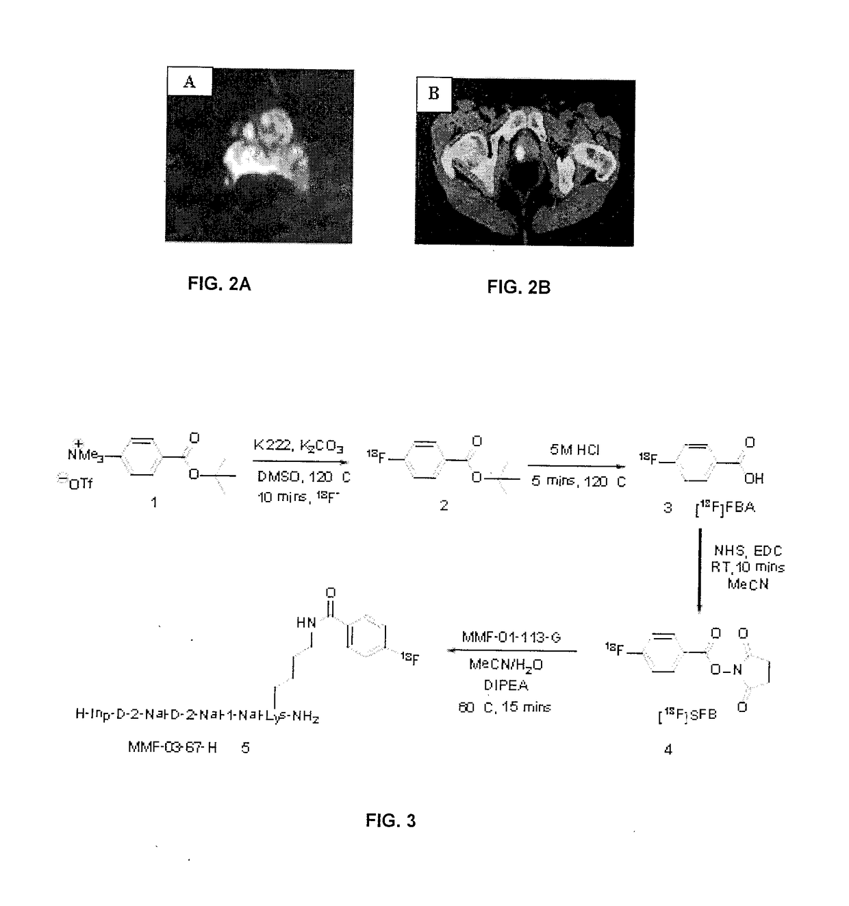
[42] Despite the usefulness of the data gathered by functional MRI, it tends to suffer from difficult and lengthy procedures (MRSI), [42] limited lesion detection in the central gland, [43] the requirement for dedicated computer software for data analysis (DCE-MRI) [42] and the inability to specify cut-off values in apparent diffusion coefficients (ADC) in DW-MRI thus making the delineation between benign and malignant tumours problematic.
In addition to these factors, there are currently no guidelines available that can specify which functional sequence is most appropriate in a particular clinical scenario.
Unfortunately, much like in the case of MRI, a number of limitations exist.
The former imaging modality alone is unable to distinguish between benign and malignant prostatic tissue, [46] thereby restricting its use in detecting primary prostate cancer.
[47, 49] In addition to this, PET has a limited ability to distinguish between benign and malignant disease, despite the use of alternative radiotracers such as [18F]fluorodihydrotestosterone (FDHT) [47], [11C]acetate, [50] [11C]Choline [51] and [11C]methionine.

Method used


Image

  • Peptidomimetics for imaging the ghrelin receptor
  • Peptidomimetics for imaging the ghrelin receptor
  • Peptidomimetics for imaging the ghrelin receptor

Examples


example 1

[0130]Materials and Methods

[0131]All reagents were obtained from commercial suppliers and used without further purification. Reaction monitoring was carried out by TLC using 60F254 silica coated plastic plates (EMD Chemicals Inc.), with visualization under a UV lamp (254 / 365 nm). Melting points were recorded on a Mel-Temp® 1101D digital melting point apparatus. Peptides were either synthesized manually or through the use of a Biotage® Syro Wave™ automated peptide synthesizer. Peptide vessels were shaken using an IKA® Vibrax VXR basic shaker with centrifugation performed on a Beckman Coulter™ Allegra X-30R or Fisher GS-6R centrifuge. In order to aid peptide dissolution, sonication of solutions was accomplished via a Bransonic® 2510R-MTH or Fisher F5-14 ultrasonic cleaner. A Fisher 2052 Isotemp® machine was used to heat up test tubes in the Kaiser Test. Peptides were cryodessicated using a Labconco® FreeZone Freeze Dry System. UV traces were obtained with a Waters 2487 UV / Vis Dual A A...

example 2

[0177]Using the compound G-7039 as a base, the novel conjugates of Table 2 were synthesized.

[0178]The synthesis of the precursor for obtaining TL-MF-3-2FP is shown in FIG. 7, while the synthesis of TL-MF-3-2FP is shown in FIG. 8.

[0179]Competitive binding assays were run on the compounds of Table 2 as described in Example 1. The results of the competitive binding are shown in FIGS. 47-48. The formulas of the compounds of Table 2 are illustrated in FIGS. 49 and 50.

TABLE 2Synthesized CompoundsAminoHRMSHRMSAcidIC50(calculated)(found)LogP (calc.)CompoundSequence(nM)([M + H]+)([M + H]+)LCE no.(ACD / Labs)TL-MF-5-H-Inp-(D-62.9959.48959.60LCE003354.34 + / − 0.902FP2-Nal)2-Ser-Phe-Lys(2-FP)-NH2TL-MF-4-H-Inp-(D-4.35959.48959.60LCE003364.34 + / − 0.902FP2-Nal)2-Phe-Ser-Lys(2-FP)-NH2TL-MF-3-H-Inp-D-2-0.283888.44888.32LCE003374.46 + / − 0.882FPNal-D-2-Nal-Tyr-Lys(2-FP)-NH2TL-MF-2-H-Inp-(D-60.3812.41812.41LCE003382.77 + / − 0.872FP2-Nal)2-Ser-Lys(2-FP)-NH2

example 3

[0180]Calcium Flux Assay

[0181]Another in vitro assay performed on the 4-fluorobenzoyl agonist [1-Nal4,Lys5(4-FB)]G-7039 and the [Lys5(2-FP)]G-7039 agonist was a calcium flux dose-response assay. This experiment allows the determination of the half-maximal effective concentration (EC50) for each agonist, a measure of the concentration of the agonist required to produce 50% of the maximum biological effect resulting from ghrelin receptor activation. In this particular case, the biological effect of the peptidomimetic [1-Nal4,Lys5(4-FB)]G-7039 and the peptidomimetic [Lys5(2-FP)]G-7039 is the release of intracellular calcium ions, a process which is detected by fluorescence using a FLIPRTETRA instrument. Endogenous ghrelin was used as a control in this assay, which was performed by EMD Millipore's GPCRProfiler® service.

[0182]The calcium flux assay was carried out for both compounds [1-Nal4,Lys5(4-FB)]G-7039 (also referred to as 4-FBG-7039 in FIG. 51 and as compound MMF-01-115-H in Table...


PUM

PropertyMeasurementUnit
Molar densityaaaaaaaaaa

Abstract

The present invention concerns compositions comprising and methods of identification and use of imaging agents. The imaging agents comprise a growth hormone secretagogues having a conjugated fluoride. The imaging agents of the present invention may be used for detection, diagnosis and / or staging of prostate or other forms of cancer, and may also be used for cardiac disease.

Description

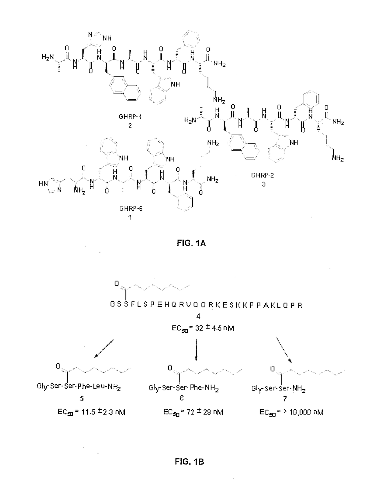
FIELD OF THE INVENTION[0001]The present invention relates to peptidomimetics designed based upon growth hormone secretagogues (GHS) and their uses in imaging the ghrelin receptor, cancer cells and cardiomyocytes.BACKGROUND OF THE INVENTION[0002]In 1977, Bowers and co-workers studied enkephalin (EK) analogues in order to gain a better understanding of small peptide structure-activity relationships (SARs) and pituitary hormone secretion. [1] During the course of their research they found that certain enkephalin derivatives (such as DTrp2-MetEKNH2 for example) released growth hormone (GH) in vitro from the pituitary glands of female rats. [1] This eventually led them to discover the hexamer H-His-D-Trp-Ala-Trp-D-Phe-Lys-NH2 in 1984, which represented the first synthetic peptide that released GH in vitro in rats and in vivo in a number of animals including lambs, rats, monkeys [2] and humans. [3] This compound was later termed growth hormone releasing peptide 6 (GHRP-6, compound 1 of FI...

Claims


Application Information

Patent Timeline
IPC IPC(8): C07K7/06C07K19/00
CPCC07K7/06C07K19/00A61K38/00A61K51/08C07K14/60C07K5/00C07K5/1027G01N33/57492G01N33/74G01N2333/71C07K5/1016
Inventor LUYT, LEONARD G.FOWKES, MILAN MRAZEK
Owner LONDON HEALTH SCI CENT RES
Learn More