Light-activated, calcium-gated polypeptide and methods of use thereof
a polypeptide and light-activated technology, applied in the field of light-activated, calcium-gated polypeptides, can solve the problems of limited use of tools, limited real-time imaging required for the use of calcium indicators, and limited field of view
- Summary
- Abstract
- Description
- Claims
- Application Information
AI Technical Summary
Benefits of technology
Problems solved by technology
Method used
Image
Examples
example 1
tems and Methods of Using the Systems
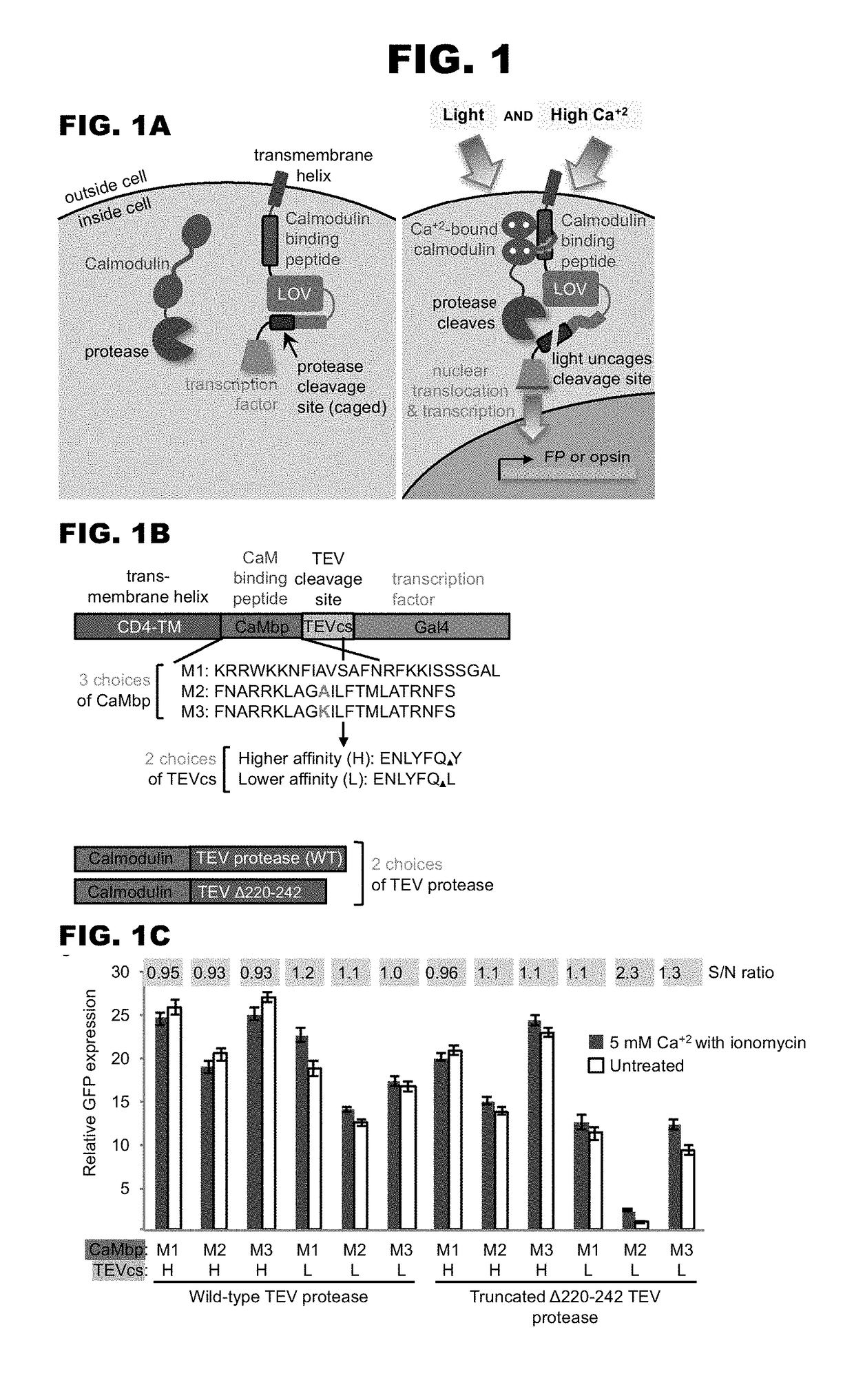
[0632]A light and calcium gated transcription factor (TF) system was designed. A schematic depiction of an example of such a system shown in FIG. 1A. In the basal state, the TF is tethered to the cell's plasma membrane, unable to activate transcription of the reporter gene located in the cell's nucleus. Upon exposure to both blue light and high calcium, however, the TF is cleaved from the membrane and translocates to the nucleus because (1) the protease recognition site is unblocked by the light-sensitive LOV domain, and (2) the protease is recruited to its recognition site via a calcium-regulated intermolecular interaction between calmodulin (CaM) and a CaM binding peptide. Importantly, high calcium alone is not sufficient to give TF release because the protease site remains blocked, and light alone is not sufficient because the protease is far away, and its affinity for its recognition site is too low to afford cleavage in the absence of induce...
example 2
ivity in Neurons
[0713]Having characterized the properties of FLARE in neuron culture, it was tested whether FLARE could be used not only to mark neurons active during defined time windows, but to manipulate them (FIG. 13A). Thus, instead of driving mCherry expression, FLARE was used to drive expression of a light-activated ion channel, Chrimson-mCherry (Chrimson from Chlamydomonas noctigama is a red light-activated channelrhodopsin (Klapoetke et al. (2014) Nat. Methods 11, 338-46)). With only a 15-minute blue light plus field stimulation time window, would opsin expression levels be sufficient to enable functional reactivation of FLARE-marked neurons? FIG. 13B shows imaging of these neurons 18 hours after blue light exposure. Opsin-mCherry expression can be seen in stimulated neurons (top row) but not in untreated neurons (bottom row). Recording of GCaMP5 fluorescence in response to pulses of opsin-activating red light shows that FLARE-marked cells can indeed be re-activated to give...
example 3
[0715]A second FLARE tool was modified and designed for use with other calcium induced protein interactions. In the basal state, the TF is tethered to the cell's plasma membrane, unable to activate transcription of the reporter gene located in the cell's nucleus. Upon exposure to both light and high calcium, however, the TF is cleaved from the membrane and translocates to the nucleus because (1) the protease recognition site is unblocked by the light-sensitive eLOV domain, and (2) the protease is recruited to its recognition site via a calcium-regulated intermolecular interaction between troponin C (TnC) and a TnC binding peptide (e.g., TnI(95-139)). Importantly, high calcium alone is not sufficient to give TF release because the protease site remains blocked, and light alone is not sufficient because the protease is far away, and its affinity for its recognition site is too low to afford cleavage in the absence of induced proximity. Also key to this design is that both calcium sens...
PUM
| Property | Measurement | Unit |
|---|---|---|
| Fraction | aaaaa | aaaaa |
| Molar density | aaaaa | aaaaa |
| Fraction | aaaaa | aaaaa |
Abstract
Description
Claims
Application Information
 - R&D
- Intellectual Property
- Life Sciences
- Materials
- Tech Scout
- Unparalleled Data Quality
- Higher Quality Content
- 60% Fewer Hallucinations
Browse by: Latest US Patents, China's latest patents, Technical Efficacy Thesaurus, Application Domain, Technology Topic, Popular Technical Reports.
© 2025 PatSnap. All rights reserved.Legal|Privacy policy|Modern Slavery Act Transparency Statement|Sitemap|About US| Contact US: help@patsnap.com


