Three-dimensional differentiation of epiblast spheroids to kidney organoids models stage-specific epithelial physiology, morphogenesis, and disease

a three-dimensional differentiation and kidney technology, applied in the field of three-dimensional differentiation of epiblast spheroids to kidney organoids models stage-specific epithelial physiology, morphogenesis, and disease, can solve the problems of difficult control comparison of different epithelia of the same genetic background, lineage restriction of conventional epithelial cell lines, and lack of genetic diversity

Active Publication Date: 2018-08-30
THE BRIGHAM & WOMEN S HOSPITAL INC
View PDF2 Cites 2 Cited by
  • Summary
  • Abstract
  • Description
  • Claims
  • Application Information

AI Technical Summary

Benefits of technology

[0009]Also described herein is a method of screening a compound for an effect on tubular organoids, including providing a quantity of tubular organoids, adding one or more compounds to the tubular organoids, determining changes to phenotype or activity of the tubular organoids, and correlating the changes with an effect of the compounds on tubular organoids, thereby screening the one or more compounds for an effect on tubular organoids.
[0010]In various embodiments, determining changes to phenotype or activity includes detecting one or more markers in the tubular organoids. In various embodiments, the one or more markers comprise kidney injury molecule (KIM-1). In various embodiments, an increase in KIM-1 expression correlates with a toxic effect of the compound. In various embodiments, the tubular organoids are kidney organoids.

Problems solved by technology

Conventional epithelial cell lines, however, are lineage-restricted and lack genetic diversity.
As a result, the 3D structures that arise are relatively simple, and it has been challenging to perform controlled comparisons of different epithelia of the same genetic background, or the same epithelia with different genetic backgrounds.
However, because those experiments were performed with mESCs cultured under conditions that do not sustain pluripotency, it remains unclear whether the observed rosettes represent ICM, epiblast, or differentiating subtypes, which can also form spheroids.

Method used


Image

  • Three-dimensional differentiation of epiblast spheroids to kidney organoids models stage-specific epithelial physiology, morphogenesis, and disease
  • Three-dimensional differentiation of epiblast spheroids to kidney organoids models stage-specific epithelial physiology, morphogenesis, and disease
  • Three-dimensional differentiation of epiblast spheroids to kidney organoids models stage-specific epithelial physiology, morphogenesis, and disease

Examples


example 1

3D Culture

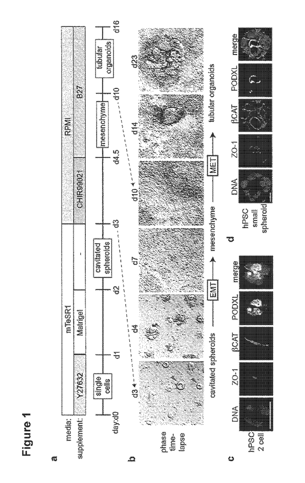
[0065]Cell lines included H9 (WA09), BJ, HDFn, hLR5, hfib2-iPS4, and hfib2-iPS5 (human) and J1, R1, and v6 (mouse). Cells were maintained feeder-free on 3% Reduced Growth Factor GeiTrex (Life Technologies) for at least one passage in media (mTeSR1 for hPSCs; N2 / 827 supplement+2i in for mESCs; hESC conditioned media (CM)+leukemia inhibitory factor (LIF)+dox for hLR5 iPSCs) and dissociated with Accutase or TrypLE. LD-iPSCs were derived from native hLR5 iPSCs by withdrawing LIF and doxycycline and substituting with FGF2. For thin gel sandwich colonies, cells were plated at 60,000 (primed) or 30,000 (native) cells / well of a 24-well plate or 4-well chamber slide pre-coated with GeiTrex in media supplemented with 10 μM Rho-kinase inhibitor Y27632 (StemGent). The following day the media was replaced with 500 μL 1.5% GeiTrex in mTeSR1. Media was changed after 24 hours. For thick gel cultures, 20,000 (epiblast-stage) or 6,000 (narve) cells / well of a 96-well plate were resuspended i...

example 2

Tubular Organoid Differentiation

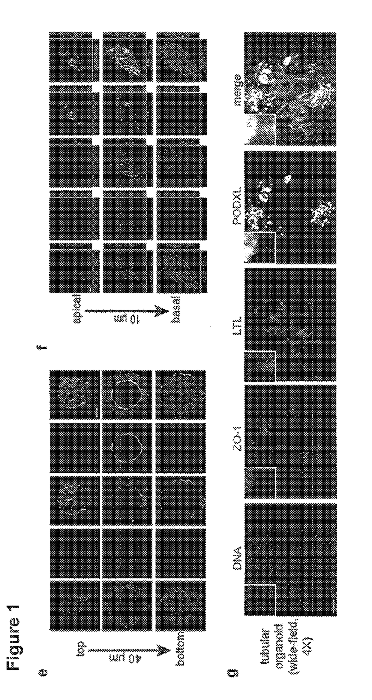
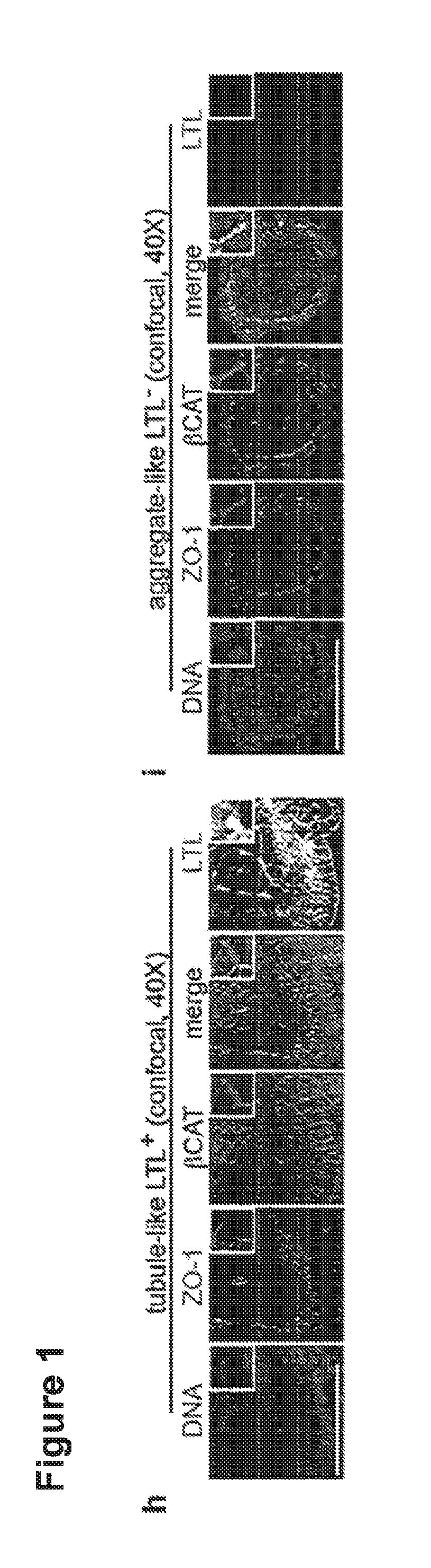
[0066]60,000-120,000 H9 hPSCs were plated, sufficient to produce scattered, isolated spheroid colonies. 48 hours after sandwiching, hPSC spheroids were treated with 12 μM CHIR for 36 hours, then changed to RB (Advanced RPMI+Glutamax+B27 Supplement) and replaced every three days thereafter. Alternatively, spheroids were treated with 12 μM CHIR in RB minus insulin (RBNI) for 24 hours, RBNI for 48 hours, 5 μM IWP2 for 48 hours, RBNI for 48 hours, and RB every three days thereafter, as described for 2D cardiomyocyte differentiation. For 2D kidney differentiation, cells were plated overnight and then treated with 8 μM CHIR in APEL media (StemCell Technologies) for 48-72 hours, 30 ng / ml FGF2+1 μg / ml heparin in APEL media for 96 hours, and subsequently cultured in APEL media for 10-15 days. For stochastic differentiation, hPSCs in 2D or 3D cultures were treated with 10% fetal bovine serum (FBS) in DMEM+P / S and observed for 19 days. Immunofluorescence and Ele...

example 3

[0068]Permeability Assays

[0069]To test permeability, media was supplemented with 20 mM HEPES plus Lucifer Yellow carbohydrazide potassium salt (Invitrogen, 38 μM) and Rhodamine-8 isothiocyanate dextran (Sigma, 0.5 μM), and imaged by confocal microscopy. For microinjection, 5 μM rhodamine-conjugated dextran solution in mTeSR1 was diluted 1:1 with Phenol Red Solution (0.5%, Sigma) for visualization. 2 nl was microinjected via a pulled glass capillary microneedle on a Nanoject-2 micromanipulator, and monitored in real-time by wide-field epifluorescence. For TEER, 50,000 hPSCs were plated on 24-well transwell plates (Corning) pre-coated with dilute Matrigel. The media was gently exchanged for 10 days until cells were completely confluent. TEER was measured using an EVOM 2 device (World Precision Instruments).


PUM

PropertyMeasurementUnit
timeaaaaaaaaaa
Timeaaaaaaaaaa
Brightfield timeaaaaaaaaaa

Abstract

Human pluripotent stem cells (hPSCs) have dual value as microphysiological laboratory models and regenerative therapeutics. hPSCs are epithelial cells, but the extent to which hPSCs and descendant epithelia can reconstitute lineage-specific functions remains poorly understood. Here the Inventors show that hPSCs in three-dimensional cultures and their differentiated descendants can functionally recapitulate tissue-specific epithelial morphogenesis, physiology, and disease.

Description

FIELD OF THE INVENTION[0001]Human stem cells can be cultured in three-dimensional cultures recapitulate tissue-specific epithelial morphogenesis, physiology, and disease.BACKGROUND[0002]Both undifferentiated stem cells and terminally differentiated somatic cells form epithelia. These can function to establish axes for differentiation in the embryo, or to perform barrier and transport roles in adult organs such as the kidney. 3D cell culture in vitro is a powerful tool for investigating epithelial morphogenesis, physiology, and disease, being readily accessible to microscopic inspection, chemical treatment, and experimental manipulation. Studies of epithelial cell lines such as Madin-Darby Canine Kidney (MOCK) cells have, for instance, revealed polarity and apoptosis pathways contributing mechanistically to lumen formation. Conventional epithelial cell lines, however, are lineage-restricted and lack genetic diversity. As a result, the 3D structures that arise are relatively simple, a...

Claims


Application Information

Patent Timeline
Patent Type & Authority Applications(United States)
IPC IPC(8): C12N5/071G01N33/50
CPCC12N5/0686G01N33/5088C12N2513/00C12N2506/03C12N2501/999C12N2500/90A61K35/22A61K35/44C12N5/0684C12N2501/727C12N2506/02C12N2506/45
Inventor FREEDMAN, BENJAMIN S.BONVENTRE, JOSEPH V.
Owner THE BRIGHAM & WOMEN S HOSPITAL INC
Why Eureka
Try Eureka