Apparatus And Method For Non-Invasive Determination Of Intracranial Pressure

a non-invasive measurement and intracranial pressure technology, applied in applications, diagnostic recording/measuring, ultrasonic/sonic/infrasonic image/data processing, etc., can solve the problem of insufficient signal quality of doppler flow signals to allow determination of icp, and the signal-to-noise ratio (snr) of acquired signals is too low, so as to improve signal fidelity and quality. the effect of quality and improved signal quality

Inactive Publication Date: 2018-10-18
BOSTON NEUROSCI INC
View PDF0 Cites 0 Cited by
  • Summary
  • Abstract
  • Description
  • Claims
  • Application Information

AI Technical Summary

Benefits of technology

[0022]There are many advantages to the present invention. The present invention results in higher quality signals, in that the detected wave reflections exhibit inherently higher SNR. Improved signal fidelity allows for the detection of lower amplitude waveform features. For example, flow waveform features during diastole, where the flow velocities diminish relative to systole, and where there may be a small amount of reverse flow, are easily buried in noise. Signals with high SNR are, however, much higher above the “noise floor”.
[0023]Further, the quality of Doppler signals depends on the location of the Doppler range gate in the vessel. It is dependent on the position of the range gate within the lumen, and also the angle between the insonation line and the flow direction. Higher SNR signals can better tolerate suboptimal range gate location and insonation line selection.
[0024]Higher signal quality thus translates, practically, to higher precision of the determined ICP, higher accuracy, higher repeatability, and fewer measurement datasets that must be discarded owing to poor signal quality. Measurements can also be performed faster, since fewer pulses are needed for analysis.
[0025]Modulation of the basic shape by the OA depends on the local elastic properties of the OA. When external pressure is applied to the EOA, this changes the local elastic properties, making the vessel wall more elastically compliant. The invention recognizes this is the primary effect of the variable applied pressure used to determine ICP. Changes in flow velocities are secondary to this change in the local mechanical properties of the wall. The OA possesses no mechanism to influence the Doppler flow velocities in its lumen except via changes in the effective material properties of its wall. In general, it is preferred to measure a primary affected property rather than a secondary, derived, property.
[0026]Also, the invention recognizes the motion of the arterial wall leads to motion of surrounding tissue. This means that the “tissue Doppler” signal may be obtained also from surrounding tissue. This makes it easier to position the transducer and range gates in order to acquire the required signals.
[0027]Another advantage is measurement of a primary quantity rather than a secondary quantity. The basic shape of the Doppler waveform in the OA is determined by the systolic ejection of the heart, as well as subsequent propagation and reflection of pressure waves through the circulatory system.

Problems solved by technology

There are potential deficiencies in the existing methods for measuring intracranial pressure.
In some cases, the quality of the Doppler flow signals obtained is not sufficient to allow determination of ICP.
This is because the signal-to-noise ratio (SNR) of the acquired signals is too low.
Thus, the ultrasound is thus required to traverse 12-16 cm, which can result in a large amount of attenuation.
Second, Doppler flow signals are 45-60 dB lower in power than wall reflection signals, which inherently leads to a low SNR.
Third, the FDA and other regulations limit the amount of power that can be used for ultrasound ophthalmic studies.
This means that the artery is difficult to locate, and the size of the ultrasound gate rate is limited, which limits SNR.
Fifth, when ultrasound traverses fluid-filled regions such as the eye, reflections and reverberations occur.
This ultrasound clutter can obscure the flow velocity signals, especially during the lower flow diastolic phase.

Method used


Image

  • Apparatus And Method For Non-Invasive Determination Of Intracranial Pressure
  • Apparatus And Method For Non-Invasive Determination Of Intracranial Pressure
  • Apparatus And Method For Non-Invasive Determination Of Intracranial Pressure

Examples


example 1

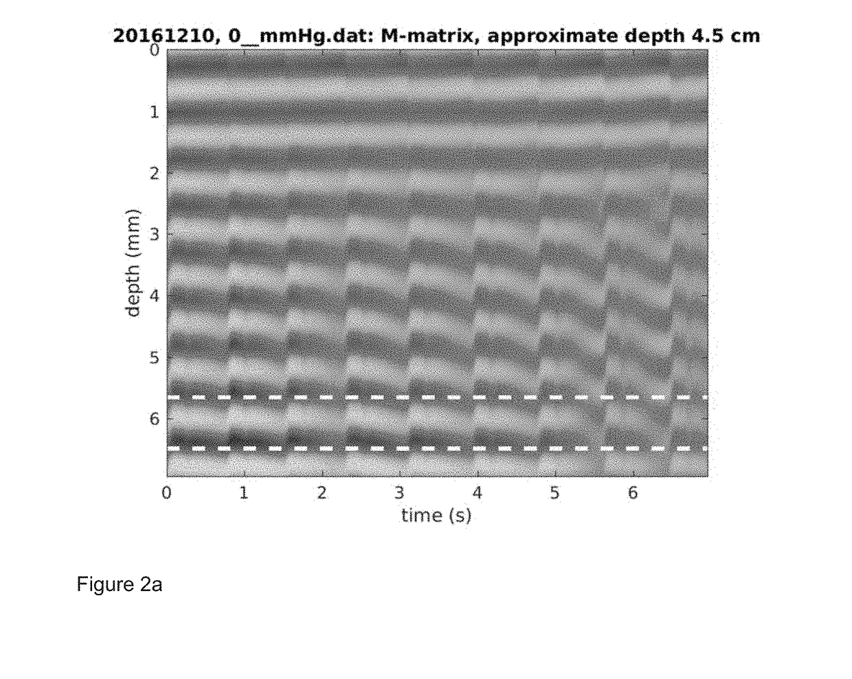
[0057]EOA measurements were taken in a subject. Clinical data were acquired using a 2 MHz single channel ultrasound system from the EOA of a subject. The external pressure was increased from 0 mmHg to 32 mmHg in steps of 4 mmHg. The M-mode matrix (ultrasound reflection amplitude-vs-depth, as a function of time) was processed using a complex correlation model to produce wall distension waveforms as described in P. J. Brands, A. P. G. Hoeks, L. A. F. Ledoux, and R. S. Reneman, “A radio frequency domain complex cross-correlation model to estimate blood flow velocity and tissue motion by means of ultrasound,” Ultrasound in Medicine & Biology, vol. 23, pp. 911-920, 1997 (“Brands”). This matrix contains as columns the raw RF echoes and differs from standard ultrasound M-mode in that the source signals are not demodulated).

[0058]FIGS. 2(a) and (b) show the M-mode matrix and the distension waveform for 0 mmHg of applied external pressure to the orbit of the eye and FIGS. 2(c) and (d) show t...

example 2

[0065]EOA and IOA measurements were taken in a second subject. The data of the Example 1 in FIGS. 2-6, suggests that the ratio of the diastolic to systolic peak amplitudes is a convenient, overall-amplitude-normalized metric that is dependent on applied pressure. The absolute pulse amplitude should be a viable measure in many cases. However, the data from Example 1 illustrates how the measured amplitude can be sensitive to, for example, the angle between the beam and the vessel, the position of the range rate within the tissue, and reverberations [clutter] in the tissue. The invention contemplates a preferred embodiment using a self-normalized metric.

[0066]To gain further data and explore the peak ratio metric, the inventors performed further measurements in the EOA and IOA of a second subject.

[0067]FIG. 7 is a chart showing the ratio of diastolic to systolic peak amplitudes measured in intracranial and extracranial ophthalmic artery of the second subject. This chart shows that the ...

example 3

[0069]Measurements were taken in a third subject using a range gate outside the OA lumen. To illustrate that it is possible to obtain potentially useful wall distension waveforms without precise positioning of the Doppler range gate within the OA lumen, the range gate was deliberately displaced so that no Doppler flow signal could be observed. FIG. 8(a) is an illustration of an M-mode matrix for 0 mmHg. FIG. 8(b) is an illustration of a distension wave form for 0 mmHg generated when using a range gate outside the OA lumen.

[0070]FIGS. 8(a) and 8(b) show that physiologically reasonable distension waveforms can be measured even though the range gate is not situated over, or even immediately adjacent to, the arterial lumen. It seems that motion of tissues nearby the OA, and ultrasound clutter, can make it easier to find useful signals, especially when a non-imaging ultrasound system is used to acquire the waveforms.

[0071]FIG. 9 shows a measurement-based model of the luminal area of the ...


PUM


Abstract

An apparatus and method for noninvasively measuring intracranial pressure of a subject using an ultrasound transducer. The transducer is used to measure arterial wall movement of an intracranial segment and an extracranial segment of the subject's ophthalmic artery as different external pressure forces are applied to the orbital area of the subject. When the waveforms of arterial wall movement between the intracranial segment and the extracranial segment are similar the intracranial pressure can be determined.

Description

CROSS-REFERENCE TO RELATED APPLICATIONS[0001]This patent application claims the benefit under Title 35 United States Code, 119(e), U.S. Provisional Patent Application No. 62 / 479,645, filed Mar. 31, 2017.FIELD OF THE INVENTION[0002]The invention relates noninvasive measurement of intracranial pressure, and more specifically to a system and methods for noninvasive measurement and monitoring of intracranial pressure using ultrasound transducers.BACKGROUND OF THE INVENTION[0003]U.S. Pat. No. 5,951,477 (the '477 patent) teaches how intracranial pressure (ICP) can be measured non-invasively by comparing blood flow velocities in the intra-cranial and extra-cranial segments of the ophthalmic artery (IOA and EOA, respectively). External pressure is exerted on the orbit of the eye using an inflatable air cuff until the flow characteristics in the IOA and EOA are equalized. A major advantage of the method described is that is produces a calibrated measure of ICP.[0004]There are potential defic...

Claims


Application Information

Patent Timeline
Patent Type & Authority Applications(United States)
IPC IPC(8): A61B8/08A61B5/03A61B5/00A61B8/00
CPCA61B8/0808A61B5/031A61B5/0053A61B8/488A61B8/461A61B8/4209A61B5/7246A61B8/5223G16H50/30
Inventor MALTZ, JONATHANRAGAUSKAS, ARMINAS
Owner BOSTON NEUROSCI INC
Why Eureka
Try Eureka