Mobilizing hiv-infected cells from lymphatic reservoirs
a technology of lymphatic reservoir and lymphatic fluid, which is applied in the direction of immunoglobulins, peptides, drug compositions, etc., can solve the problems of complete eradication of hiv from an infected individual, and achieve the effect of promoting movement reducing retention of hiv+ t cells
- Summary
- Abstract
- Description
- Claims
- Application Information
AI Technical Summary
Benefits of technology
Problems solved by technology
Method used
Image
Examples
example 1
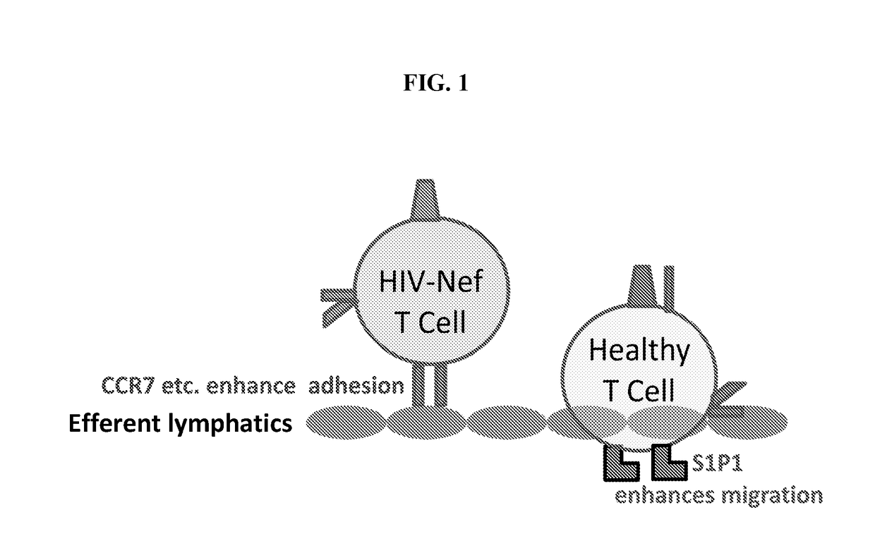
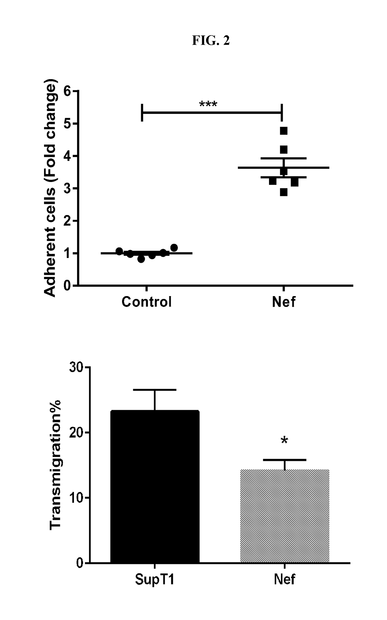
Adhesion to Lymphatic Endothelium is Increased in the Presence of HIV-Nef
[0075]Nef was activated in Nef-ER cells by culturing them with RPMI1640+10% FBS (fetal bovine serum) and 1 μM 4-hydrotamoxifen for at least 6 hours or overnight at 37° C. For inhibitor use, the Nef-ER cells were pre-incubated for at least 12 hours with 10 μM atorvastatin. Other inhibitors may require shorter pre-incubations.
[0076]Approximately 100K HUVECS were seeded per well in a collagen-coated 6-well plate. Cells were incubated overnight in EGM2(LONZA) medium. Nef-ER cells were stained using 5 μM Calcein AM (Ref C3100MP, Fischer Scientific) in 1 mL of staining solution and incubated for 30 minutes at 37° C. The cells were centrifuged at 300 g for 5 min, and the cell pellet was resuspended in 2 mL RPMI+10% FBS. Cells were centrifuged at 300 g for 5 min and the supernatant was removed.
[0077]After staining, 1 μL of 1 mM 4-Hydrotamoxifen per ml of EGM2(LONZA) medium was added for final concentration of 1 μM. Sta...
example 2
Transmigration Assay
[0079]Approximately 20,000 endothelial cells (HUVECs lung microsvascular / lymphatic endothelial cells) were seeded 2 days prior to each experiment in a collagen-coated 24-well transwell plate. Culture medium was changed after overnight incubation, and the cells were allowed to form tight junctions for another 24 hours before each transmigration assay.
[0080]5×10⁵ SupT1 Control cells or SupT1 Nef-ER cells were labeled with 2.5 μM Calcein AM in serum-free RPMI for 30 minutes. The labeled cells were then added to endothelial cells as described above for the adhesion assay.
[0081]For the transmigration assay, EGM2 growth media was aspirated from HUVEC monolayer. HUVEC monolayer cells were washed with PBS, and then 200 μL of SupT1 control / SupT1 Nef-ER cells were added (approximately 100,000 cells / well). 300 μL of phenol red-free DMEM+10% FBS were placed in the bottom well. HUVEC monolayer+SupT1 cells-containing transwell was placed in a well containing DMEM+10% FBS and i...
example 3
Nef-Mediated Downregulation of S1P1
[0083]SUP-T1 cells (a T-cell lymphoblastic lymphoma cell line) stably expressing tamoxifen-inducible Nef-ER and control SupT1 were treated for 16 hours with 1μM tamoxifen. Sample protein lysates (20 μg / lane) were separated by 4-20% gradient Tris / Glycin SDS-PAGE and blotted onto FL-PVDF membranes. S1P1 was detected with anti-human CD363 (S1P1R) e-fluor 660 (E-bioscience). Blot signals were detected and quantified on an Licor Odyssey scanner. Samples from two experiments were analyzed.
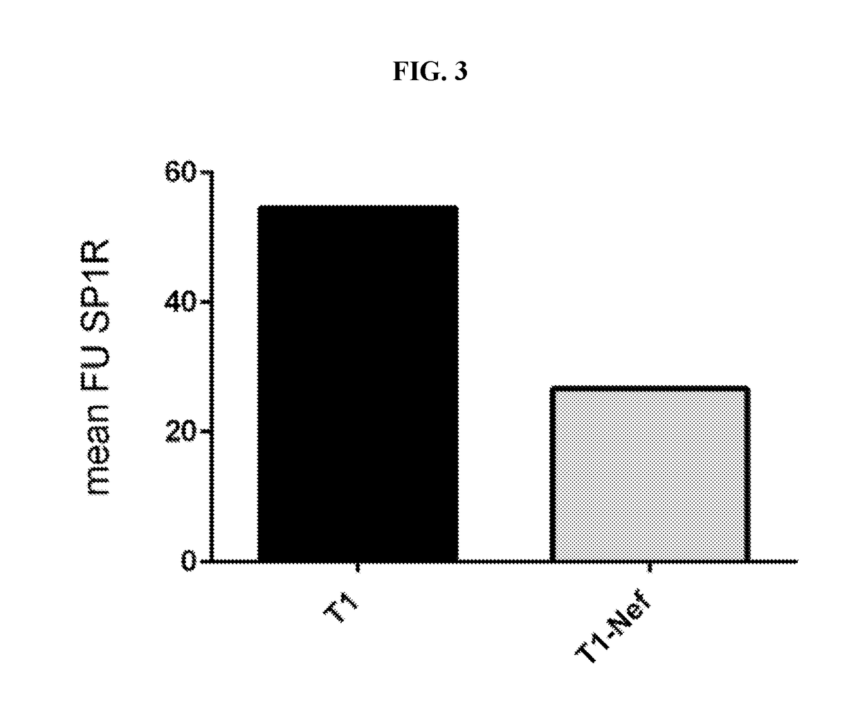
[0084]FACS analysis of S1P1: Nef-ER or control T1 cells were activated with 1 μM 4-hydrotamoxifen in RPMI1640+10% FBS for 16 hours, stained with anti-human S1P1R coupled E-fluor 680 from E-BioScience for one hour and then analyzed in a FACS Calibur. 30,000 cells were counted per sample.
[0085]As shown in FIGS. 3 and 4, the surface receptor S1P1 is down-regulated by HIV-Nef-induced cell signaling as determined using FACS analysis. These data demonstrate that the expressi...
PUM
| Property | Measurement | Unit |
|---|---|---|
| dissociation constant KD | aaaaa | aaaaa |
| dissociation constant KD | aaaaa | aaaaa |
| dissociation constant KD | aaaaa | aaaaa |
Abstract
Description
Claims
Application Information
 - R&D
- Intellectual Property
- Life Sciences
- Materials
- Tech Scout
- Unparalleled Data Quality
- Higher Quality Content
- 60% Fewer Hallucinations
Browse by: Latest US Patents, China's latest patents, Technical Efficacy Thesaurus, Application Domain, Technology Topic, Popular Technical Reports.
© 2025 PatSnap. All rights reserved.Legal|Privacy policy|Modern Slavery Act Transparency Statement|Sitemap|About US| Contact US: help@patsnap.com


